6
the respiratory and circulatory systems
The elegant way that insects obtain oxygen from the air and pump fluids around their bodies has had a profound influence on the direction of their evolution. In some ways this has been a limiting factor, most notably on body size, but it also creates opportunities not available to larger air-breathing animals.
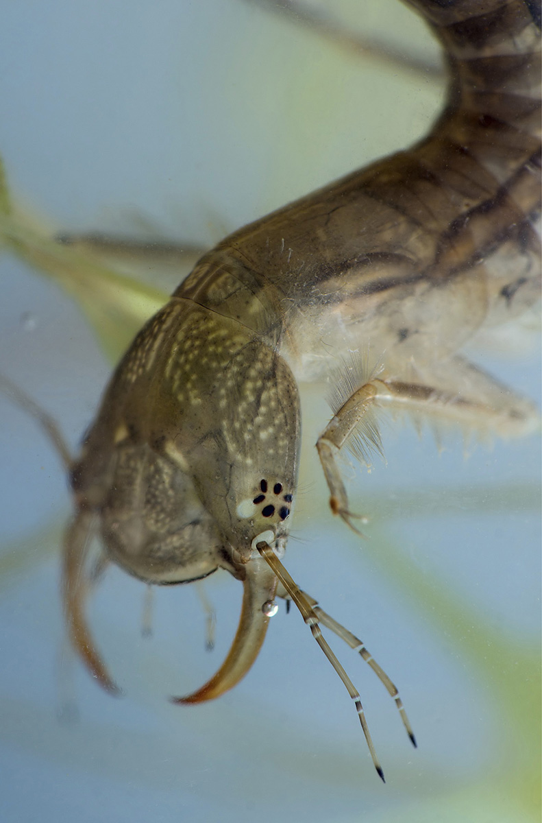
Diving beetles and their fierce, huge-jawed larvae have a range of respiratory and other bodily adaptations to suit their life underwater.
breathing system
Insects have a breathing system enormously different from ours—one that offers various advantages but also imposes certain curious limitations that we do not experience.
Like other animals, insects need to take in oxygen to drive energy-freeing chemical reactions in their cells. They also need to lose the carbon dioxide that is produced by the same process. But while we can only take in and get rid of these gases through our mouths and noses, insects have multiple air intakes over a wide area of their bodies.
In most insects, the middle and last thoracic segments and the first eight or nine abdominal segments each have a pair of spiracles—round openings that sit one on either side of the segment. Spiracles are lined with hairs to trap dust and keep in water vapor to prevent dehydration. They can be fully closed with a valve, keeping the insect from drowning if it falls into water.
The spiracles lead into a system of tubes (tracheae) that branch into smaller passageways, extending through the whole body, delivering air directly to the tissues (rather than via the blood, as in vertebrates). The tracheae are supported and held rigid by rings of chitin, the same material that forms the insect’s cuticle. In some insects, the tracheal system also includes air sacs, to hold extra air.
Air enters the spiracles passively, though larger-bodied insects can help draw air in through contractions of the abdominal muscles. When the muscles relax after contraction, air is pulled in. This movement can be seen if you look closely—the abdomen of a honeybee, for example, will seem to flatten and puff up in a rhythmic motion.

These spiracles are on the body-segments of a beetle.
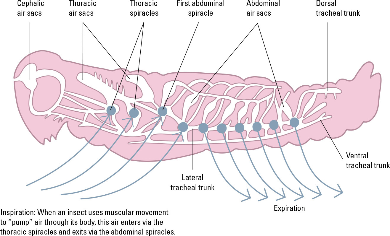
The air system of some insects includes air sacs—spaces where a supply of air can be held, should the spiracles (in blue) need to be closed.
keeping it small
The way insects take on air is one of the reasons that they must have a small body size. Even with their large number of spiracles, and with assistance from the abdominal muscles, they cannot take in air very quickly. A larger body, with a higher volume-to-surface-area ratio, would need much larger spiracles and bigger muscles to pump the body. This would have related effects—less space available for other body parts, more time needed to molt, much thicker tracheae needed to ensure they could remain open just through air pressure without being squashed by the weight of the body, and so on.
On prehistoric Earth in the Carboniferous Period, when the atmosphere was much more oxygen-rich, much larger insects did exist. However, body size is also limited by the insects’ lack of bones—an exoskeleton strong enough to support a much higher body-weight would be impractically thick and render its wearer too slow and awkward to survive.

A Giant Weta, heaviest of modern insects: Insect body size is limited by the amount of oxygen in the atmosphere.
gas exchange
Earth’s atmosphere is 78 percent inert nitrogen, but also contains two gases vital to life—oxygen and carbon dioxide. Animals need oxygen for survival, and plants utilize the carbon dioxide that animals produce.

Air entering the spiracles in the cuticle passes along branching tracheoles, which reach the cells forming internal body tissues, and allow direct gas exchange.
Insects, as we have seen, take in air from the atmosphere via the paired spiracles on each of their abdominal segments. Inside their bodies, the air passes through a narrowing and branching system of tubes—the tracheae. The smallest, terminal branches, the tracheoles, are tiny enough to deliver air directly to the cells.
The ends of tracheoles, where they contact the body cells, are fluid-filled, and gases dissolve into this fluid in both directions, with oxygen passing from the air in the tracheole into the fluid, and carbon dioxide passing from the body cells into the fluid. The relative concentrations of the two gases dictate the direction of movement—each diffuses from an area of high concentration to an area of low concentration. Because oxygen is taken in via the tracheae and consumed by the body cells, its concentration is lower in the cells so it diffuses in that direction. Carbon dioxide is produced by the cells, so it diffuses outward, leaving the body via the network of tracheae. The cells also produce water vapor as they consume oxygen, and some of this will also leave the body in the same way, though the insect can recapture it if needed by closing the spiracles.
the respiration reaction
As with other animals, insects primarily derive their energy from glucose, which comes directly from their diet or from their body’s stores of glycogen molecules (which are, effectively, long chains of glucose molecules). Oxygen is required for the chemical reaction that breaks down glucose molecules to release energy. The reaction takes place inside the cells and is as follows:
1 glucose molecule + 6 oxygen molecules (O2) produces 6 water molecules (H2O) + 6 carbon dioxide molecules (CO2) + energy
The carbon dioxide released when this occurs contributes to atmospheric carbon dioxide, which helps drive the photosynthesis reaction in plants. However, when carbon dioxide is too abundant in the atmosphere, the increased photosynthesis rate means that plants produce a surfeit of glucose. This can weaken their defenses against plant-eating insects, and cause an imbalance in plant and insect populations.

A beefly (family Bombyliidae) feeding from a flower: Hovering flight is extremely energy-demanding.

One of the world’s largest insects, the mantis Rhombodera basalis can grow to 4¾ inches (12cm) long. The practicalities of their passive gas exchange system impose upper limits on insect body size.
circulatory system
In vertebrates, blood vessels carry gases between the lungs and the body tissues. Insects carry out gas exchange quite differently, but still possess a fluid circulatory system to distribute other substances around their bodies.
The other most obvious difference between the insect’s circulatory system and our own is that the insect’s body has an open system. Its equivalent of blood, its hemolymph, is not contained within a system of vessels but flows freely through its whole body cavity (hemocoel). However, there is a pump in the insect’s body to keep the hemolymph moving, just as our bodies have a heart to pump our blood. This structure, the abdominal section of the dorsal vessel, is often described as the insect’s heart, because its function is similar, even though its structure is very different.
The dorsal vessel is a tube that runs the length of the insect’s body, along its back (about where the spine is in a vertebrate animal), but only that part within the abdomen works as a pump. Like many other insect body structures, it is modular—there are repeated expandable sections in each segment. These have one-way, valved openings called ostia, which connect to the hemocoel and allow hemolymph to enter. Muscles around the vessel contract to squeeze the hemolymph upward toward the head, and when they relax the vessel expands and hemolymph enters through the ostia.
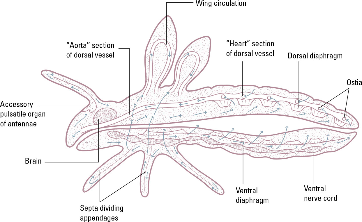
An insect’s circulatory system: The pumping of the dorsal vessel and accessory pulsatile organs pushes hemolymph through the body, and septa and diaphragms in certain parts of the body help guide the flow.
The section of the dorsal vessel in the thorax is sometimes known as the aorta, as (like the vertebrate aorta) it is a large tube that carries blood away from the “heart.” From the aorta, the hemolymph enters the head end of the hemocoel. Some insects also discharge some hemolymph from their hearts into adjacent muscles via additional ostia that let fluid out rather than inward. Some also have additional pumps (accessory pulsatile organs) at narrow points in the hemocoel, to push hemolymph to particular body parts, such as the antennae.
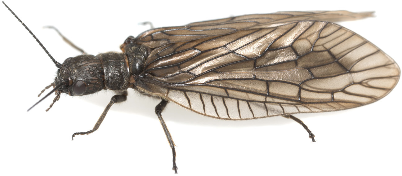
All winged insects have a network of veins supporting the wing membranes, and in some cases (such as the alderflies, family Sialidae) they are very prominent.
the hemocoel
The hemocoel is an extensive space that occupies much of the head, thorax, and particularly the abdomen. Although the body cavity also contains many structures, the space between these structures is continuous, allowing for free flow of hemolymph to all the body’s tissues. In some body parts there are septa—dividing walls—and valves, which control the flow of hemolymph, keeping it moving one way, and hemolymph also flows unidirectionally through the wings by means of their system of veins. The nature of this circulatory system—having an open hemocoel rather than narrow, contained blood vessels—is one of the factors that places an upper size limitation on insect bodies, as the flow of hemolymph would be increasingly impacted by the action of gravity as body size increased.
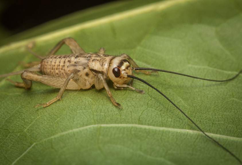
Insects like crickets possess antennal accessory pulsatile organs, which function like miniature hearts to pump the hemolymph into the antennae.
hemolymph
Hemolymph—the yellow or green fluid that leaks from an insect’s body when it is injured—pervades all body tissues and has many vital functions.
Our blood is given its red color by iron-rich, oxygen-carrying red blood cells. Take these cells away and the remaining fluid (plasma) is yellowish, much like insect hemolymph. It also has a similar function—transporting water, nutrients, salts, waste material, and hormones around the body, as well as being involved in the body’s immune response and in temperature regulation. Hemolymph also has an important part to play when a larval insect molts its cuticle.
Some insects can release hemolymph intentionally as a defensive mechanism. They include the blister beetles (family Meloidae), whose hemolymph contains cantharidin, a toxin that causes stinging burns. The gall aphid Pemphigus spyrothecae lives in colonies that include “soldiers” that sacrifice themselves to protect the colony. They release sticky hemolymph that causes their bodies to stick to predators, immobilizing them.
The main constituents of hemolymph, besides water, are salts of chlorine, potassium, and other metals, and organic molecules including amino acids, proteins, glucose, larger carbohydrates, and fats. The hemolymph also carries the waste products of digestion, such as uric acid. In some species, there are antifreezing agents present, to help the insect survive hibernation through subzero temperatures. In a very small number of insects—for example, the stonefly Perla marginata—the hemolymph contains molecules called hemocyanins, which (like hemoglobin in vertebrates’ red blood cells) can bind to and transport oxygen. Hemocyanins are common in some other arthropod groups, but in insects the tracheal breathing system has largely replaced them.
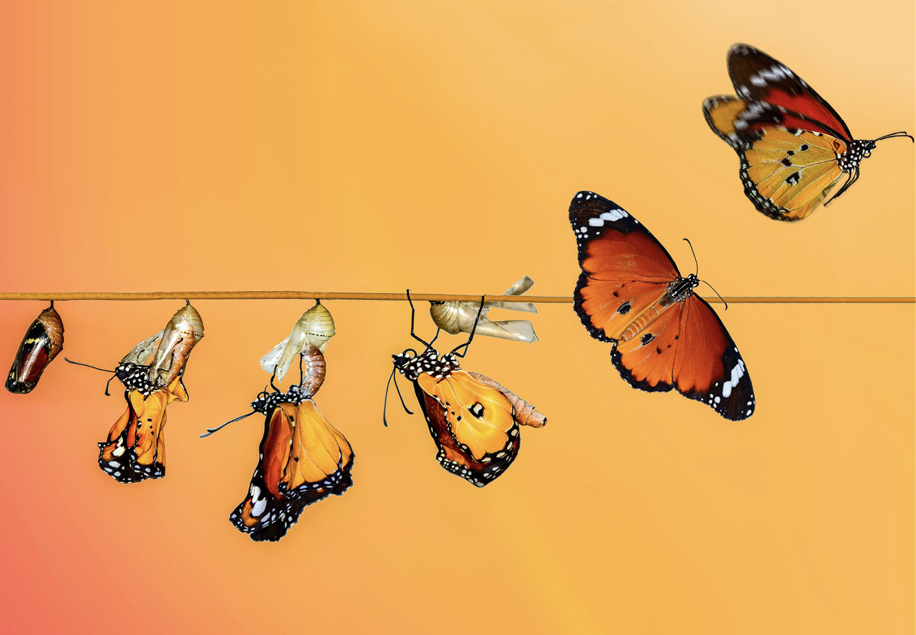
When an insect emerges from its pupa, it needs to pump hemolymph through its wing veins to expand and flatten the wings before it can fly.

The process of phagocytosis: A phagocyte cell engulfs a pathogen and destroys it, and in the process gains increased sensitivity to that type of pathogen.
When a larva is ready to molt, it may retain extra water in its hemolymph to help fracture its cuticle. After molt, before the new cuticle hardens (becomes sclerotized), the insect may pump hemolymph into certain body-sections to allow them to grow bigger. The final molt from larva (or pupa) to adult involves a period of extensive redistribution of hemolymph, to prepare the insect’s body for this new and often very different life stage.
hemocytes
Hemocytes are cells that are carried in the hemolymph. The majority of them are phagocytic, meaning that they can engulf and break down unwanted cells in the hemolymph, such as bacteria and other pathogens, but also dead cells from the insect’s own body. Some larval insects have hemocytes that are adapted to attack and destroy the eggs laid by parasitic wasps. See Chapter 11 for a detailed look at the different types of hemocytes and their function in the insect immune system.
unusual adaptations
The great diversity of insects, in both body type and lifestyle, has naturally given rise to various specializations in terms of how their circulatory and respiratory systems function.
In insect larvae of certain species, especially those that live in aquatic or very humid environments, hemolymph can make up nearly 50 percent of the body-weight. In these soft-bodied larvae, the cuticle is thin to allow the body to be highly pliant. However, this means the exoskeleton is not very strong, so instead the body’s high fluid content provides much more bodily support. The prolegs on a caterpillar, several pairs of which sit behind the six true legs, have very limited musculature and are primarily powered by the flow of hemolymph.
Several insects, including certain moths, dragonflies, and true flies, have a sophisticated hemolymph flow system to help them regulate their body temperature. They have a muscular sheet on the underside of the body (the ventral diaphragm), which can be used to pump hemolymph from the thorax (where much of the body heat is generated, by the action of the flight muscles) into the abdomen to help the body lose heat.
Insects that live at high altitudes with lower oxygen concentration tend to have a smaller body size than related species that live at or near sea level. A smaller body size means that heat retention is more difficult (which is why, in vertebrates, the trend is for cold-climate species to be bigger, rather than smaller), and so relatively few insect species occur in high-altitude habitats. However, experiments on fruit flies show that larvae reared in a low-oxygen atmosphere can compensate developmentally by growing additional tracheal branches.

A caterpillar’s prolegs, on the rear half of its body, are held rigid with the aid of hemolymph flow, enabling them to grip strongly.
breathing underwater
There are several ways that underwater insects obtain their oxygen. Some, such as the “rat-tailed maggot” larvae of certain hoverflies, have a long, narrow tube or siphon at their rear ends, which projects out of the water into the air. Diving beetles may trap air under their elytra. The larvae of mayflies have feathery gill-like structures through which oxygen can diffuse directly out of the water into the body tissues, while dragonfly larvae have internal gills that absorb oxygen sucked into the body via the rectum. Absorbing oxygen from water is only possible if the water is moving, so mayfly larvae keep a constant flow of water over their gills by fanning them constantly.
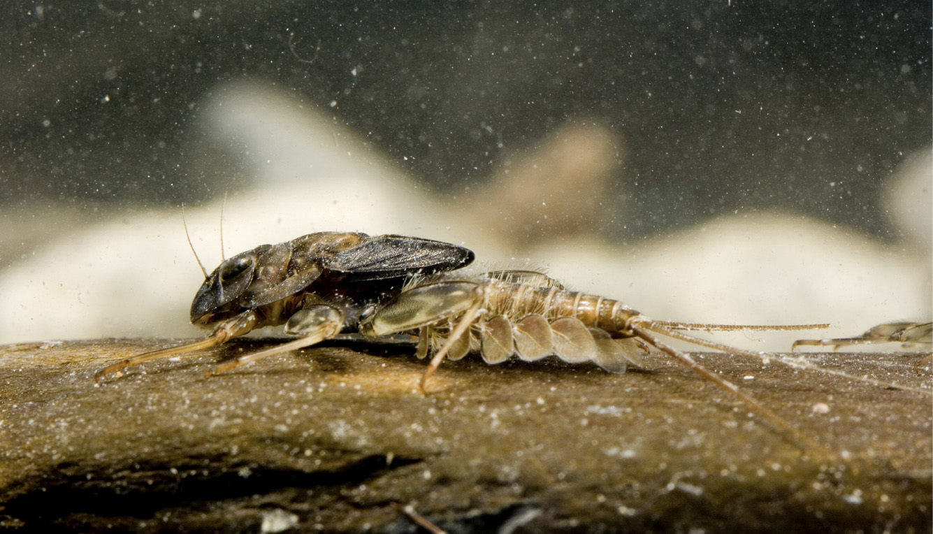
This mayfly larva has prominent gills on its abdominal segments.
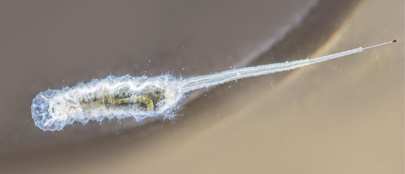
The underwater-dwelling larva or “rat-tailed maggot” of a hoverfly, of the genus Eristalis, shows its long breathing tube.
hormones
Many of the complex processes that take place in an insect’s body as it grows and matures are regulated by the action of hormones, which circulate through the body via the hemolymph.
Hormones are sometimes described as “chemical messengers.” They are molecules that, when they reach their target cells, cause some physiological change to occur. For example, two hormones (prothoracicotropic hormone and ecdysone) work together to trigger the bodily changes that initiate the process of molt in larvae. The presence of juvenile hormone in larvae ensures that each molt is from one larval stage to the next. Juvenile hormone diminishes as the larva matures—when it is no longer present, the next molt is from larva to pupa. In insects that can develop in various different adult forms (for example, worker and soldier “castes” in some species of ants and termites), hormones guide them along their particular developmental pathways.
The various hormones and the body tissues that secrete them are collectively known as the endocrine system. Hormones are produced by specialized structures (glands) or specialized regions in other body parts (especially the brain), and released into the hemolymph, in due course binding to the cell membranes in their target tissues. Some hormones are proteins, others are lipids, and some are terpenes—aromatic molecules made of hydrogen and carbon chains.
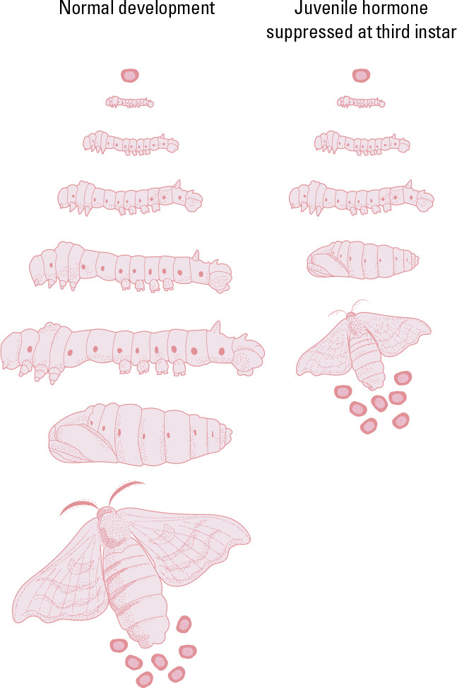
If juvenile hormone (JH) is suppressed in the caterpillar of the Silk Moth (Bombyx mori), at its next molt the caterpillar will pupate rather than continuing to grow, and a miniature adult will result. It is perfectly formed and, if female, will lay normal-sized eggs.

Processes such as secreting pheromones to attract a mate are under hormonal control.
hormonal alterations
Insect development can be disrupted by interfering with hormone production. For example, if a young moth caterpillar has its juvenile hormone-secreting glands removed, it will pupate at its next molt rather than molt to its next larval stage, and from the pupa a miniature adult will emerge. Manipulation of hormones as a means of pest control is a burgeoning field of research. Certain chemicals can mimic the action of natural hormones, and cause insects to die or fail to reach maturity. As a means of pest control, hormones have the advantage of not harming nontarget species (or human workers), but at present their synthesis is costly and their use is difficult (as they deactivate quickly in the environment).
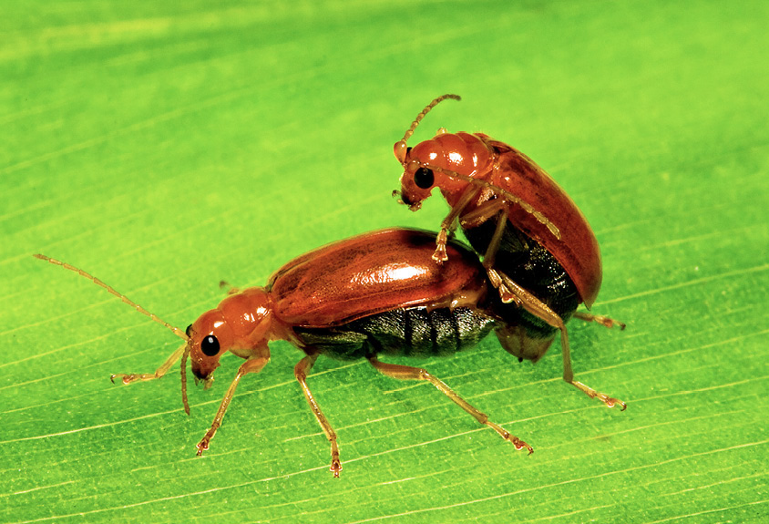
Hormonal activity in adult insects triggers mating behavior.