Link the regions of the forebrain to their functions
Describe the laterality of brain–body communication and the role of hemispheric dominance
Describe the functions of the four lobes of the cerebral cortex:
The forebrain is the most “modern” portion of the brain, and—in humans—forms the largest
portion of the brain by weight and volume. The forebrain contains regions derived
from the diencephalon, such as the thalamus, hypothalamus, posterior pituitary, and
pineal gland; it also includes derivatives of the telencephalon, such as the cerebral
cortex, basal ganglia, and limbic system.
Thalamus
The thalamus is a structure within the forebrain that serves as an important relay station for
incoming sensory information, including all senses except for smell. After receiving
incoming sensory impulses, the thalamus sorts and transmits them to the appropriate
areas of the cerebral cortex. The thalamus is therefore a sensory “way station.”
Hypothalamus
The hypothalamus, subdivided into the lateral hypothalamus, ventromedial hypothalamus, and anterior
hypothalamus, serves homeostatic functions, and is a key player in emotional experiences
during high arousal states, aggressive behavior, and sexual behavior. The hypothalamus
also helps control some endocrine functions, as well as the autonomic nervous system.
The hypothalamus serves many homeostatic functions, which are self-regulatory processes
that maintain a stable balance within the body. Receptors in the hypothalamus regulate
metabolism, temperature, and water balance. When any of these functions are out of
balance, the hypothalamus detects the problem and signals the body to correct the
imbalance; for example, osmoreceptors in the hypothalamus may trigger the release
of antidiuretic hormone to increase water reabsorption as part of fluid balance. The
hypothalamus is also the primary regulator of the autonomic nervous system and is
important in drive behaviors: hunger, thirst, and sexual behavior.
Mnemonic
Functions of the Hypothalamus—The Four Fs:
Feeding
Fighting
Flighting
(Sexual) Functioning
The lateral hypothalamus (LH) is referred to as the hunger center because it has special receptors thought to
detect when the body needs more food or fluids. In other words, the LH triggers eating
and drinking. When this part of the hypothalamus is destroyed in lab rats, they refuse
to eat and drink and would starve to death if not force-fed through tubes.
Mnemonic
When the Lateral Hypothalamus (LH) is destroyed, one Lacks Hunger.
The ventromedial hypothalamus (VMH) is identified as the “satiety center,” and provides signals to stop eating. Brain
lesions to this area usually lead to obesity.
Mnemonic
When the VentroMedial Hypothalamus (VMH) is destroyed, one is Very Much Hungry.
The anterior hypothalamus controls sexual behavior. When the anterior hypothalamus is stimulated, lab animals
will mount just about anything (including inanimate objects). In many species, damage
to the anterior hypothalamus leads to permanent inhibition of sexual activity. The
anterior hypothalamus also regulates sleep and body temperature.
Mnemonic
When the Anterior hypothalamus is destroyed, one is Asexual.
Real World
In the early 1920s, researchers first discovered the hypothalamus’s role in rage and
fighting through classic experiments conducted with cats. When researchers removed
the cat’s cerebral cortex but left the hypothalamus in place, the cat displayed a
pattern of pseudoaggressive behavior that was called “sham rage”—lashing of the tail,
arching of the back, clawing, and biting—except that rage was spontaneous or triggered
by the mildest touch. The researchers concluded that the cortex typically inhibits
this type of response. When the researchers removed the cat’s cortex and hypothalamus
together, the outcome was very different. The cat no longer showed any signs of sham
rage, and much rougher stimulation was required before the cats showed any defensive
behavior at all.
Other Parts of the Diencephalon
The diencephalon also differentiates to form the posterior pituitary gland, pineal
gland, and connecting pathways to other brain regions. The posterior pituitary is comprised of axonal projections from the hypothalamus and is the site of release
for the hypothalamic hormones antidiuretic hormone (ADH, also called vasopressin) and oxytocin. The functions of these hormones are described in Chapter 5 of MCAT Biology Review. The pineal gland is the key player in several biological rhythms. Most notably, the pineal gland secretes
a hormone called melatonin, which regulates circadian rhythms. The pineal gland receives direct signals from
the retina for coordination with sunlight.
Basal Ganglia
In the middle of the brain are a group of structures known as the basal ganglia. The
basal ganglia coordinate muscle movement as they receive information from the cortex and relay
this information (via the extrapyramidal motor system) to the brain and the spinal
cord. The extrapyramidal
system gathers information about body position and carries this information to the central nervous system, but does not function directly through motor neurons. Essentially, the basal ganglia help make our movements smooth and our posture steady. Parkinson’s disease is one chronic illness associated with destruction of portions of the basal ganglia.
It is characterized by jerky movements and uncontrolled resting tremors. The basal
ganglia may also play a role in schizophrenia and obsessive–compulsive disorder.
Limbic System
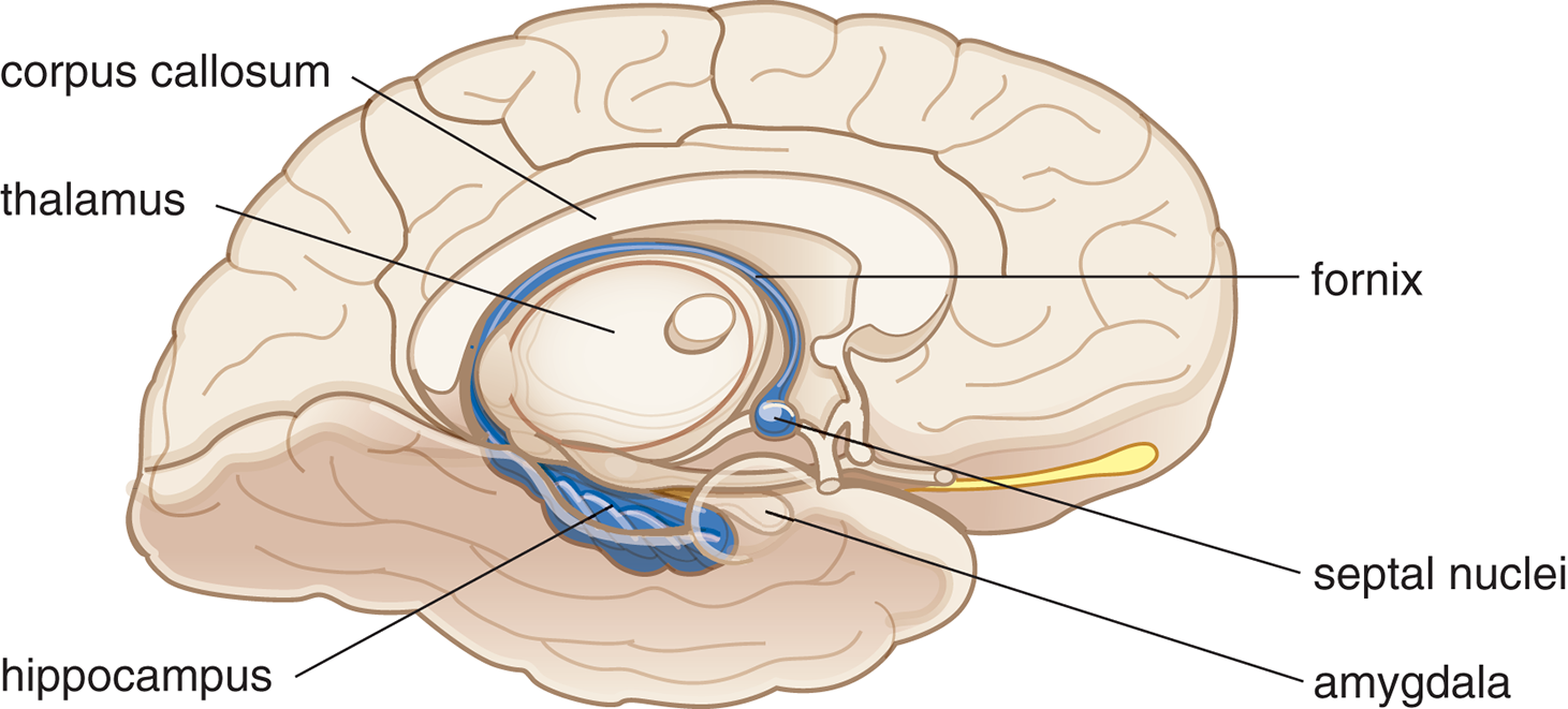
The limbic system, diagrammed in Figure 1.8, comprises a group of interconnected structures looping
around the central portion of the brain and is primarily associated with emotion and
memory. Its primary components include the septal nuclei, amygdala, and hippocampus.
In Chapter 5 of MCAT Behavioral Sciences Review, we will also explore the roles of the thalamus, hypothalamus, and cortex in the
limbic system.
Figure1.8.The Limbic System
Septal Nuclei
The septal nuclei contain one of the primary pleasure centers in the brain. Mild stimulation of the
septal nuclei is reported to be intensely pleasurable; there is an association between
these nuclei and addictive behavior.
Real World
James Olds and Peter Milner discovered the association between the septal nuclei and
addictive behavior in the 1950s. They demonstrated that when rats could stimulate
their septal regions at will by pushing a lever, they found it so pleasurable that
they preferred it to eating or any other activities, even after going 24 hours without
food or sleep.
Amygdala
The amygdala is a structure that plays an important role in defensive and aggressive behaviors,
including fear and rage. Researchers base this observation on studies of animals and
humans with brain lesions. When the amygdala is damaged, aggression and fear reactions
are markedly reduced. Lesions to the amygdala result in docility and hypersexual states.
Real World
Heinrich Klüver and Paul Bucy performed studies that linked the amygdala with defensive
and aggressive behavior in monkeys. When the amygdala of the Rhesus monkey was removed,
they noted increased sexual behavior, decreased fear responses, and hyperorality,
or the examination of inanimate or animate objects by mouth. These symptoms are now
referred to as Klüver–Bucy syndrome.
Hippocampus
The hippocampus plays a vital role in learning and memory processes; specifically, the hippocampus helps consolidate information to form long-term memories, and can redistribute remote memories to the cerebral cortex. The hippocampus communicates with other portions of the limbic system through a long projection called the fornix. Researchers originally discovered the connection between memory and the hippocampus
through a famous patient named Henry Molaison (known as H.M. in the scientific literature
until his death in 2008). Parts of H.M.’s temporal lobes—including the amygdala and
hippocampus—were removed in an effort to control epileptic seizures. After surgery,
H.M.’s intelligence was largely intact but he suffered a drastic and irreversible
loss of memory for any new information. This kind of memory loss is called anterograde amnesia and is characterized by not being able to establish new long-term memories, whereas
memory for events that occurred before brain injury is usually intact. The opposite
kind of memory loss, retrograde amnesia, refers to memory loss of events that transpired before brain injury.
Bridge
Learning and memory are discussed thoroughly in Chapter 3 of MCAT Behavioral Sciences Review.
Mnemonic
Lobes of the brain: F-POT
Frontal
Parietal
Occipital
Temporal
Cerebral Cortex
The outer surface of the brain is called the cerebral cortex. The cortex is sometimes called the neocortex, a reminder that the cortex is the most recent brain region to evolve. Rather than
having a smooth surface, the cortex has numerous bumps and folds called gyri and sulci, respectively. The convoluted structure of the brain provides increased surface area.
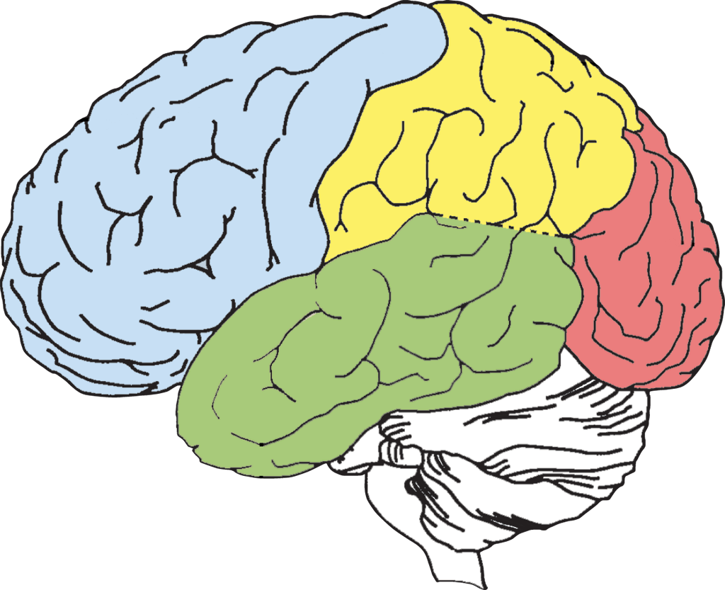
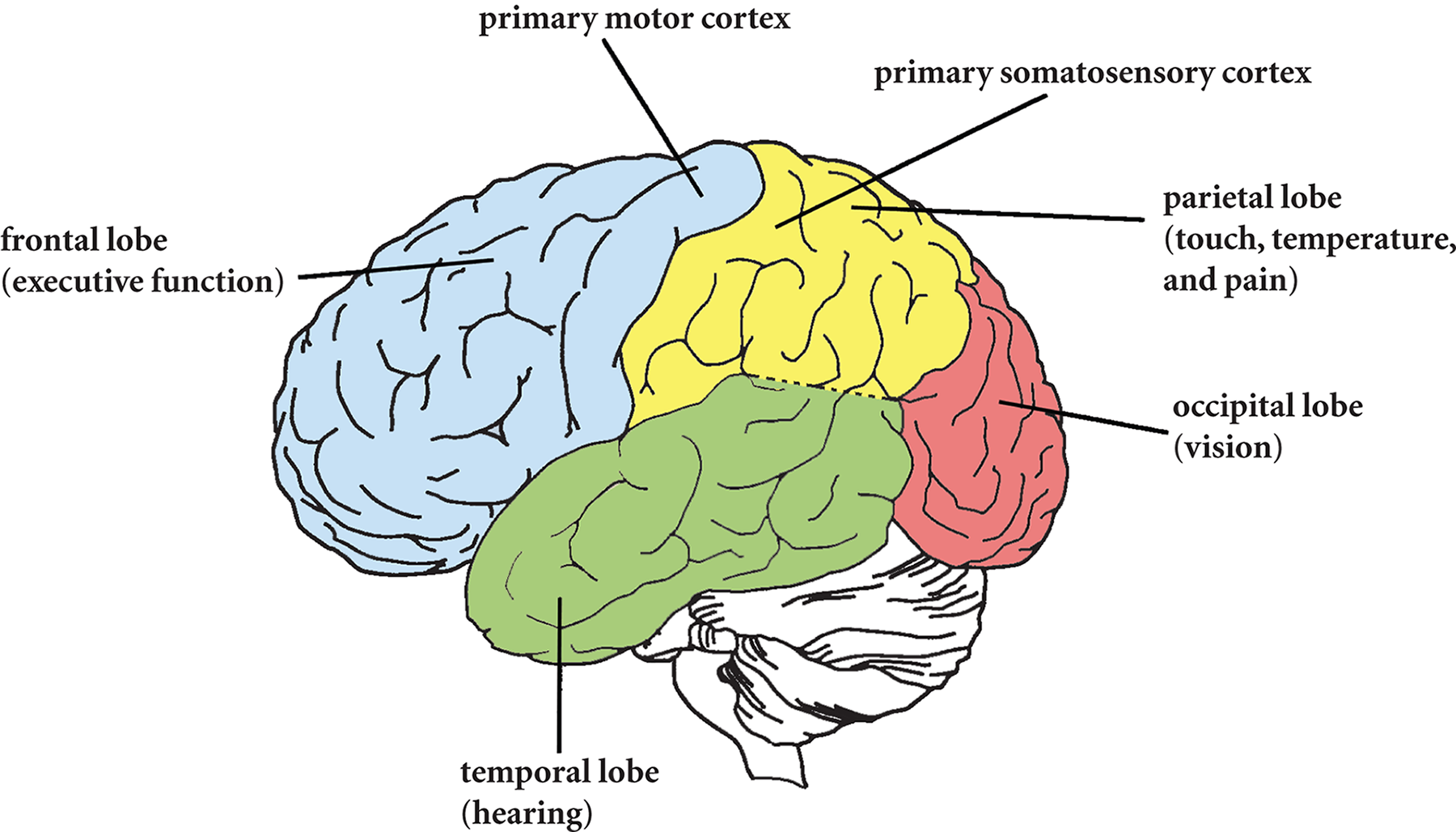
The cerebrum is divided into two halves, called cerebral hemispheres. The surface of the cortex is divided into four lobes—the frontal lobe, parietal
lobe, occipital lobe, and temporal lobe. These lobes are identified in Figure 1.9,
which shows a side view of the left cerebral hemisphere.
Figure1.9.Lobes of the Brain
Frontal Lobe
The frontal lobe is comprised of two basic regions: the prefrontalcortex and the motor cortex. The
prefrontal cortex manages executive function by supervising and directing the operations of other brain
regions. This region supervises processes associated with perception, memory, emotion,
impulse control, and long-term planning. In memory, for instance, the role of the
prefrontal cortex is not to store any memory traces, but rather to remind the individual
that he or she has something to remember at all. To regulate attention and alertness,
the prefrontal cortex communicates with the reticular formation in the brainstem,
telling an individual either to wake up or relax, depending on the situation.
Because it integrates information from different cortical regions, the prefrontal
cortex is a good example of an association area: an area that integrates input from diverse brain regions. For example, multiple
inputs may be necessary to solve a complex puzzle, to plan ahead for the future, or
to reach a difficult decision. Association areas are generally contrasted with projection areas, which perform more rudimentary or simple perceptual and motor tasks. Examples of
projection areas include the visual cortex, which receives visual input from the retina,
and the motor cortex, which sends out motor commands to the muscles.
Damage to the prefrontal cortex impairs its overall supervisory functions. A person
with a prefrontal lesion may be more impulsive and generally less in control of his
or her behavior, or depressed. It is not unusual, for instance, for someone with a
prefrontal lesion to make vulgar and inappropriate sexual remarks, or to be apathetic.
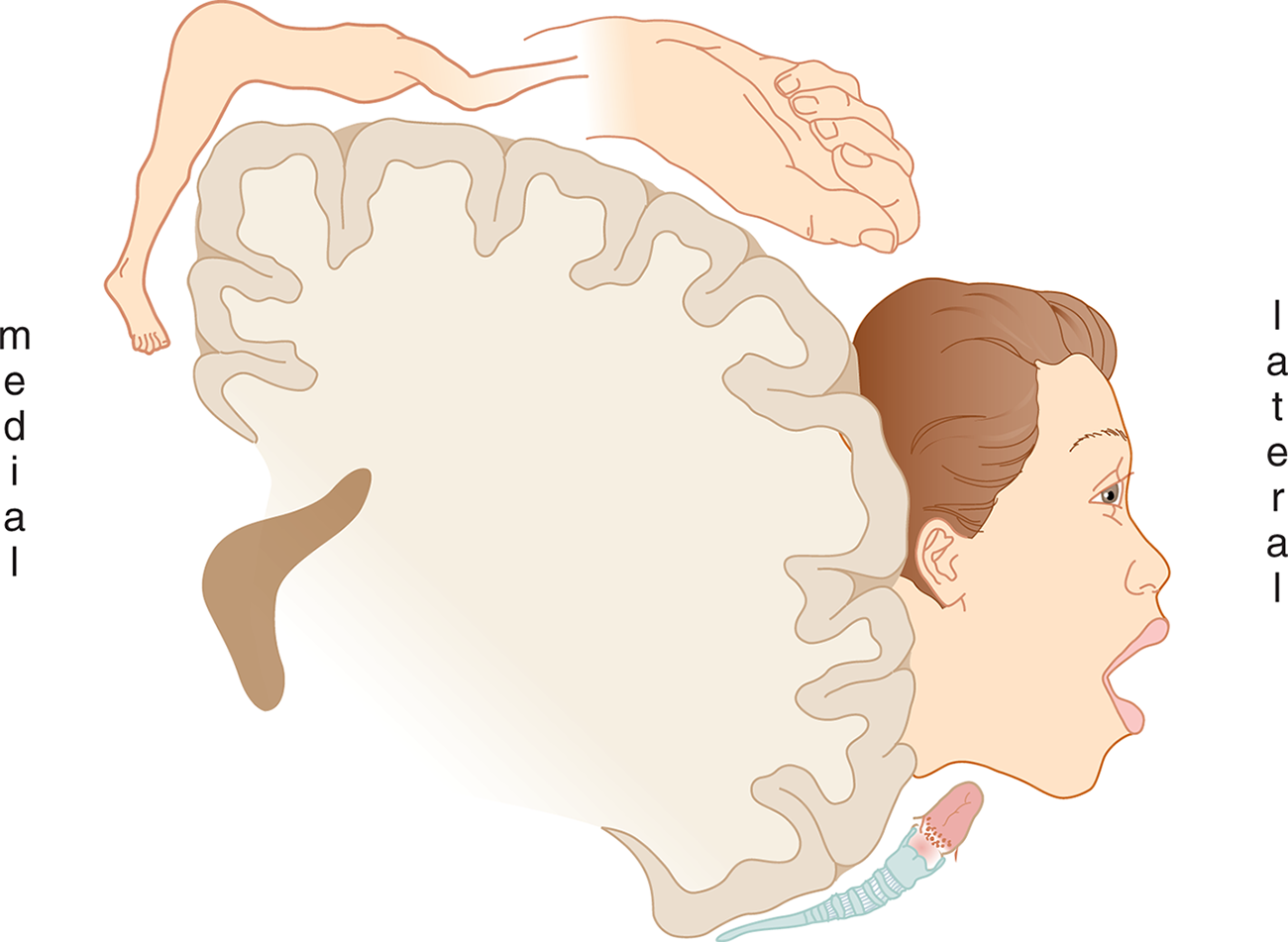
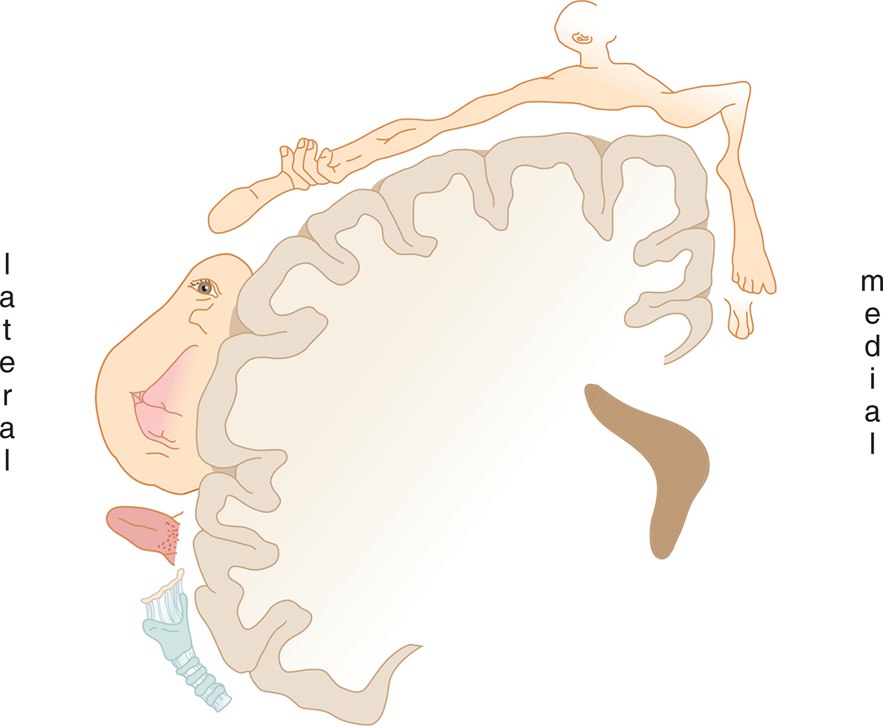
The primary motor cortex is located on the precentral gyrus (just in front of the central sulcus that divides the frontal and parietal lobes), and initiates voluntary motor movements
by sending neural impulses down the spinal cord toward the muscles. As such, it is
considered a projection area in the brain. The neurons in the motor cortex are arranged
systematically according to the parts of the body to which they are connected. This
organizational pattern can be visualized through the motor homunculus, as shown in Figure 1.10. Because certain sets of muscles require finer motor control
than others, they take up additional space in the cortex relative to their size in
the body.
Figure1.10.Motor Homunculus on the Precentral Gyrus of the Frontal Lobe
A third important part of the frontal lobe is Broca’s area, which is vitally important for speech production. Broca’s area is usually found
in only one hemisphere, the so-called “dominant” hemisphere; for most people—both
right- and left-handed—this is the left hemisphere.
Parietal Lobe
The parietal lobe is located to the rear of the frontal lobe. The somatosensory cortex is located on the postcentral gyrus (just behind the central sulcus) and is involved in somatosensory information processing.
This projection area is the destination for all incoming sensory signals for touch,
pressure, temperature, and pain. Despite certain differences, the somatosensory cortex
and motor cortex are very closely related. In fact, they are so interrelated they
sometimes are described as a single unit: the sensorimotor cortex. The somatosensory
homunculus is shown in Figure 1.11.
The central region of the parietal lobe is associated with spatial processing and manipulation. This region makes it possible to orient oneself and other objects in
three-dimensional space, to do spatial manipulation of objects, and to apply spatial
orientation skills such as those required for map-reading.
Figure1.11.Somatosensory Homunculus on the Postcentral Gyrus of the Parietal Lobe
Occipital Lobe
The occipital lobes, at the very rear of the brain, contain the visual cortex, which is sometimes called the striate cortex. Striate means furrowed or striped, which is how the visual cortex appears when examined under
a microscope. The visual cortex is one of the best-understood brain regions, owing
to the large amount of research that has been done on visual processing. Sensation
and perception of visual information is discussed thoroughly in Chapter 2 of MCAT Behavioral Sciences Review. Areas in the occipital lobe have also been implicated in learning and motor control.
Temporal Lobe
The temporal lobes are associated with a number of functions. The auditory cortex and Wernicke’s area
are located in the temporal lobe. The auditory cortex is the primary site of most sound processing, including speech, music, and other
sound information. Wernicke’s area is associated with language reception and comprehension. The temporal lobe also functions
in memory processing, emotion, and language. Studies have shown that electrical stimulation
of the temporal lobe can evoke memories for past events. This makes sense because
the hippocampus is located deep inside the temporal lobe. It is important to note
that the lobes, although having seemingly independent functions, are not truly independent
of one another. Often, a sensory modality may be represented in more than one area.
Cerebral Hemispheres and Laterality
In most cases, one side of the brain communicates with the opposite side of the body.
In such cases, we say a cerebral hemisphere communicates contralaterally. For example, the motor neurons on the left side of the brain activate movements
on the right side of the body. In other cases (for instance, hearing), cerebral hemispheres
communicate with the same side of the body. In such cases, the hemispheres communicate
ipsilaterally.
We distinguish between dominant and nondominant hemispheres. The dominant hemisphere
is typically defined as the one that is more heavily stimulated during language reception
and production. In the past, hand dominance was used as a proxy for hemispheric dominance;
that is, right-handed individuals were assumed to have left-dominant brains and left-handed
individuals were assumed to have right-dominant brains (because the brain communicates
contralaterally with the hand). However, this correlation has not held up under scrutiny;
95 percent of right-handed individuals are indeed left brain dominant, but only 18
percent of left-handed individuals are right brain dominant.
The dominant hemisphere (usually the left) is primarily analytic in function, making it well-suited for managing
details. For instance, language, logic, and math skills are all located in the dominant
hemisphere. Again, language production (Broca’s area) and language comprehension (Wernicke’s
area) are primarily driven by the dominant hemisphere.
Real World
The corpus callosum connects and shares information between the two cerebral hemispheres;
its function was discovered in epileptic patients whose corpora callosa were severed
in a last-ditch effort to limit their convulsive seizures. In these “split-brain”
patients, in whom the corpus callosum has been severed, each hemisphere has its own
function and specialization that is no longer accessible by the other. Thus, an object
felt only by the left hand (which projects to the right hemisphere) could not be named
(because language function is usually in the left hemisphere).
The nondominant hemisphere (usually the right) is associated with intuition, creativity, music cognition, and
spatial processing. The nondominant hemisphere simultaneously processes the pieces
of a stimulus and assembles them into a holistic image. The nondominant hemisphere
serves a less prominent role in language. It is more sensitive to the emotional tone
of spoken language, and permits us to recognize others’ moods based on visual and
auditory cues, which adds to communication. The dominant hemisphere thus screens incoming
language to analyze its content, and the nondominant hemisphere interprets it according
to its emotional tone. The roles of the dominant and nondominant hemispheres are summarized
in Table 1.2; remember that the left hemisphere is the dominant hemisphere in most
individuals, regardless of handedness.
Table 1.2.Comparison of Dominant and Nondominant Hemispheres’ Functions