
After Chapter 2.3, you will be able to:
The ear is a complex organ responsible not only for our sense of hearing, but also for both rotational and linear acceleration (vestibular sense). These senses are critically important to our ability to get around in the world, and their associated structures are encased in some of the densest bone of the body to protect them from damage.
The ear is divided into three parts, as shown in Figure 2.7: the outer, middle, and inner ear. A sound wave first reaches the cartilaginous outside part of the ear, called the pinna or auricle. The main function of the pinna is to channel sound waves into the external auditory canal, which directs the sound waves to the tympanic membrane (eardrum). The membrane vibrates in phase with the incoming sound waves. The frequency of the sound wave determines the rate at which the tympanic membrane vibrates: it moves back and forth at a high rate for high-frequency sounds and more slowly for low-frequency sounds. Louder sounds have greater intensity, which corresponds to an increased amplitude of this vibration.
The tympanic membrane divides the outer ear from the middle ear. The middle ear houses the three smallest bones in the body, called ossicles. The ossicles help transmit and amplify the vibrations from the tympanic membrane to the inner ear. The malleus (hammer) is affixed to the tympanic membrane; it acts on the incus (anvil), which acts on the stapes (stirrup). The baseplate of the stapes rests on the oval window of the cochlea, which is the entrance to the inner ear. The middle ear is connected to the nasal cavity via the Eustachian tube, which helps equalize pressure between the middle ear and the environment.
Remember that sound is a longitudinal wave carried through air (or another medium), which causes displacement of particles parallel to the axis of sound propagation. In other words, when a sound wave hits your eardrum, it literally causes it to oscillate back and forth because of moving air particles. Sound is discussed in Chapter 7 of MCAT Physics and Math Review.
The inner ear sits within a bony labyrinth, which contains the cochlea, vestibule, and semicircular canals, as shown in Figure 2.8. These structures are continuous with each other and are mostly filled by the membranous labyrinth, which is bathed with a potassium-rich fluid called endolymph. The membranous labyrinth is suspended within the bony labyrinth by a thin layer of another fluid called perilymph. Perilymph simultaneously transmits vibrations from the outside world and cushions the inner ear structures.

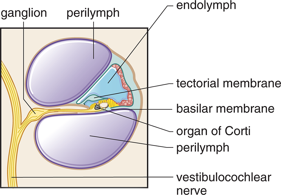
The cochlea is a spiral-shaped organ divided into three parts called scalae, as shown in Figure 2.9. All three scalae run the entire length of the cochlea. The middle scala houses the actual hearing apparatus, called the organ of Corti, which rests on a thin, flexible membrane called the basilar membrane. The organ of Corti is composed of thousands of hair cells, which are bathed in endolymph. On top of the organ of Corti is a relatively immobile membrane called the tectorial membrane. The other two scalae, filled with perilymph, surround the hearing apparatus and are continuous with the oval and round windows of the cochlea. Thus, sound entering the cochlea through the oval window causes vibrations in perilymph, which are transmitted to the basilar membrane. Because fluids are essentially incompressible, the round window, a membrane-covered hole in the cochlea, permits the perilymph to actually move within the cochlea. Like the rods and cones of the eye, the hair cells in the organ of Corti convert the physical stimulus into an electrical signal, which is carried to the central nervous system by the auditory (vestibulocochlear) nerve.
The junction between the stapes and the oval window is extremely similar to a thermodynamic gas–piston system, as described in Chapter 3 of MCAT Physics and Math Review. However, fluids are not as compressible as gases; therefore, the round window must be present to allow the perilymph in the cochlea to actually move back and forth with the stapedial footplate.
The vestibule refers to the portion of the bony labyrinth that contains the utricle and saccule. These structures are sensitive to linear acceleration, so are used as part of the balancing apparatus and to determine one’s orientation in three-dimensional space. The utricle and saccule contain modified hair cells covered with otoliths. As the body accelerates, these otoliths will resist that motion. This bends and stimulates the underlying hair cells, which send a signal to the brain.
While the utricle and saccule are sensitive to linear acceleration, the three semicircular canals are sensitive to rotational acceleration. The semicircular canals are arranged perpendicularly to each other, and each ends in a swelling called an ampulla, where hair cells are located. When the head rotates, endolymph in the semicircular canal resists this motion, bending the underlying hair cells, which send a signal to the brain.
The auditory pathways in the brain are a bit more complex than the visual pathways. Most sound information passes through the vestibulocochlear nerve to the brainstem, where it ascends to the medial geniculate nucleus (MGN) of the thalamus. From there, it projects to the auditory cortex in the temporal lobe for sound processing. Some information is also sent to the superior olive, which localizes the sound, and the inferior colliculus, which is involved in the startle reflex and helps keep the eyes fixed on a point while the head is turned (vestibulo–ocular reflex).
The lateral geniculate nucleus (LGN) is for light; the medial geniculate nucleus (MGN) is for music.
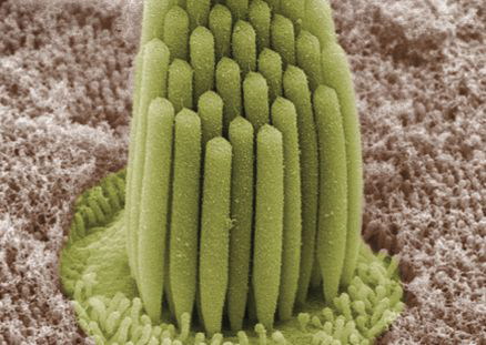
Hair cells are named for the long tufts of stereocilia on their top surface, shown in Figure 2.10. As vibrations reach the basilar membrane underlying the organ of Corti, the stereocilia adorning the hair cells begin to sway back and forth within the endolymph. The swaying causes the opening of ion channels, which cause a receptor potential. Certain hair cells are also directly connected to the immobile tectorial membrane; these hair cells are involved in amplifying the incoming sound.
The basilar membrane changes thickness depending on its location in the cochlea. The accepted theory on sound perception is place theory, which states that the location of a hair cell on the basilar membrane determines the perception of pitch when that hair cell is vibrated. The highest-frequency pitches cause vibrations of the basilar membrane very close to the oval window, whereas low-frequency pitches cause vibrations at the apex, away from the oval window. Thus, the cochlea is tonotopically organized: which hair cells are vibrating gives the brain an indication of the pitch of the sound.