CHAPTER FOUR
OUR THREE BRAINS
AND MORE
In proportion to our body mass, our brain
is three times as large as that of our nearest relatives.
This huge organ is dangerous and painful to give birth to,
expensive to build, and, in a resting human, uses about
20 percent of the body’s energy even though it is just
two percent of the body’s weight. There must be
some reason for all this evolutionary expense.
—SUSAN BLAKEMORE
American author Kurt Vonnegut, in his novel Galapagos, uses a refrain to express his disdain for the “so-called” advances in human progress and social and political evolution. He writes, “Thanks a lot, big brain.”
While Vonnegut writes of his unhappiness with war, poverty, violence, and so on—results of what our brains produce—many of us don’t share his cynicism. When Vonnegut spoke of the “big brain,” he didn’t mean it literally. Weighing some three pounds and making up about two percent of our body weight, the human brain is six times larger, relative to body size, than that of any other living mammal, with the exception of dolphins. Human and dolphin brains are very close in proportion to body size. But the dolphin brain has not significantly developed or changed in the last 20 million years.
A mystery of human brain evolution has long puzzled many biologists and paleontologists. As animal species evolved, their brain mass enlarged in the same ratio as the lungs, liver, stomach, and the rest of the body’s physical structures. About 250,000 years ago, most mammals reached the height of their evolution in brain complexity and mass. Just in the last 250,000 to 300,000 years, as the mammalian brain reached its zenith in size and efficiency, the evolution of our human species diverged from other mammals in several quite unpredictable ways. For one thing, early humans should have reached a plateau in brain development, as other mammals did during the same period. Instead, the human neocortex underwent an enormous leap in overall mass and complexity within a short amount of time.
The Quandary of Brain Growth
Recent findings show that when the human midbrain reached its present-day level of evolutionary complexity (250,000 to 300,000 years ago), our ancestors at that time experienced a 20 percent increase in actual mass of the neocortex, the thinking, reasoning area of the human brain.1 This sudden acceleration in the volume and density of brain mass appears to have occurred spontaneously and unexplainably, as opposed to the normal, linear course of evolution. Our rapid 20 percent outgrowth of gray matter is responsible for the superiority of the human brain. What caused this explosive brain development, which gave us a neocortex so much larger and denser than that of any other species, remains a mystery.
Also unlike other mammals, when the density of the human neocortex enlarged by 20 percent, the size of the human body increased only by 16 percent. To put this another way, the human body’s size increased only 80 percent in proportion to the expansion in mass of the brain, which is quite a deviation from the mammalian body-brain ratio.
Another interesting question comes to mind. Why did the brain expand to such a great degree, while the size of the head, both generally and in relation to the growth of the rest of the body, did not keep pace? The overall volume of the human skull did enlarge to some degree but not proportionally, the way animal evolution would predict. Scientists believe that if the human head had grown at the same rate of increase as the brain, the female pelvis could not have accommodated an infant’s enlarged head circumference during birth. Even today, the human birthing process remains risky and difficult due to fetal head size. Back then, an increase in fetal head size without an increase in pelvic size would have accelerated infant and maternal mortality, and humans would have been eliminated as a species. One possible solution that Mother Nature rejected was merely to increase the size of the female pelvis to allow a larger fetal head circumference. We can only imagine what shape females would have evolved into, had there been this increase in the size of the head. With such an increase in pelvic capacity, this probably would have forced early female humans back on four legs.
The Nerf Ball Brain
Nature’s solution to the need for a larger brain without a corresponding increase in skull size was simple and elegant. The brain folded in on itself, so that about 98 percent of the neocortex is hidden within the folds. Just as a Japanese fan, when folded, hides its floral patterns beneath the surface, the new, enfolded brain hides most of its gray matter and material. This design, which greatly resembles a walnut, is an efficient way to pack more material into a smaller space.
A few years ago, I was helping my daughter with a school assignment on the brain. We were discussing how the brain’s numerous folds maximize mass and minimize the use of space. She was having difficulty understanding the general idea. After she went to school the next morning, I bought 10 Nerf balls that were four inches in diameter. I also found a one-gallon glass jar with a large opening. That evening I asked her to put just two balls in the jar. They took up most of the jar’s volume. “No folds, right?” I asked. She nodded. “That’s what the brain would look like if it had no folds in it,” I said. Then I asked her to stuff all 10 Nerf balls into the jar and put the lid on it. As she did, she began to grin, then to laugh. The contents of the jar now looked like the folds of the brain.
A vital part of the brain’s evolutionary leap 250,000 years ago, the brain’s folding gradually increased to the level we see today. As my daughter can now tell you, the brain’s folding in on itself was an adaptation that gave early humans crucial advantages over other species in their environment. By increasing the potential for early humans to grow in intelligence and in their ability to learn, without compromising the body in other ways, brain folding gave us an evolutionary edge that improved our species’ chances for survival.
Brain folding and the evolution of the new brain also gave humankind a potential for mental growth that we have barely tapped, even today. Present-day humans still have almost the same proportional brain mass as we did 250,000 to 300,000 years ago. Once we became a new species of humans with an enlarged new brain, we were no longer limited to traveling the long, linear evolutionary road that the rest of the planet’s creatures had to follow. Clearly, however, our species as a whole is not using the full capacity of the new brain.
The Brain: Evolution’s Time Capsule
If you want to trace the evolutionary development of humanity, a good place to start is at the very top. The brain serves as a kind of time capsule illustrating humankind’s evolutionary development, and evolution has a long memory. We hold the entire course of our evolution inside our skull. If our brain were different today, the history of our species would be different as well.
According to research pioneered by Paul MacLean, M.D., the human brain has three formations, each with a different shape, size, chemistry, structure, and pattern of function that reflect our development during distinct eras. In essence, the human brain consists of three separate sub-brains. MacLean’s research suggests that the three brains amount to three interconnected biological computers. Each possesses its own intelligence, its own individual subjectivity, its own sense of time and space, and its own memory, as well as other functions.2
The original names given to the three substructures were the archipallium (also referred to as the reptilian brain; the R-complex or reptilian complex; and the brainstem in conjunction with the cerebellum or hindbrain), the paleopallium (the midbrain, mammalian brain, or limbic brain), and the neopallium (the new brain, neocortex, cerebral cortex, or forebrain). For simplicity, we refer primarily to the brainstem and cerebellum together as the first brain, the midbrain as the second brain, and the neocortex as the third brain or new brain. At times, I use the different names assigned to each of our three brain systems interchangeably throughout the book. Look at Figure 4.1; this drawing is taken from MacLean’s book, The Triune Brain in Evolution. You can compare it to the present-day human brain in Figure 3.7. Although each sub-brain does work independently, in humans the whole brain works together to make the sum greater than the parts.
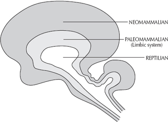
Figure 4.1
The Triune Brain
The hierarchal order of these three brains tells us important information about our evolution and the brain’s functions. First to evolve, more than 500 million years ago, was the brainstem, the junction where the spinal cord connects to the base of the brain. The most primitive brain area, it makes up the majority of brain matter in reptiles and lizards. Scientists of old called this the reptilian brain, because it resembles the entire brain of a reptile.
Attached directly behind the brainstem, the cerebellum evolved roughly 500 to 300 million years ago. This part of the first brain is responsible for coordination, proprioception (the unconscious perception of movement and spatial orientation), and body movement, both gross and fine. Recent studies suggest that the cerebellum performs additional functions. For example, the cerebellum is closely connected to the frontal lobe, the area of the neocortex responsible for intentional planning.3 In addition, the cerebellum has been shown to play a dynamic role in complex emotional behaviors.4 The neurons in the cerebellum are the most densely connected nerve cells in the entire brain. This heightened interconnectivity enables the cerebellum to control many functions without our having to impose our conscious awareness on them.
The midbrain appeared somewhere between 300 to 150 million years ago. This second brain is sometimes called the mammalian brain, because it is most highly evolved in mammals. Wrapping around the brainstem, the midbrain underwent its greatest increase in complexity and development in the last 3 million years and reached the height of its development about 250,000 years ago. This area is home to our involuntary, autonomic nervous system.
Finally, beginning about 3 million years ago, the new brain—with its most important component, the neocortex (neo means new or modified) or cerebral cortex—molded itself around the first two brains. That makes this outer shell (which looks like the skin of an orange) the most recent layer and the most advanced brain area to evolve in primates and humans. The seat of our conscious awareness, the new brain houses our free will, our thinking, and our capacity to learn, reason, and rationalize. Figure 4.2 is a cross section of the brain (ear to ear) demonstrating the thickness and size of the neocortex. The gray matter (neurons) as well as white matter (glial cells) of the third brain are also visualized.
The First Brain to Develop: The Brainstem and Cerebellum
The brainstem primarily supports the basic life functions, including the maintenance and control of heart rate and breathing. These life functions are common to all species of animals. The brainstem also has the job of regulating our various levels of wakefulness and sleep. Both wakefulness and levels of alertness are controlled by the brainstem to a greater extent than by the higher centers of the neocortex.

Figure 4.2
Ear-to-Ear Cross Section of the Brain
The cerebellum, or little brain, is also part of our first, or reptilian, brain. Its wrinkles and folds give it a distinct appearance. Relatively large compared to other brain structures, it is a three-lobed structure attached to the brainstem at the very back of the skull, underneath the hindmost area of the neocortex.
Recent functional brain scans reveal that the cerebellum is the brain’s most active area.5 Scientists believe the cerebellum is responsible for balance, coordination, proprioception, and the execution of controlled movements. In coordinating movement, the cerebellum performs both a motor (excitatory) function as well as a braking (inhibitory) function.
Certain types of simple actions and responses are learned, coordinated, memorized, and stored in the cerebellum. For example, once a person learns how to crochet, or even ride a bicycle, it requires very little conscious memory to perform this action. After a skill is learned and memorized—wired to the cerebellum—our body can perform the action automatically with very little conscious thought. Hardwired attitudes, emotional reactions, repeated actions, habits, conditioned behaviors, unconscious reflexes, and skills that we have mastered are all connected to and memorized in the cerebellum.
As we have learned, in the neocortex, the average number of connections per neuron is about 40,000. Remarkable as that is, in the cerebellum those neurons called Purkinje’s cells process between 100,000 to 1 million connections per neuron. The cerebellum is the most densely packed area of gray matter in the brain. More than half of all the neurons that make up the human brain are contained in the cerebellum. In fact, the cerebellum is one of the few areas of the brain where brain cells continue to reproduce long after birth. Interestingly, when a baby is rocked or cuddled, impulses are directed to the cerebellum, which actually stimulates his development. This benefit of rocking continues until around age two.
The Second Brain to Develop: The Midbrain
The second brain area to evolve is called the midbrain, because the structures that make up this particular region are located directly in the middle of the brain. One of many terms for this area is the limbic system; limbus means forming a border around an edge or ring and pertains to something that is marginal or at a junction between structures. The term mammalian brain also applies, because this region is the most highly developed and specialized in mammals. Situated just above the brainstem, the midbrain in an adult human is about the size of an apricot. As a reminder, check out Figure 3.7 to examine the midbrain’s location and size. Also see Figure 4.3, which illustrates and labels most of the brain regions related to our discussion in this chapter.
Regulatory Functions of the Midbrain
Although the midbrain occupies only one-fifth of the volume of the brain, its influence on behavior is extensive, which is why it is also known as the emotional brain. The midbrain is sometimes called the chemical brain as well, because it is responsible for regulating many different internal states.
It is our midbrain that performs all those marvels that we usually take for granted, automatically maintaining and controlling body temperature, blood sugar levels, blood pressure, digestion, hormone levels, and innumerable other processes. The midbrain also adjusts and maintains our internal state to compensate for changes in our external world. Without the midbrain, our metabolism would be like that of a cold-blooded reptile because we could not maintain a sustained internal state to counter environmental temperature changes.
The Midbrain’s Four Fs
In addition to these kinds of regulatory functions, the midbrain is responsible for what we can describe as the four Fs: fighting, fleeing, feeding, and fornicating.
Fight-or-flight. We know the midbrain’s first two roles as our fight-or-flight response. As you’ll recall from chapter 3, the autonomic nervous system originates in the midbrain and includes the sympathetic (fight-or-flight) nervous system, which kicks in when you feel threatened or scared. Imagine that you are taking the garbage out, and you see a bear in the bushes. The moment your neocortex (conscious brain) perceives the threat, this fear-producing external stimulus activates the autonomic nervous system. (In fact, we now know that certain parts of the midbrain sense the external threat even before you are conscious of it.) In turn, your autonomic nervous system automatically triggers your fight-or-flight response to prepare you for activity. This initiates a sequence of automatic internal events. An instant burst of adrenalin prepares your body to flee. Blood flow is directed away from your internal organs to your arms and legs, maximizing your ability to move so that you have the best odds of escaping.
In threatening situations, the midbrain controls your vital functions for preservation of life. These reflexive responses seem to be universal among all mammals because we all share that portion of the brain called the mammalian brain. In other words, when faced with fearful situations, human beings respond physiologically and biochemically almost exactly as would a rabbit or a dog.

Figure 4.3
Overview of the Brain
The spinal cord acts as a “fiberoptic” cable that conveys impulses from the brain to other parts of the body and relays messages from the body back to the brain.
The brainstem helps regulate primitive functions like breathing, swallowing, blood pressure, levels of wakefulness, and respiratory rate.
The cerebellum is responsible for balance, posture, and the body’s position in space. It also coordinates movements and facilitates automatic hardwired memories and behaviors.
The midbrain acts as the chemical brain, where automatic internal regulation occurs and chemical balance is maintained. It also helps organize signals from the external world with our internal world.
The thalamus acts as a junction box to integrate all incoming sensory information (except smell) to various regions of our conscious thinking brain.
The hippocampus is responsible for formulating experiences with associated emotional memories, for processing vital information during learning, and for encoding long-term memories.
The amygdala works with the hippocampus to generate primary emotions from external perceptions and internal thoughts. It helps to emotionally charge experiences and to warn us about vital sensory information.
The hypothalamus chemically regulates the body’s internal environment so that homeostasis is maintained. Conditions like body temperature, blood sugar levels, hormone levels, and emotional reactions are regulated here.
The pituitary gets its orders from the hypothalamus to secrete hormones in the form of peptides that circulate throughout the bloodstream and activate different glands, tissues, and organs in the body.
The pineal gland chemically regulates levels of sleep as well as cyclic rhythms of procreation and mating.
The corpus callosum is a band of fibers that connects the two hemispheres of the brain so that they can exchange information.
The cerebral cortex is the seat of our conscious awareness and is responsible for carrying our sophisticated functions like learning, remembering, creativity, invention, and voluntary behavior.
The midbrain is also intrinsically involved in emotional reactions that have to do with the survival of the physical body.
Feeding. When you sit down to have a meal, your parasympathetic nervous system relaxes you, conserves your energy, and prepares your body for digestion and metabolism.
Fornicating. If you’re interested, when you engage in the fourth F, both the parasympathetic and the sympathetic components of your autonomic nervous system come into play. The former helps you get in the mood (you probably wouldn’t feel too sexually aroused if that bear was chasing you), and the latter turns on when you have an orgasm.
To progress your understanding of the limbic brain a little more, let’s add a couple more Fs and see how they all relate to the sympathetic and parasympathetic nervous systems. The sympathetic system has its own four Fs: fight, flight, fright, and fornicating (orgasm). The parasympathetic system is responsible for a few Fs as well: feeding, fixing (growth and repair), and fornicating (getting in the sexual state of mind). One system utilizes, releases, and mobilizes energy, while the other system conserves, builds, and stores energy.
STRUCTURES OF THE MIDBRAIN
The midbrain is primarily composed of the thalamus, hypothalamus, pituitary, pineal gland, hippocampus, amygdala, and basal ganglia.
Thalamus. The thalamus is the meeting point for almost all nerves that connect one part of the brain to another part of the brain, the body to the brain, and the brain to the body. The thalamus, the name of which is derived from the Greek word meaning “inner chamber,” is the oldest and largest part of the midbrain. A collection of nerve cell nuclei that meet at a central junction point, it is made up of two distinct thalamic centers, one on each side of the midbrain. Think of the thalamus as a switchboard or air traffic control tower that can connect any part of the brain and the body. There is not one signal from the environment that does not pass through the thalamus. The sensory organs (ears, eyes, skin, tongue, nose) send messages to the thalamus, which relays them to their final destination in the neocortex/conscious brain.
At the same time, the thalamus can send signals to other areas of the brain so as to alert or inhibit different brain systems. In this way, the thalamus processes sensory information from the external world, identifies and sorts the input into the appropriate category, and transmits this data to the many conscious centers in the cerebral cortex. Depending on the nature of the sensory information or the type of stimulation from the environment, the data is then passed in many different directions throughout the brain (the midbrain, the brainstem, and so on) and body. The thalamus is also the relay system between the neocortex and the brainstem. Thus, this part of the midbrain allows the entire brain to receive a multitude of important data from the external world all at once, so that the brain can readily be introduced to vital information.
Hypothalamus. This area of the midbrain is a chemical factory that regulates your body’s internal environment and balances your systems with the external world. The hypothalamus (which translates literally to “under the thalamus”) is the most important and fascinating part of the midbrain, because it generates the chemical messengers for the entire body. The oldest part of the limbic system, it can affect any organ or tissue in the body.
Unlike the thalamus, which monitors external stimuli, the main job of the hypothalamus is to make chemicals called neuropeptides that keep the internal affairs of the body in balance with reference to the external world. The hypothalamus regulates many bodily functions necessary for survival through the process of homeostasis, the automatic self-righting mechanism that, like a thermostat, regulates and maintains the body’s chemical balance and internal order. The hypothalamus controls and manages bodily functions such as appetite, thirst, sleep, wakefulness, blood sugar levels, body temperature, heart rate, blood pressure, chemical balance, hormonal balance, sex drive, immune system reactions, and metabolism. It also plays the most important role in your experience of emotions. This is the part of the brain that manufactures the chemicals that allow you to feel the way you were just thinking or how you were reacting.
Let’s get back to our hypothetical survival situation, an encounter with a bear, to see how the thalamus and the hypothalamus would come into play. When your sensory organs pick up the sight and the sound of an approaching bear, those important messages are sent to the thalamus. The thalamus quickly orients your brain to the danger, ensuring that the warning sensory signals arrive throughout the entire brain nearly at the same time. The thalamus then coordinates your entire body for immediate action. It sends information to the neocortices (the higher conscious brain centers within the neocortex), which make decisions, plan actions, observe the surroundings for quick exits, and the like.
The thalamus also signals the hypothalamus to chemically prepare your fight-or-flight body functions, so that your body has the energy and resources to respond to the threat. For instance, the hypothalamus ensures that your legs are physiologically ready to run, jump, and turn quickly, based on the decision of the conscious brain. On the other hand, you don’t need any blood flow to the digestive organs during this imminent threat, so the hypothalamus manages your body’s internal state for action rather than for digestion—that is, for fighting and fleeing, but not for feeding (or fornicating).
Pituitary gland. The pituitary gland secretes chemicals that activate your body’s hormones. Briefly, glands are organs or specialized groups of cells that separate certain elements from the blood and secrete them in a form the body can use or eliminate easily. Hormones are complex chemicals, produced in one part or organ of the body, that initiate or regulate the activity of an organ or group of cells in another part of the body. The different glandular tissues of the body that secrete various hormones are organs such as the adrenal glands, the thyroid gland, and the reproductive organs, to name a few.
The pituitary is often called the master gland, because it governs and controls many vital processes in the body. This pear-shaped gland, which hangs off the hypothalamus like a piece of fruit, helps in manufacturing most of the hormonal signals created by the hypothalamus to communicate with the body’s major glands. The hypothalamus sends both chemical and electrical signals to the pituitary so that it can make certain chemicals that turn on various chemical/hormonal states.
Pineal gland. The pineal gland, a tiny, pine cone–shaped structure, sits in the back of the midbrain, above the cerebellum. (A common misconception is that, in humans, the pineal is embedded in the brain just above the eyes. Hence it has been termed our third eye.) The pineal chemically regulates our cycles of sleep and wakefulness. Think of the pineal gland as the brain’s internal clock—it chemically controls patterns of sleep and wakefulness. Photoreceptors in the eyes sense levels of daylight or darkness, then transmit that information to the hypothalamus and, in turn, to the pineal gland. The pineal in humans (and in many other non-nocturnal mammals) then secretes different neurotransmitters that are directly influenced by the amount of light the eyes receive.
Two neurotransmitters are produced in the highest quantities in the human body by the pineal gland. Serotonin, the so-called daytime neurotransmitter, prepares the brain to be awake during the hours of daylight. Melatonin, the nighttime neurotransmitter, prepares the body to experience restorative sleep during the hours of darkness and plays a role in causing the brain to dream. Thus, if you are reading this book late at night and experiencing sleepiness, the reason (I sincerely hope) is biological. The fact that your eyes’ photoreceptors are no longer sensing daylight prompts your pineal gland to transmute serotonin into melatonin.
Unlike its embedded location in humans and other primates, the pineal gland sits close to the surface of the skull in many lower life forms, including amphibians, reptiles, fish, birds, and certain mammals. This placement enables the pineal gland to sense the changing amounts of sunlight and darkness that these animals are exposed to during different times of the year, as well as different times of the day.
Thus in many animal species, the pineal directly influences biological cycles that are dependent on the changing seasons, such as migratory patterns, circadian rhythms, reproductive cycles, seasonal bearing of offspring, and even mating rituals.
How does the pineal gland prompt animals to birth their young at certain times of the year? Take, for example, animals that hibernate during the winter, such as bears. During the darker winter months, their pineal glands secrete more of the nighttime neurotransmitter, melatonin, into the bloodstream and the fluid of the brain. Some of this melatonin is absorbed by the pituitary gland. The pituitary responds by producing neurohormones that suppress the activity of the sex organs, decreasing the animals’ drive to procreate.
The pineal gland also alters melatonin into a neurohormone called 5-methoxytryptamine, which eliminates sex drive and decreases appetite in some species of hibernating mammals. Their altered brain chemistry also produces a slowdown in metabolic and other body functions, which causes them to sleep throughout the winter.
When spring brings the stimulation of increasing light levels, this heightens production of serotonin and other neurotransmitters, prompting these animals to once again become sexually active and have an increased appetite. As a result, they deliver and raise their offspring during the warmer months, when food supply and other environmental conditions favor their survival.
Hippocampus. The hippocampus makes long-term memories. It gets its name from the Greek word for seahorse, which this brain region resembles. We learn from new experiences and form memories thanks to this area of the midbrain.
A sort of clearinghouse for memory, the hippocampus classifies incoming information as having either short-term or long-term importance, and files it accordingly. Memories that move into short-term storage pertain to information that we need immediately but can then forget. Shopping lists, phone numbers that we will call only once, and directions that we will probably never need again are good examples of information that is stored in short-term memory.
In long-term memory, the hippocampus stores information that we may need to access repetitively or at will in the future. Obvious examples are our address, our spouse’s name, what type of car we own, and so on. At our annual office party, we may meet numerous people whose names we won’t need to remember tomorrow, but it would be wise to store the name of our employer’s spouse in long-term memory. The hippocampus stores long-term memories that are involved mostly with our experiences, based on the various types of information our five senses provide.
The type of memory encoding that takes place in the hippocampus is called associative learning or associative memory. For example, imagine that a child throws rocks at a beehive, and then has the novel experience of receiving multiple stings. In the future, the child will associate bee-provoking behavior, such as rock-throwing, with the sight of the agitated bees pouring out of the hive, the sound of their angry buzzing, the place he stood when he was repeatedly stung, and the feel of those painful stings. The hippocampus will facilitate storage of this sensory information as long-term memory throughout different regions of the neocortex, so that the experience can be encoded as wisdom. With any luck, this child won’t have to repeat the experience to make the message clear. The evolution of the hippocampus has permitted many species to repeat behaviors that improve their chances of survival and to avoid repeating actions that threaten their survival.
Let’s explore how the hippocampus accomplishes this feat. It keeps a log of facts associated with people, places, things, time, and events. Humans tend to remember experiences better when they are somehow connected to one of these items. The hippocampus creates a memory of personal events associated with things that happen to us at a particular time and place.6 In this example, people might be the neighbor whose hobby is beekeeping; the place might be the neighbor’s property; things might include the rocks the child threw, the bees, and beehives; the time could be a midsummer day; and events would certainly include the throwing of the rocks, the consequence of being stung, and perhaps any subsequent first-aid treatment.
Whenever we have a new experience, the hippocampus, through the combination of all our senses (seeing, smelling, tasting, feeling, hearing), allows us to create a new memory. By connecting all this incoming sensory information, the hippocampus will associate a person with a thing, a place with a time, a person with an event, and so on. The child in our example will file his experience into long-term memory by associating the neighbor (people) with bees (things), beehives (things) with the first-aid lotion his mother applied (smell), the neighbor’s property (place) with the experience of getting stung (event), the pain of the stings (feeling) with rocks (things), and so on. Later on, experiencing one of these elements again (smelling the first-aid lotion, for example) will trigger a flood of memories of this experience. But this happens only after about age four. The reason we cannot remember many conscious memories as a very young child is that the hippocampus is not fully developed until after we are four years old.
Associative memories allow us to use what we already know in order to understand or learn what we don’t know; in other words, to use what is familiar to us to understand something that is unfamiliar. These memories are the building blocks for us to arrive at greater understandings. When we take new information relating to people, places, things, time, and events and we associate this information with our log of past events that we have already experienced through our five senses, we build an associative memory.
One primary function of the hippocampus is closely related to our search for novelty. This is the part of the brain that is responsible for making unknowns, known. For example, if the hippocampus is destroyed in laboratory animals, and then they are given the opportunity to explore new environments, they will ignore unfamiliar areas and return repeatedly to familiar areas of their cage. In fact, new research suggests that our ideas about what motivates learning may not be very accurate. Some scientists are reevaluating their long-held models involving conditioned behavior, in which reward or punishment (pleasure or pain) appeared to provide inducements for animals to learn. Perhaps the animals in such studies, rather than learning, were being trained. Many studies involving the hippocampus suggest that for several different animal species, learning new things is a reward in itself.7
Amygdala. The amygdala, which means “almond-shaped,” is a structure of the midbrain that is responsible for alerting the body in survival situations. It also stores the four highly charged primitive emotions: aggression, joy, sadness, and fear. The amygdala also helps to attach different emotional charges to our long-term memories.
When a life-threatening situation exists, the amygdala gives a rapid, action-oriented assessment of the external environment. It is the most important fear-generating region of the brain. In fact, the amygdala is the part of the midbrain that activates the body to respond even before you are consciously aware of the danger, so we sometimes call this a precognitive response. This is why the amygdala is so important for the survival of our species, as well as many animals. It processes incoming sensory information that is vital to survival in a crisis situation and instantaneously alerts the body, bypassing other circuits.
For example, imagine that you are riding your bicycle in the park while listening to your MP3 player, mesmerized by a melody. In an instant, a young child darts out of the bushes and begins to cross your path, right in front of the bike. Your amygdala receives vital information that bypasses your neocortex, causing you to hit your brakes even before you are conscious of your actions. This enhanced precognitive reaction may make the difference between life and death. Because the midbrain is a more primitive area than the neocortex, it makes sense that this mechanism was probably hardwired into our species millions of years ago, long before the development of our newer thinking, reasoning neocortex.
When activated, the amygdala also creates emotions of rage and aggression to help us protect ourselves in potentially threatening situations. Thus, a mother will aggressively defend her offspring or risk her life in any harmful situation, even though the odds are against her.
Recent studies also indicate that the amygdala is associated with the storage of emotional memories and with the perception of certain situations based on those memories. The amygdala brands survival situations as emotionally fearful, so that memories of threatening circumstances can help us avoid similar situations. In humans, highly charged emotional experiences involving anger, fear, sadness, and even joy are encoded by the amygdala for long-term memory. However, the amygdala does not assign any specific region of nerve cells to store memories of these primitive hardwired feelings in order to create or facilitate memory of any single, specific emotion. Researchers cannot point to a particular region of the brain and say that it is where sadness, for example, resides. Similarly, studies involving primates have found no specific areas of the amygdala that produce joy, sadness, rage, or fear.
In an intriguing new study, scientists at the University of Wales worked with a blind patient who seems to possess a sixth sense that allows him to recognize sad, angry, or happy faces. Patient X, age 52, cannot see after having two different strokes, which damaged the brain areas that process visual signals. However, brain scans reveal that when he looks at faces expressing emotion, another part of his brain besides the visual cortex is activated—the amygdala. This small structure responds to nonverbal facial signs (or memories) depicting anger and fear.8
Dr. Alan Pegna, in the School of Psychology at the University of Wales, Bangor, headed the research team with colleagues in north Wales and at Geneva University Hospital. They found that Patient X was unable to identify shapes like circles and squares. Furthermore, he could not identify the sex of “deadpan” male and female faces, nor tell the difference between “normal” and jumbled faces. But when the subject was asked to identify the emotions of an angry or happy human face, he accurately did so 59 percent of the time. (Most blindfolded subjects in this type of test are usually successful 50 percent of the time, give or take a percentage point.) This success rate is statistically quite a bit higher than what would be expected by chance, and it consistently applied as well when he was asked to distinguish between sad and happy, or fearful and happy, faces.
From this experiment, the researchers concluded that emotions displayed on a human face are registered not in the visual cortex but in the right amygdala, which sits deep within the brain’s temporal lobe. “This discovery is … interesting for behavioral scientists as the right amygdala has been associated with subliminal processing of emotional stimuli in clinically healthy individuals,” said Dr. Pegna. “What Patient X has assisted us with establishing is that this area undoubtedly processes visual facial signals connected with all types of emotional facial expressions.”9 Having memory stored in this area of the brain, which also triggers instantaneous responses, could explain much about the sensitivity of some individuals.
Basal ganglia. The basal ganglia integrate thoughts and feelings with physical actions. Basal ganglia are intricate bundles of neurological networks that are interconnected with the neocortex; they are situated in each hemisphere of the midbrain, directly under the neocortex and above the midbrain’s deeper structures.
To illustrate how the basal ganglia function, recall a time when you were learning a skill that involved muscle movements, such as riding a bike. In the beginning, you had to think consciously about what you were doing. Every time you practiced, you reinforced neural circuits in your brain that relayed commands to your body relating to balance, coordination, and so on. After much repetition, these neural networks became hardwired, and your movements in pedaling the bike and keeping your balance became automatic.
At that point, your basal ganglia, along with your cerebellum, took over the coordination of those automatic movements. As you rode, the basal ganglia received sensory information from your environment via the neocortices, plus commands from your neocortex to move your muscles and orchestrate your actions. Your basal ganglia integrated your thoughts and feelings with your physical actions, smoothed out your fine motor movements, and suppressed your body from making random, involuntary movements. In addition to that role, the basal ganglia allows us to control our impulses, to set our idle speed for anxiety, and to contribute to our feelings of pleasure and ecstasy.
To get a clearer picture of the important roles basal ganglia play, consider what can happen when they malfunction. In people with Tourette’s syndrome, the basal ganglia fire improperly and fail to coordinate their thoughts and feelings with their actions. These people often lose inhibitory control over their impulses, feel overly anxious, and display uncontrolled behaviors such as erratic motions, twitches, eye blinks, head jerks, and so on.
At one time or another, most of us have been in a situation in which our basal ganglia receive so much input from the neocortex that the threshold of electrochemical charge is too high for the basal ganglia to process. When this happens, the stimulus causes the basal ganglia to act like a breaker in a fuse box and throw the main circuit, so to speak, putting the body into a temporary state of disruption. For example, when we are scared, we may freeze; when we are embarrassed or intimidated, we sometimes become speechless; when we try to speak with someone we find very attractive, our mind occasionally goes blank. (Not that I know any of this from personal experience, I’m just saying …)
Just as some cars idle faster than others, some people have overactive basal ganglia. These people are frequently anxious or nervous. Without good cause, they constantly evaluate their environments, anticipate risks, and prepare for potential danger. Their basal ganglia operate in a heightened state—not high enough to throw the body’s circuit breaker, but higher than seen in most people. As a result, these people tend to be easily overwhelmed by minor stresses in their lives.
On the other hand, according to the latest functional brain scans, so-called doers usually have basal ganglia that function at a slightly more active level than in most people. Their increased basal ganglia activity does what it is supposed to—it processes thought and emotion into immediate action—but doing becomes the means for these people to keep their basal ganglia from reaching overload. The increased activity of their basal ganglia produces excess energy, which they release by taking action. If they stop doing, they can experience an energy overload, and the byproduct is nervous anxiety. A simple example of this is when we are sitting with a group of people and someone cannot stop bouncing his leg up and down—his basal ganglia is slightly overactive and is discharging anxious energy.10
The Third and Most Recent Brain to Evolve: The Neocortex
The neocortex is the seat of our awareness and of our creativity as a species. It is our thinking, reasoning brain that allows us to learn and remember everything that we experience from our external world, and then modify our actions to do something better, or different, or to repeat an action the next time, if it had a positive result.
When our brain is actively performing one of the so-called higher functions—reasoning, planning, intellectualizing, learning, remembering, creating, analyzing, verbally communicating, among a host of others—our neocortex is at work. Without the neocortex, our senses would still be able to alert us to the fact that we are cold, but we could not proceed further. The neocortex is what allows you to interpret the sensation of being cold and choose among multiple options—remain cold, close the window, put on a sweater (and choose from among a number of sweater options), or turn up the thermostat—and your neocortex would also remember a time you camped in the winter at Mt. Rainier National Park and nearly got frostbite.
HOW DO MALE AND FEMALE BRAINS COMPARE?
Generally, the male brain is larger than the female brain by more than 100 cubic centimeters, about the size of a small lemon. Does this difference have direct cognitive effects? Not necessarily. Although there is still a gender difference in brain volume after scientists compensate for body size, studies attribute some of the variation to an individual’s physical dimensions. In a very specific MRI study, which paid equal attention to both brain and body size parameters, Michael Peters and associates, at the University of Guelph, Ontario, Canada, showed that the difference in brain volume between the sexes dropped by two-thirds after height was included as an additional covariant.11
Differences in brain volume between the sexes are distributed quite evenly throughout the major lobes of the brain. The proportions of the four major lobes of the neocortex are similar. In both sexes, the frontal lobe comprises about 38 percent of the neocortex (ranging from 36 to 43 percent); the parietal lobe, 25 percent (21 to 28 percent); the temporal lobe, 22 percent (19 to 24 percent); and the occipital lobe encompasses about 9 percent (ranging from 7 to 12 percent) of the neocortex.
This means that there is no sex-specific brain region that contributes to an additional share in total brain volume, and that it will be difficult to find a functional sex difference that correlates with differences in total brain volume. In simple terms, if we were to look at the brains of two individuals, one male and the other female, we would not be able to tell them apart, aside from the size difference, because male and female brains share similar proportions.
In terms of differences between males and females, the brain structure that has probably drawn the most attention over the years is the corpus callosum. This band of white matter connects the right and left hemispheres, and some early research suggested that it might be larger in women than in men. When that was first suggested in the early 1980s, many scientists speculated that the larger band in women meant that females had a greater degree of communication between the two hemispheres. This idea seemed to support the myth that in women, the emotional right side of the brain and the analytical left side were more connected and integrated with each other.
It is now known that women do not have a larger corpus callosum than men do. The corpus callosum is actually about 10 percent larger in men than in women, probably because men have larger brains due to their larger body size. There is no substantial anatomical evidence for greater functional connectivity between the hemispheres (as the stereotype would have it) in either men or women.
The source of this myth may be that the corpus callosum does account for a significantly greater percentage of the total white matter in women (2.4 percent in females versus 2.2 percent in males). This fact just might mean that women are able to process the two types of thoughts (emotional and analytical) between the two hemispheres of the brain a lot faster than men. If the greater distribution of total fatty myelin, or white matter, in the female corpus callosum does account for speedier neurological transmission between the brain hemispheres, this may explain why men are often dumbfounded when observing women’s problem-solving abilities in action.
Evolution’s most sophisticated achievement to date, as we discussed earlier, the new brain appeared when mammals began their climb up the ladder of evolution. Highly developed in mammals, the new brain reached its greatest level of complexity in humans. Since our new brain is proportionately larger and more complex than that of any other species—comprising two-thirds of our total brain area—it affords us unique characteristics that distinguish us from reptiles, other mammals, and fellow primates.
For the sake of simplicity, I will describe the new brain as having an inner, supporting layer, and an outer layer. The inner layer of the brain is like the meat of an orange, while the outer layer, called the cortex, is like the rind or skin of the orange. The word cortex literally means “bark.” As discussed, most of the brain is structured in convoluted folds rather than simple layers. But as my purpose is to build a mental model for understanding the brain, I will occasionally overlook some of the brain’s complexities.
Wrapping around the midbrain is that portion of the new brain called the white matter, made mostly of nerve fibers insulated by fatty myelin sheathes, as well as glial cells, which are neural cells that primarily have a connective tissue supporting function in the central nervous system (see chapter 3). Several types of glial cells exist, performing different functions in the various components of the nervous system. The most important thing to remember about glial cells is that they help facilitate the forming of synaptic connections; that may explain their large numbers. In other words, every time you learn something new and make a new synaptic connection in the brain, a specific type of glial cell called an astrocyte is always present, helping with the process. Every neuron has the possibility of making an incredible number of connections to other neurons, and nature may have provided humans with an abundance of glial cells to facilitate so many potential synaptic connections. Researchers have found evidence that glial cells have their own independent communication system, separate from neurons.12
The part of the new brain that we will refer to most often is the outer layer, the neocortex or cerebral cortex, also called our gray matter. Although it is only about 3 to 5 millimeters thick (1/7 to 1/4 of an inch), this layer is so rich in neurons that, aside from the cerebellum, the neocortex has more nerve cells than any other brain structure.
Like the midbrain, the neocortex is composed of several parts.
THE CORPUS CALLOSUM
The corpus callosum is a “fiberoptic” bridge comprised of hundreds of millions of neurons that connect the two hemispheres of the new brain.
As most people are aware, the new brain is divided anatomically into two distinct sections that mirror each other in some degree of anatomical symmetry. If you drew an imaginary line from the middle of the forehead over the top of the head to the center of the base of the skull, you would be dividing the new brain into its two halves. These are commonly known as the left and the right cerebral hemispheres. These twin neocortices literally encapsulate the midbrain and the brainstem. Each hemisphere is responsible for controlling the opposite side of the body.
The cerebral hemispheres are not completely separate structures. This thick band of nerve fibers called the corpus callosum joins the two halves of the new brain. Figure 4.4 provides a view of the corpus callosum. The corpus callosum is the largest fibrous pathway of neurons in the entire body, totaling approximately 300 million nerve fibers. This large band of white matter possesses the greatest number of nerve bundles anywhere in the brain or the body. Scientists postulate that the corpus callosum evolved along with the new brain, so that its two separate houses could communicate with each other through this bridge. Nerve impulses constantly travel back and forth across the corpus callosum, giving our new brain the specialized ability to observe the world from two different points of view.
THE FOUR NEOCORTICAL LOBES
The two cerebral hemispheres are further subdivided into four separate regions known as lobes. Thus as part of the neocortex, we have two frontal lobes, two parietal lobes, two temporal lobes, and two occipital lobes. Each of these areas processes different sensory information, motor abilities, and mental functions and is assigned to perform different tasks.
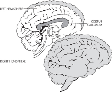
Figure 4.4
The corpus callosum and how it connects the two hemispheres of the neocortex.
In general, the frontal lobes are responsible for intentional action as well as for focusing our attention, and they coordinate nearly all the functions in the rest of the brain (the motor cortex and language center are part of the frontal lobe). The parietal lobes deal with sensations related to touch and feeling (sensory perception), visual-spatial tasks, and body orientation, and they also coordinate some language functions. The temporal lobes process sounds, perception, learning, language, and memory, and they are the centers that process smell. This lobe also includes a region that facilities our ability to choose which thoughts to express. The occipital lobes manage visual information and are often called the visual cortex. Take a minute, if you will, to examine the four lobes of the cerebral cortex in Figure 4.5.
For purposes of building our understanding in a logical way, I’m going to go out of sequence here, and describe the parietal, temporal, and occipital lobes first, then conclude with the most recent achievement of our evolution, the frontal lobes.
The parietal lobes. The parietal lobes are located just above each ear, and they extend to the top center of the head, reaching the midline of the brain. This is the feeling/sensing region of the cortex. The parietal lobes process what we feel with our hands and with our bodies, also called tactile and somatosensory perceptions. Somatosensory by definition is the information we receive from the body (somato) that we feel (sensory) in the brain. Features such as pressure, temperature, vibration, pain, pleasure, light touch, two-point discrimination, and even the awareness of where our body parts are located without looking at them (proprioception) are all integrated in the somatosensory cortex of the parietal lobes.
The parietal lobes process information from the body received by our peripheral nerves, mainly from our external environment and to a lesser degree from our internal environment. Remember, peripheral nerves are those long nerves that act like communication wires, transmitting information from the brain to the body and from the body to the brain. In particular, we are discussing the peripheral nerves that are sensory in nature, which receive and process billions of bits of information every second, from all areas of our body, and send it to the brain. These peripheral nerves converge from different areas of the body (hands, arms, legs, toes, feet, lips, tongue), and then connect to the spinal cord, which is the “fiberoptic” cable that passes all the incoming information to the brain—specifically, the somatosensory cortex.
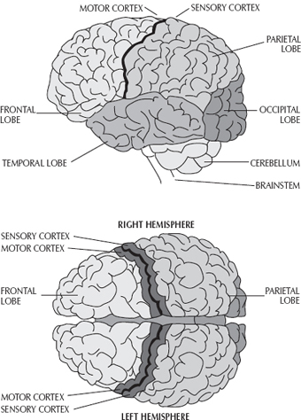
Figure 4.5
A side and top view of the different lobes of the neocortex.
When you have a rock in your shoe, feel a warm breeze on your face, receive a relaxing massage, or have a stomachache, it is the parietal lobe that gathers all that sensory information and determines how it feels and what you should do about it. First, this lobe interprets what type of stimulus it is receiving. Then, it evaluates how a stimulus feels—whether you like it or whether it is a threat to the body. The somatosensory cortex is the region that primarily gauges how you consciously feel under different environmental conditions. Once the sensory cortex processes the information, other regions such as the frontal lobe take over to carry out the brain’s primary goal—taking care of the body’s survival and maintenance.
Here is an example. The subtlety of a fly landing on your arm instantly catches your attention. The sensory receptors of the arm send an immediate message along the peripheral nerves to the spine, entering through the cervical vertebrae and on to the somatosensory cortex in the side of the brain opposite to the arm. Once your brain interprets the stimulus, the message is then forwarded to the frontal lobe, where it is processed for motor responses. At this point, the whole brain may or may not be involved. You may respond automatically by using your motor cortex to move your arm, shooing the fly away. Or you might think for a moment what to do. Maybe you’ll get up, look for some ice cream in the freezer, and get the flyswatter.
The parietal lobes are subdivided and organized into several areas that relate to different regions of sensory experience in the body. Every inch of the body’s surface area has a corresponding point on this somewhat narrow slice of cortical neurons. The somatosensory area is like a map of individual clusters of neurons that are somewhat compartmentalized into specific sensory regions that relate to different parts of the body.
In the mid 1900s, a few scientists were learning about how to map these regions by studying animals. Researchers used touch to stimulate different parts of their bodies, identifying the activated neurons in the brain corresponding with the particular region of the body being touched. The initial work using animals to explore the sensory cortex was performed on rats and monkeys by Vernon Mountcastle at Johns Hopkins University.
In humans, these particular sensory areas of the parietal lobes are classically known as the representation zones, named during this same period by Canadian neurosurgeon Wilder Penfield.13 Penfield conducted several experiments using human subjects to determine the precise sensory correlations between particular parts of the brain and specific areas of the body. While performing brain surgery on conscious human patients under local anesthesia, Penfield used a tiny electrode to stimulate different regions of the somatosensory cortex. As he excited the exposed surface of their cortex, he asked the patients what they were feeling. In every case the patients were quick to report particular sensations in the hands, fingers, feet, lips, face, and tongue as well as other body parts. In this fashion, Penfield was able to explore and name the regions of sensory input within the somatosensory cortex.
As Penfield discovered, the entire body surface is outlined or laid out along the sensory cortex in humans and all mammals. There are regions specified for the lips, hands, feet, tongue, genitalia, face, fingers, and so on. In humans, this area has been affectionately called the homunculus or “little person.” Figure 4.6 shows the homunculus and it illustrates how somatosensory feelings are mapped in the human brain.
Curiously, however, the body as mapped in the sensory cortex looks nothing like an actual human body. Not only is this map peculiarly compartmentalized, but it is also not in direct correlation to the anatomical layout and proportions of the human body. For example, the representation zone for the face is located next to the hand and fingers. Penfield also discovered that the feet are neighbors to the genitals. In the cortex, the tongue area exists outside the mouth area, under the chin. At that time he had no idea why the cortical map is so structurally odd.
Presently, there are two working models that, together, explain this obscure presentation.14 The first model pertains to the locations of the representation zones. During prenatal growth, the fetus has its arms bent so that the hands are touching the face, and its legs are folded so that the feet are touching its genitalia. Developmentally, the recurring contact in utero between these body parts might produce repetitive firing of sensory neurons within different regions in the developing cortex. This sensory activation of the cortical neurons might trick the parietal lobe into organizing its sensory regions as if these body parts are side by side, when they are merely in constant contact. Thus the first impressions of cortical mapping may lay the foundation for where different sensory regions will ultimately exist within the somatosensory template.

Figure 4.6
An ear-to-ear slice of the neocortex demonstrating a view of both the sensory and motor cortices. The shaded areas are the representation zones that illustrate how the entire body is mapped as a little distorted man called the homunculus.
The second working model may explain distortions in the size of individual sensory areas, as compared to normal human anatomy. According to the sensory map, the “little person” lying along the sensory cortex has a huge face with large lips, big hands and fingers with extra-large thumbs, and oversized sexual organs. What is the explanation for this? We can look to these oversized areas of the cortical map for the answer. When I was a child and felt sick, my mother accurately gauged my body temperature by putting her lips on my forehead. This makes sense since human lips are so highly specialized; they possess numerous, densely packed sensory receptors. Similarly, the neurons sensitive to touch on the fingertip of the index finger are 15 times as dense as the touch-sensitive receptors on the leg. There is an immense amount of sensory receptors in the genitals of human beings.
During evolution, the acute sensitivity of our lips, tongue, hands, and sexual organs has been crucial to supporting the survival of our species. In humans, not only are more sensory receptors located in these parts of the body, but additional territory is allocated to them in the brain. The amount of cortical tissue designated to a specific body part reflects not the size of that particular part of the body, but its sensitivity. In simple terms, larger regions are mapped in the sensory cortex because we feel more with those parts of the body. As a result, the body parts of the homunculus appear in a hierarchal order that is directly proportional to how specialized each area of the body is, with regard to sensation, and how much we use that body part to feel.
The same principle holds true for other mammals. In cats, the sensory cortex is regionalized differently than in humans. A feline has a huge cortical area mapped for its nose and whiskers, because those structural organs are associated with its primary means of information processing. Therefore the cat, which explores the world primarily with its nose and whiskers, will have a “catunculus”—a different map of the somatosensory cortex than our human one.
So, the areas of the human body having the densest amount of sensory nerves will have larger real estate in the somatosensory cortex. That is why, comparatively, more territory in the sensory cortex is assigned to the lips than to the back, and more cortical space is designated to the fingers than the entire leg. Thus, you can better tune your brain into feeling with your hands, lips, and fingers than with other body parts.
Here, too, is a clear demonstration as to why we humans are so driven by sexuality. The map of the feeling body in the sensory cortex of the brain has more real estate devoted to the genitalia than to the entire surface of the chest, abdomen, back, shoulders, and arms put together. We are literally mapped for procreation to ensure the propagation of our species. Interestingly, when epileptic seizures originate in these areas of the sensory cortex, they are usually preceded by intense sexual sensations.
What is most important to remember at this point is that an entire map of how the body feels can be charted in the sensory cortex of the brain, specifically in the somatosensory areas located in the parietal lobes.
The temporal lobes. The temporal lobes are just under the surface of and slightly above each ear. They are responsible for auditory perception—that is, how we process what we hear. The auditory lobes are primarily positioned in this quadrant to process all types of sounds. Within these lobes there seem to be thousands of colonies of neurons related to specific aspects of how we process sounds. Because what we hear is so intricately tied to language, we will define language as a series of specific sounds that are produced for intentional communication, and then comprehensively understood. In other words, what arrives at your ears is a stream of continuous sounds carrying an intention or meaning that is called language.
The eardrum vibrates as a result of sound waves hitting it, which produce electrical signals that travel along the auditory nerve to individual compartments in the temporal lobes. The temporal lobes deal with language comprehension, decoding sound into meaning. This trait is assigned mostly to the diversified regions on the left side of the neocortex, unless we are learning a new word or sound or language, and then it is the right temporal lobe that takes over.
There are different clusters of neurons in the auditory cortex that apply to every single phoneme, or minimal unit of sound that we use to interpret language. For example, when we hear the sounds baah, moo, or su, individual modules or compartments within the auditory complex are assigned to process these specialized sounds. As human infants develop through interactions with the environment, different noises that we hear are stored as geographically mapped patterns of diversified sounds, ready for us to access and to process as language. The infant’s brain is also busy pruning away unnecessary synaptic connections, to make meaning out of sounds from its environment.
Our brains are nonlinear enough that when we hear a series of sounds, we can immediately understand what is being verbally communicated. Remarkably, as electrical signals from the eardrum activate multiple clusters of neurons in the temporal lobes to fire simultaneously, the combination and sequence, as well as locations of these neural circuits, allow us to gain meaning from the auditory stimuli. There are hundreds of neuron clusters within specific compartments in the temporal lobes that are doing this as we listen to music, watch television, have a conversation at dinner, and even talk to ourselves, out loud or internally.
The temporal lobes are intricately involved in storage of some types of memory and facilitate the making of long-term memories. As we know, this takes place through the hippocampus. When there is damage to both the temporal lobes and the hippocampus, many people cannot form new memories. Scientists who experiment on the temporal lobes using low-voltage electrical stimuli have reported that their subjects experience immediate sensations of déjà vu (an uncanny sense of familiarity and memory), jamais vu (a feeling that a familiar person or place is unfamiliar), heightened spontaneous emotions, and/or strange spiritual reveries or insights.
The temporal lobes also have a visual association center that links what we see to our emotions and memories. It is the storehouse of many of our visual emotional memories. Once we see something in the external world, our brain uses this association area to process what we see with what we remember and how it may feel emotionally. In other words, the temporal lobes process visual symbols with meaningful feelings.
When this part of the temporal lobes is electrically stimulated, subjects report vivid visual imagery, equally as real to them as their external surroundings. We use the stored database of the temporal lobes when we associate what we know to better understand what we are attempting to learn that is new and unknown. The temporal lobes also help us recognize familiar stimuli that we have already experienced.
For example, let’s say I told you that a special type of white blood cell chases and attacks foreign agents, and then ingests them just like a little Pac-Man (if you can remember the old video game from the 1980s). The visual association center in your temporal lobes would bring up the visual memory of the Pac-Man video game, so that you could identify this new concept with what you already have stored in your brain as memory. It would flash pictures representing your accumulated memories of those little munching Pac-Man creatures, and then assemble a three-dimensional memory to help you understand the new idea about white blood cells. Most of the millions of learned associations that you have experienced in your lifetime are stored in the temporal lobes’ association cortex, to be activated as needed.
Thus, the temporal lobes are responsible for language, hearing (processing sounds), conceptual thinking, and associative memories. The temporal lobes associate most of what we have learned and experienced via our senses throughout our lifetime to people, places, things, time, and past events in the form of memories. We can associate what we hear, see, feel, taste, and smell, and it is the temporal lobes that facilitate this skill.
The occipital lobes. The occipital lobes are the vision centers. The visual cortex, as it is sometimes described, has six distinct regions that process data from the outside world in order for us to see coherently. This complexity stands to reason, because vision is the sense that human beings rely on the most in order to function in the world.
If we were to start at the very back of the brain at the occipital lobe and slice it with a knife like a loaf of bread six times to the temporal lobe, this would give us a good idea of how the visual cortex is organized. These regions are functionally separated so as to process different sensory data about what and how the brain is seeing. Six distinct layers are allocated to interpret visual qualities like light, movement, form, shape, depth, and color.
The primary visual cortex (V1) is the first slice of brain tissue located farthest back in the brain. This area of the visual cortex encounters visual information that our eyes see and we consciously process. V1 is organized in such a way that nerve cells are divided up to process different parts of a whole picture. Therefore, when only one small area of V1 is damaged, we have a visual blind spot, because the nonfunctioning neurons cannot process their part of the picture. When this area is completely damaged, normal sight as we know it is lost. Amazingly, when scientists began studying individuals who were blind in the V1 area, these subjects not only perceived movement, but could also perceive the shape of an object.
A completely different area of the visual cortex is organized to exclusively process movement (V5). The nerve cells in this area cannot detect a stationary object; they are stimulated only when an object moves across one’s visual field. These cells were discovered when blind people were found to be able to see movements. The first subjects who were ever recorded as having an ability to perceive moving objects without seeing them were soldiers in World War II. Some soldiers who had lost their sight from combat injuries could still dodge grenades and rockets, even though they could not consciously see them. This phenomenon was appropriately termed blindsight.15
Distinct geographical locations within the visual cortex process other aspects of sight. Some clusters of neurons perceive only color. Generalized forms and edges are perceived in one area, while specific shapes and patterns (such as the shape of a hand) are recognized in another neural region. Still other nerve cells respond to depth perception, angles, and dimension.
As visual information passes from the eyes to the occipital lobe, it is processed in a cascade of nerve reactions from the back of the brain to the front, through these six different regions. This is why a blindsighted person could still interpret reality through his visual field. The information that made it to his primary visual cortex was passed to the adjacent areas, which were activated for further processing. Thus while he could not consciously see an object, he could perceive movement, shape, the direction from whence the object came, and other aspects of vision.
When visual stimuli are all integrated, a picture appears as a “hologram” of what we are seeing. How does this take place? As sensory information is transmitted through the different regions of the visual cortex, there is a hierarchy of data processing, layer by layer. By the time the information has passed through these layers of specialized neurons, which make sense out of light, movement, form, shape, depth, and color, a continuous picture has been created. This image is then distributed to the appropriate associated areas of the brain’s temporal lobe, which participate with the visual cortex to make meaning out of the incoming data.
The frontal lobes. If you are asked, “Where do you, as a conscious being, think, dream, feel, focus, concentrate, and imagine?” you will most likely point to the area on your forehead just above the bridge of your nose—the frontal lobe.
The frontal lobe is the resting place of conscious awareness. When we are the most conscious and the most aware, our frontal lobe is at the height of activity. Although the visual cortex, the temporal lobes, and the parietal lobes can serve to create a picture, a concept, or an idea, it is the frontal lobe that willfully keeps an idea on our mind, calling it to the stage for an extended review.
The frontal lobe is also where self-awareness is born. The most highly evolved area in the brain, it is the place where the self can express itself. Because of the frontal lobe, we break from the outmoded view that a human being is merely the byproduct of accumulated sensory experiences. Instead, the frontal lobe allows us to take our emotions and define them into meaning. The prefrontal cortex is the laboratory where we paste together thoughts with their associations to derive new meaning from what we are learning. The frontal lobe gives us the privilege to gain meaning from the external world.
Free will is a major keyword we use to describe the frontal lobe. The seat of our free will and self-determination, the frontal lobe allows us to choose our every thought and action and, in so doing, control our own destiny. When this lobe is active, we focus on our desires, create ideas, make conscious decisions, assemble plans, carry out an intentional course of action, and regulate our behavior. The evolution of the human frontal lobe bestowed on humans a focused, intentional, creative, willful, decisive, purposeful mind, if we will only put it to use.
The frontal lobes are regionally divided into subsections that are responsible for myriad related functions. The back part of the frontal lobes is home to the motor cortex, which exists as a neighboring slice of cortical tissue right in front of the sensory cortex. The motor cortex and the sensory cortex are at the dividing line between the parietal lobe and the frontal lobe. If you return to Figure 4.5, you will see the division between the two cortical regions marked by the sensory and motor cortex. (Some references refer to the sensory-motor cortex as one region of the neocortex; however, for the sake of simplicity, I discuss them separately.)
The motor cortex activates all of the voluntary muscles in the body and participates in all our voluntary movements and actions. We activate the motor cortex when we need to take determined actions and control purposeful movements.
Just as the sensory cortex has areas allocated by sensitivity and function, the motor cortex is divided into territories according to structure and function. And like the sensory cortex, the neurological map of the motor cortex displays a quite distorted homunculus. In this homunculus, the face has the hand exiting the crown of the head, and the arm, the shoulder, the trunk, the leg, and the feet are formatted in a disproportional layout, out of sequence with the normal human anatomy. Figure 4.6 shows the diversified subdivisions of the motor cortex parceled into bodily regions. The individual size of compartments is based on necessity, much like the sensory cortex.
In the motor cortex, for example, the real estate apportioned for hand movement is enormous, when compared to the area allocated to moving the neck. As a matter of fact, the hand and fingers take up more space in the motor cortex than the combined areas of the wrist, the elbow, the shoulder, the thigh, and the knee. What is the reason? We use our hands and fingers more than these other body parts, because their specialized structure permits us to be more functionally skilled in our environment. The brain provides enlarged domains to handle the considerable motor demands placed on our hands and fingers.
The frontal cortex also extends all the way back to the temporal lobes, where intentional speech is initiated in the language centers. Thus the frontal lobe is intrinsically connected to voluntary speech articulation, which is seamlessly encoded in the area farthest back in the frontal lobe toward the rest of the brain.
Just in front of the motor cortex is an area called the premotor cortex or the supplementary motor area (SMA), which is responsible for mentally rehearsed intentional actions—before those actions are actually carried out. This is the planning center for our future actions.
The prefrontal cortex is a cortical region related to the crowning achievement of our abilities in the areas of consciousness and awareness. This is the brain area that is most active during our important periods of conscious, deliberate concentration. It is in this compartment that our true uniqueness as human beings exists.
This area allows us to supersede the stimulus-response, action-reaction, cause-effect patterns we unconsciously live by day to day. For example, all the automatic, repetitive programs that have been hardwired in the brain such as brushing our teeth, driving, dialing familiar phone numbers, combing our hair, and so on, are of no interest to the prefrontal cortex. These predictable, recurring behaviors, which stem from what we constantly see, smell, taste, hear, and feel, can be performed quite nicely without the allegiance of the prefrontal cortex.
Test Driving the New, Improved Neocortex
With its enlarged size, the cerebral cortex is what separates us from other species, in our ability to consciously learn and remember by processing data derived from our senses. The neocortex is the seat of your executive mind, your identity, your personality, and your higher brain functions. At this very moment, you comprehend the information on this page by using many different regions of your neocortex. Mapped within the neocortex are the capacities for rational thought, reasoning, problem solving, freewilled decision-making, planning, organization, verbal communication, language processing, and computation, to name just a few.
Scientists have to put their collective neocortices to work to better understand the neocortex. We do know that its development made possible our highly advanced level of adaptability in the world. Early humans with the new, expanded neocortex would have learned faster than other species, and would have had a greater capacity for invention, reason, and ingenuity with which to outsmart predators or overcome other dangerous situations. The neocortex gives us the intellect to create new ideas, develop new behaviors and skills, and invent new tools and technologies. Because of its enormous size, it gathers volumes of learned or remembered information (that is, known information) and it naturally creates new models, ideas, and archetypes to explore or invent—both in the physical world and in our imagination. Thus, we are not limited to evolving in a lengthy, linear fashion. Instead, we can change the course of our species with even one new theory or invention.
Moreover, the advancements the neocortex makes possible are not limited to the need to ensure our survival in the face of harsh or changing environments. Via the neocortex, we create and appreciate music, art, and literature, and we strive to explore and understand both the external world and our internal world. The creative neocortex gives each of its owners an individual, unique personality, and enables humans to live as great thinkers and fabulous dreamers.
How is the human head able to accommodate not only the reptilian brain and the mammalian brain but also the new brain? To reiterate our computer analogy, when our new bio-computer evolved, we gained the world’s most powerful processor, the most advanced operating system, the largest hard drive, and the greatest amount of memory. As we mentioned, neurons themselves should never be thought of only as wires that connect to each other. Instead, each neuron should be seen as a complete, individual super-processor system that performs millions of functions daily. By connecting billions of neurons to each other, we now have billions of computer systems working as one incredibly huge computer network, having outstanding memory, storage abilities, and superhigh speed, as well as other amazing capabilities. Remember that the number of potential synaptic connections in the human brain is virtually unlimited. When the size of the new brain expanded during evolution, we managed to compress all this processing capability into a bio-computer that is no bigger than a cantaloupe. We have all the machinery to express unlimited potential.
Why do human beings as a whole seem to use only a small fraction of our potential? In our defense, Homo sapiens sapiens is a relatively young species, and we have only had a few hundred thousand years to start learning how to use our new brain efficiently. Perhaps we are still novices, and we have barely begun to take our new brain out for a test drive. It’s my hope that by reading this book, you’ll be better able to push the limits of reality’s engine: your brain.