THE KNEES
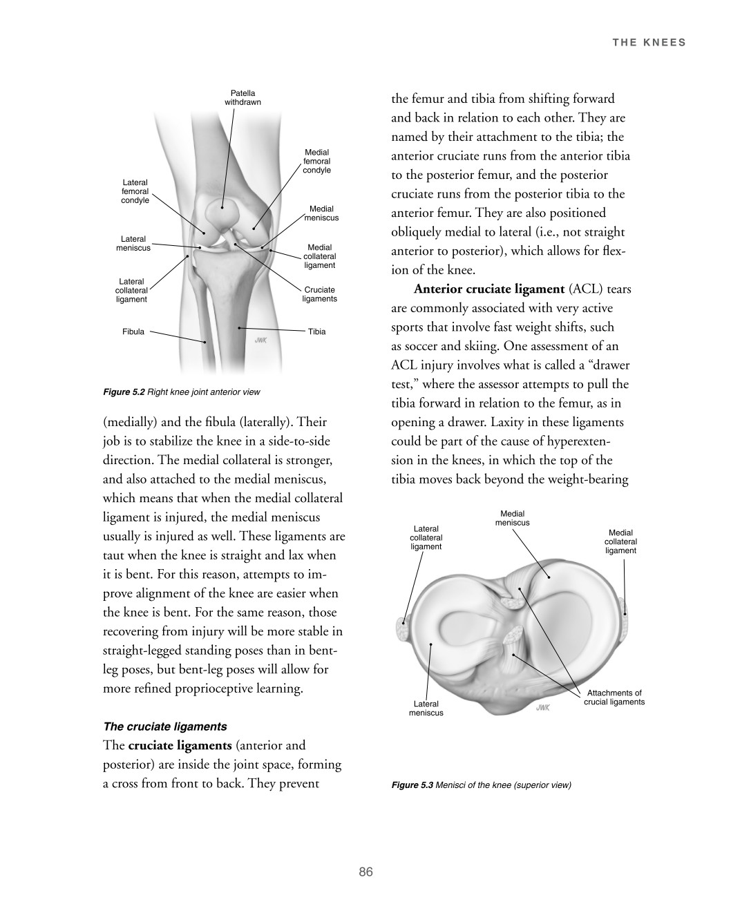
Patella withdrawn Medial femoral condyle Lateral femoral condyle Medial meniscus Lateral meniscus Lateral collateral ligament Fibula Medial collateral ligament Cruciate ligaments Tibia the femur and tibia from shifting forward and back in relation to each other. They are named by their attachment to the tibia; the anterior cruciate runs from the anterior tibia to the posterior femur, and the posterior cruciate runs from the posterior tibia to the anterior femur. They are also positioned obliquely medial to lateral (i.e., not straight anterior to posterior), which allows for flex- ion of the knee. Anterior cruciate ligament (ACL) tears Figure 5.2 Right knee joint anterior view (medially) and the fibula (laterally). Their job is to stabilize the knee in a side-to-side direction. The medial collateral is stronger, and also attached to the medial meniscus, which means that when the medial collateral ligament is injured, the medial meniscus usually is injured as well. These ligaments are taut when the knee is straight and lax when it is bent. For this reason, attempts to im- prove alignment of the knee are easier when the knee is bent. For the same reason, those recovering from injury will be more stable in straight-legged standing poses than in bent- leg poses, but bent-leg poses will allow for more refined proprioceptive learning. The cruciate ligaments The cruciate ligaments (anterior and posterior) are inside the joint space, forming a cross from front to back. They prevent are commonly associated with very active sports that involve fast weight shifts, such as soccer and skiing. One assessment of an ACL injury involves what is called a “drawer test,” where the assessor attempts to pull the tibia forward in relation to the femur, as in opening a drawer. Laxity in these ligaments could be part of the cause of hyperexten- sion in the knees, in which the top of the tibia moves back beyond the weight-bearing Medial meniscus Lateral collateral ligament Medial collateral ligament
Lateral meniscus Attachments of crucial ligaments
Figure 5.3 Menisci of the knee (superior view)
86