10. Porphyrins and the Basis of Life
I see little hope to be able to explain the subtle difference between a normal and a sick cell as long as we do not understand the basic difference between a cat and a stone.
ALBERT SZENT-GYÖRGYI
STRANGELY ENOUGH, “porphyrin” is not a household word. It is not a sugar, fat, or protein, nor is it a vitamin, mineral, or hormone. But it is more basic to life than any other of life’s components, because without it we would not be able to breathe. Plants could not grow. There would not be any oxygen in the atmosphere. Wherever energy is transformed, wherever electrons flow, there look for porphyrins. When electricity alters nerve conduction, or interferes with the metabolism of our cells, porphyrins are centrally involved.
As I write this chapter, a dear friend has just died. For the last seven years she had had to live without electricity, hardly ever seeing the sun. She seldom ventured out in the daytime; when she did, she covered herself from head to foot in thick leather clothing, a broad-brimmed leather hat hiding her face, and glasses bearing two layers of dark lenses concealing her eyes. A former dancer who loved music, nature, and the outdoors, Bethany was virtually abandoned by a world in which she no longer belonged.
Her condition, probably caused by her years of work for a computer company, was a classic example of an illness that has been known to medicine only since 1891, its emergence at that time being one of the side effects of the sudden worldwide expansion of electrical technology. Its connection with electricity was discovered a century later. Although it is now considered an extremely rare genetic disease, affecting as few as one person in fifty thousand, porphyria was originally thought to affect as many as ten percent of the population. Its supposed rarity is due in large part to the ostrich-like behavior of the medical profession after World War II.
In the late 1940s, medical practitioners were staring at an impossible contradiction. Most synthetic chemicals were known poisons. But one of the legacies of the war was the ability to manufacture products from petroleum, easily and cheaply, to substitute for almost every consumer product imaginable. Now, thanks to the fledgling petrochemical industry, bringing us “Better Living Through Chemistry,” synthetic chemicals were going to be literally everywhere. We were going to be wearing them, sleeping on them, washing our clothes, our hair, our dishes, and our homes with them, bathing in them, insulating our houses with them, carpeting our floors with them, spraying our crops, our lawns, and our pets with them, preserving our food with them, coating our cookware with them, packing our groceries in them, moisturizing our skin with them, and perfuming our bodies with them.
The medical profession had two choices. It could have attempted to study the health effects, singly and in combination, of the hundreds of thousands of new chemicals that were kaleidoscoping over our world, a virtually impossible task. The attempt itself would have put the profession on a collision course with the mushrooming petrochemical industry, threatening the banning of most new chemicals and the strangling of the economic boom of the next two decades.
The other alternative was for the profession to bury its collective head in the sand and pretend that the world’s population was not actually going to become poisoned.
Environmental medicine was born as a medical specialty in 1951, founded by Dr. Theron Randolph.1 It had to be created: the scale of the poisoning was too great to go completely ignored. The sheer numbers of sickened patients, abandoned by mainstream medicine, produced an urgent need for practitioners trained to recognize at least some of the effects of the new chemicals and to treat the resulting diseases. But the specialty was ignored by the mainstream as though it didn’t exist, its practitioners ostracized by the American Medical Association. When I attended medical school from 1978 to 1982, environmental medicine wasn’t even on the curriculum. Chemical sensitivity, the unfortunate name that has been given to the millions of poisoned patients, was never mentioned in school. Neither was porphyria, arguably a more appropriate name. It still isn’t mentioned, not in any medical school in the United States.
Heightened sensitivity to chemicals, we recall, was first described by New York physician George Miller Beard, who considered it a symptom of a new disease. The initial electrification of society through telegraph wires brought with it the constellation of health complaints known as neurasthenia, two of which were a tendency to develop allergies and a drastically reduced tolerance for alcohol and drugs.
By the late 1880s, insomnia, another prominent symptom of neurasthenia, had become so rampant in western civilization that the sale of sleeping pills and potions became big business, with new formulations coming on the market almost every year. Bromides, paraldehyde, chloral, amyl hydrate, urethane, hypnol, somnal, cannabinon, and other hypnotics flew off pharmacists’ shelves to satisfy the frustrated urge to sleep—and the addiction that so often followed the long term use of these drugs.
In 1888, one more drug was added to the list. Sulfonal was a sleeping medication that had a reputation for its prompt effect, its non- addictive nature, and its relative lack of side effects. There was just one problem, which only became widely known after three years of its popularity: it killed people.
But its effects were quirky, unexpected. Nine people could take sulfonal, even in large doses and for a long time, with no untoward effects, but the tenth person, sometimes after only a few or even one small dose, would become critically ill. He or she would typically be confused, so weak as to be unable to walk, constipated, with pain in the abdomen, sometimes with a skin rash, and reddish urine often described as the color of port wine. The reactions were idiosyncratic, liable to affect almost any organ, and the patients were apt to die of heart failure without warning. Between four and twenty percent of the general population were reported to be subject to such side effects from taking sulfonal.2
During the ensuing decades the chemistry of this surprising disease was worked out.
Porphyrins are light-sensitive pigments that play pivotal roles in the economy of both plants and animals, and in the ecology of planet Earth. In plants a porphyrin bound to magnesium is the pigment called chlorophyll, that makes plants green and is responsible for photosynthesis. In animals an almost identical molecule bound to iron is the pigment called heme, the essential part of hemoglobin that makes blood red and enables it to carry oxygen. It is also the essential part of myoglobin, the protein that makes muscles red and delivers oxygen from our blood to our muscle cells. Heme is also the central component of cytochrome c and cytochrome oxidase, enzymes that are contained in every cell of every plant, animal and bacterium, that transport electrons from nutrients to oxygen so that our cells can extract energy. And heme is the main component of the cytochrome P-450 enzymes in our liver that detoxify environmental chemicals for us by oxidizing them.
In other words, porphyrins are the very special molecules that interface between oxygen and life. They are responsible for the creation, maintenance, and recycling of all of the oxygen in our atmosphere: they make possible the release of oxygen from carbon dioxide by plants, the extraction of oxygen back out of the air by both plants and animals, and the use of that oxygen by living things to burn carbohydrates, fats, and proteins for energy. The high reactivity of these molecules, which makes them transformers of energy, and their affinity for heavy metals, also makes them toxic when they accumulate in excess in the body, as happens in the disease called porphyria—a disease that is not really a disease at all, but a genetic trait, an inborn sensitivity to environmental pollution.
Our cells manufacture heme from a series of other porphyrins and porphyrin precursors in a series of eight steps, catalyzed by eight different enzymes. Like workers on an assembly line, each enzyme has to work at the same rate as all the others in order to keep up with the demand for the final product, heme. A slowdown by any one enzyme creates a bottleneck, and the porphyrins and precursors that accumulate behind the bottleneck get deposited all over the body, causing disease. Or if the first enzyme is working harder than the rest, it produces precursors faster than the enzymes down the line can handle, with the same result. Their accumulation in the skin can cause mild to disfiguring skin lesions, and mild to severe light sensitivity. Their accumulation in the nervous system causes neurological illness, and their accumulation in other organs causes corresponding illness. And when excess porphyrins spill into the urine, it takes on the color of port wine.
Because porphyria is assumed to be so rare, it is almost always misdiagnosed as some other disease. It is fairly called “the little imitator” because it can affect so many organs and mimic so many other conditions. Since patients usually feel so much sicker than they look, they are sometimes wrongly thought to have psychiatric disorders and too often wind up on mental wards. And since most people don’t carefully examine their own urine, they usually fail to notice its reddish hue, particularly since the color may be evident only during severe disabling attacks.
The enzymes of the heme pathway are among the most sensitive elements of the body to environmental toxins. Porphyria, therefore, is a response to environmental pollution and was indeed extremely rare in an unpolluted world. Except for one severe, disfiguring congenital form, of which only a few hundred cases are known in the world, porphyrin enzyme deficiencies do not normally cause disease at all. Human beings are genetically diverse, and in times past most people with relatively lower levels of one or more porphyrin enzymes were simply more sensitive to their environment. In an unpolluted world this was a survival advantage, allowing the possessors of this trait to easily avoid places and things that might do them harm. But in a world in which toxic chemicals are inescapable, the porphyrin pathway is to some degree always stressed, and only those with high enough enzyme levels tolerate the pollution well. Sensitivity has become a curse.
Because of the way it was discovered, and the lack of synthetic chemicals in the environment at that time, porphyria became known as a rare disease that was triggered in genetically susceptible people by certain drugs, such as sulfonal and barbiturates, which these patients had to avoid. It was not until another century had passed, in the early 1990s, that Dr. William E. Morton, professor of occupational and environmental medicine at Oregon Health Sciences University, realized that because ordinary synthetic chemicals were far more widespread in the modern environment than pharmaceuticals, they had to be the most common triggers of porphyric attacks. Morton proposed that the controversial disease called multiple chemical sensitivity (MCS) was in most cases identical with one or more forms of porphyria. And when he began testing his MCS patients he found that, indeed, 90 percent of them were deficient in one or more porphyrin enzymes. He then investigated a number of their family trees, looking for the same trait, and succeeded in demonstrating a genetic basis for MCS—something no one had attempted before because MCS had never before been connected to a testable biological marker.3 Morton also found that most people with electrical sensitivity had porphyrin enzyme deficiencies, and that electrical and chemical sensitivities appeared to be manifestations of the same disease. Porphyria, Morton showed, is not the extremely rare illness it is currently thought to be, but has to affect at least five to ten percent of the world’s population.4
Morton was courageous, because the rare-disease world of por-phyria had come to be dominated by a handful of clinicians who controlled virtually all research and scholarship in their small, inbred field. They tended to diagnose porphyria only during acute attacks with severe neurological symptoms and to exclude cases of milder, smoldering illness. They generally would not make the diagnosis unless porphyrin excretion in urine or stool was at least five to ten times normal. “This makes no sense,” wrote Morton in 1995, “and would be analogous to restricting the diagnosis of diabetes mellitus to those who have ketoacidosis or restricting the diagnosis of coronary artery disease to those who have myocardial infarction.”5
The higher numbers reported by Morton agree with the numbers reported over a century ago—the proportion of the population that became ill when they took the sleeping medication sulfonal. They are consistent with the finding, in the 1960s, of “mauve factor,” a lavender-staining chemical, not only in the urine of patients diagnosed with porphyria, but in the urine of five to ten percent of the general population.6 Mauve factor was eventually identified as a breakdown product of porphobilinogen, one of the porphyrin precursors.7 Morton also found, in agreement with recent reports from England, the Netherlands, Germany, and Russia, that persistent neurological problems occur during the chronic, smoldering phase of every type of porphyria—even those types which were previously supposed to cause only skin lesions.8
Hans Günther, the German doctor who, in 1911, gave porphyria its name, stated that “such individuals are neuropathic and suffer from insomnia and nervous irritability.”9 Morton has brought us back to the original view of porphyria: it is not only a fairly common disease but exists most often in a chronic form with comparatively mild symptoms. And its principal cause is the synthetic chemicals and electromagnetic fields that pollute our modern environment.
Porphyrins are central to our story not only because of a disease named porphyria, which affects a few percent of the population, but because of the part porphyrins play in the modern epidemics of heart disease, cancer, and diabetes, which affect half the world, and because their very existence is a reminder of the role of electricity in life itself, a role which a few courageous scientists have slowly elucidated.
As a child, Albert Szent-Györgyi (pronounced approximately like “Saint Georgie”) hated books and needed a tutor’s help to pass his exams. But later, having graduated from Budapest Medical School in 1917, he went on to become one of the world’s greatest geniuses in the field of biochemistry. In 1929 he discovered Vitamin C, and during the next few years he worked out most of the steps in cellular respiration, a system now known as the Krebs cycle. For these two discoveries he was awarded the Nobel Prize in Physiology or Medicine in 1937. He then spent the next two decades figuring out how muscles function. After emigrating to the United States and settling at Woods Hole, Massachusetts, he received the Albert Lasker Award of the American Heart Association in 1954 for his work on muscles.
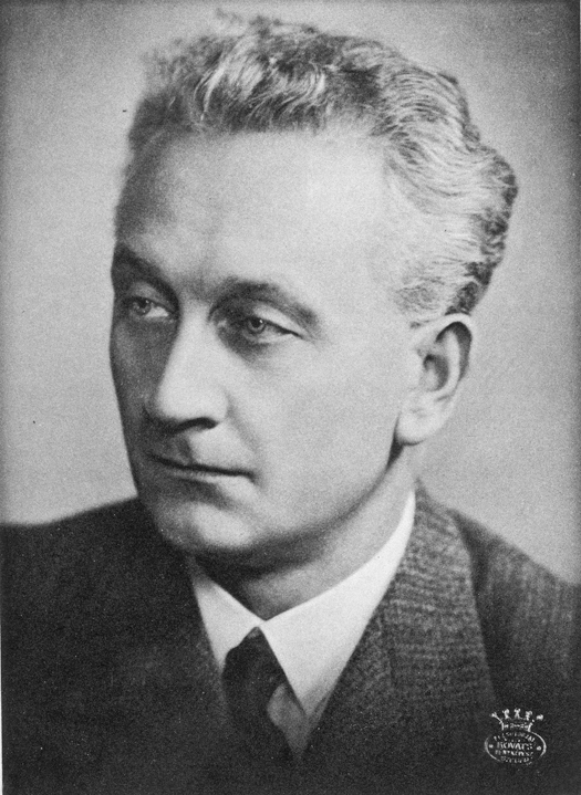
Albert Szent-Györgyi, M.D., Ph.D. (1893-1986)
But perhaps his greatest insight is one for which he is least known, although he devoted almost half his life to the subject. For on March 12, 1941, in a lecture delivered in Budapest, he boldly stood up before his peers and suggested to them that the discipline of biochemistry was obsolete and should be brought into the twentieth century. Living organisms, he told them, were not simply bags of water in which molecules floated like tiny billiard balls, forming chemical bonds with other billiard balls with which they happened to collide. Quantum theory, he said, had made such old ideas invalid; biologists needed to study solid state physics.
In his own specialty, although he had worked out the structures of the molecules involved in muscular contraction, he could not begin to fathom why they had those particular structures, nor how the molecules communicated with one another to coordinate their activities. He saw such unsolved problems everywhere he looked in biology. “One of my difficulties within protein chemistry,” he bluntly told his colleagues, “was that I could not imagine how such a protein molecule can ‘live.’ Even the most involved protein structural formula looks ‘stupid,’ if I may say so.”
The phenomena that had forced Szent-Györgyi to face these questions were the porphyrin-based systems of life. He pointed out that in plants, 2,500 chlorophyll molecules form a single functional unit, and that in dim light at least 1,000 chlorophyll molecules have to cooperate simultaneously in order to split one molecule of carbon dioxide and create one molecule of oxygen.
He spoke about the “enzymes of oxidation”—the cytochromes in our cells—and wondered, again, how the prevailing model could be correct. How could a whole series of large protein molecules be arranged geometrically so that electrons could wander directly from one to the other in a precise sequence? “Even if we could devise such an arrangement,” he said, “it would still be incomprehensible how the energy liberated by the passing of an electron from one substance to the other, viz., from one iron atom to the other, could do anything useful.”
Szent-Györgyi proposed that organisms are alive because thousands of molecules form single systems with shared energy levels, such as physicists were describing in crystals. Electrons don’t have to pass directly from one molecule to another, he said; instead of being attached to only one or two atoms, electrons are mobile, belong to the whole system, and transmit energy and information over large distances. In other words, the stuff of life is not billiard balls but liquid crystals and semiconductors.
Szent-Györgyi’s sin was not that he was incorrect. He wasn’t. It was his failure to respect the old animosity. Electricity and life were long divorced; the industrial revolution had been running full bore for a century and a half. Millions of miles of electric wires clothed the earth, exhaling electric fields that permeated all living things. Thousands of radio stations blanketed the very air with electromagnetic oscillations that one could not avoid. Skin and bones, nerves and muscles were not allowed to be influenced by them. Proteins were not permitted to be semiconductors. The threat to industry, economics, and modern culture would be too great.
So biochemists continued to think of proteins, lipids, and DNA as though they were little marbles drifting in a watery solution and colliding with one another at random. They even thought of the nervous system this way. When forced to, they admitted parts of quantum theory, but only on a limited basis. Biological molecules were still only permitted to interact with their immediate neighbors, not to act at a distance. It was okay to acknowledge modern physics only that much, like opening a small hole in a dam for knowledge to leak through one drop at a time, while the main structure is reinforced lest a flood demolish it.
Old knowledge about chemical bonds and enzymes in a water solution must now coexist with new models of electron transport chains. It was necessary to invent these to explain phenomena that were most central to life: photosynthesis and respiration. Large porphyrin-containing protein molecules no longer had to move and physically interact with one another in order for anything useful to happen. These molecules could stay put and electrons could shuttle between them instead. Biochemistry was becoming that much more alive. But it still had a long way to go. For even in the new models, electrons were constrained to move only, like little messenger boys, between one protein molecule and its immediate neighbor. They could cross the street, so to speak, but they couldn’t travel down a highway to a distant town. Organisms were still pictured essentially as bags of water containing very complex solutions of chemicals.
The laws of chemistry had explained a lot about metabolic processes, and electron transport now explained even more, but there was not yet an organizing principle. Elephants grow from tiny embryos, which grow from single brainless cells. Salamanders regenerate perfect limbs. When we are cut, or break a bone, cells and organs throughout our body mobilize and coordinate their activities to repair the damage. How does the information travel? How, borrowing Szent-Györgyi’s words, do protein molecules “live”?
Despite Szent-Györgyi’s sin, his predictions have proven correct. Molecules in cells do not drift at random to collide with one another. Most are firmly anchored to membranes. The water inside cells is highly structured and does not resemble the free-flowing liquid that sloshes around in a glass before you drink it. Piezoelectricity, a property of crystals that makes them useful in electronic products, that transforms mechanical stress into electrical voltages and vice versa, has been found in cellulose, collagen, horn, bone, wool, wood, tendon, blood vessel walls, muscle, nerve, fibrin, DNA, and every type of protein examined.10 In other words—something most biologists have been denying for two centuries—electricity is essential to biology.
Szent-Györgyi was not the first to challenge conventional thinking. It was Otto Lehmann, already in 1908, who, noticing the close resemblance between the shapes of known liquid crystals and many biological structures, proposed that the very basis of life was the liquid crystalline state. Liquid crystals, like organisms, had the ability to grow from seeds; to heal wounds; to consume other substances, or other crystals; to be poisoned; to form membranes, spheres, rods, filaments and helical structures; to divide; to “mate” with other forms, resulting in offspring that had characteristics of both parents; to transform chemical energy into mechanical motion.
After Szent-Györgyi’s daring Budapest lecture, others pursued his ideas. In 1949, Dutch researcher E. Katz explained how electrons could move through a semiconducting chlorophyll crystal during photosynthesis. In 1955, James Bassham and Melvin Calvin, working for the U.S. Atomic Energy Commission, elaborated on this theory. In 1956, William Arnold, at Oak Ridge National Laboratory, confirmed experimentally that dried chloroplasts—the particles in green plants that contain chlorophyll—have many of the properties of semiconductors. In 1959, Daniel Eley, at Nottingham University, proved that dried proteins, amino acids, and porphyrins are indeed semiconductors. In 1962, Roderick Clayton, also at Oak Ridge, found that photosynthetic tissues in living plants behave like semiconductors. In 1970, Alan Adler, at the New England Institute, showed that thin films of porphyrins do also. In the 1970s, biochemist Freeman Cope, at the United States Naval Air Development Center in Warminster, Pennsylvania, emphasized the importance of solid state physics for a true understanding of biology, as did biologist Allan Frey, the most active American researcher into the effects of microwave radiation on the nervous system at that time. Ling Wei, professor of electrical engineering at the University of Waterloo in Ontario, stated baldly that a nerve axon is an electrical transmission line and that its membrane is an ionic transistor. He said that the equivalent circuitry “can be found in any electronics book today,” and that “one can easily derive the nerve behavior from semiconductor physics.” When he did so, his equations predicted some of the properties of nerves that were, and still are, puzzling to physiologists.
In 1979, a young professor of bioelectronics at the University of Edinburgh published a book titled Dielectric and Electronic Properties of Biological Materials. The earlier work of Eley and Arnold had been criticized because the activation energies they had measured—the amount of energy necessary to make proteins conduct electricity—seemed to be too large. Supposedly there was not enough energy available in living organisms to lift electrons into the conduction band. Proteins might be made to conduct electricity in the laboratory, said the critics, but this could not happen in the real world. Eley and Arnold, however, had done all their work on dried proteins, not living ones. The young professor, Ronald Pethig, pointed out the obvious: water is essential to life, and proteins become more conductive if you added water to them. In fact, studies had shown that adding only 7.5 percent water increased the conductivity of many proteins ten thousandfold or more! Water, he proposed, is an electron donor that “dopes” proteins and turns them into good semiconductors.
The electronic role of living water had already been noted by others. Physiologist Gilbert Ling, realizing that cell water is a gel and not a liquid, developed his theory of the electronic nature of cells in 1962. More recently, Gerald Pollack, professor of bioengineering at the University of Washington, has taken up this line of investigation. He was inspired by Ling when they met at a conference in the mid-1980s. Pollack’s most recent book, The Fourth Phase of Water: Beyond Solid, Liquid, and Vapor, was published in 2011.
The late geneticist Mae-Wan Ho, in London, has clothed Szent-Györgyi’s ideas in garments that all can see. She developed a technique using a polarizing microscope that displayed, in vivid color, the interference patterns generated by the liquid crystalline structures that make up living creatures. The first animal she put under her microscope was a tiny worm—a fruit fly larva. “As it crawls along, it weaves its head from side to side flashing jaw muscles in blue and orange stripes on a magenta background,” she wrote in 1993 in her book, The Rainbow and the Worm: The Physics of Organisms. She and many others have urged that the liquid crystalline properties of our cells and tissues not only teach us about our chemistry, but have something special to tell us about life itself.
Włodzimierz Sedlak, pursuing Szent-Györgyi’s ideas in Poland, developed the discipline of bioelectronics within the Catholic University of Lublin during the 1960s. Life, he said, is not only a collection of organic compounds undergoing chemical reactions, but those chemical reactions are coordinated with electronic processes that take place in an environment of protein semiconductors. Other scientists working at the same university are continuing to develop this discipline theoretically and experimentally today. Marian Wnuk has focused on porphyrins as key to the evolution of life. He states that the principal function of porphyrin systems is an electronic one. Józef Zon, head of the Department of Theoretical Biology at the University, has focused on the electronic properties of biological membranes.
Oddly enough, the use of porphyrins in electronic products instructs us about biology. Adding thin films of porphyrins to commercially available photovoltaic cells increases the voltage, current, and total power output.11 Prototype solar cells based on porphyrins have been produced,12 as have organic transistors based on porphyrins.13
The properties that make porphyrins suitable in electronics are the same properties that make us alive. As everyone knows, playing with fire is dangerous; oxidation releases tremendous energy quickly and violently. How, then, do living organisms make use of oxygen? How do we manage to breathe and metabolize our food without being destroyed in a conflagration? The secret lies in the highly pigmented, fluorescent molecule called porphyrin. Strong pigments are always efficient energy absorbers, and if they are also fluorescent, they are also good energy transmitters. As Szent-Györgyi taught us in his 1957 book, Bioenergetics, “fluorescence thus tells us that the molecule is capable of accepting energy and does not dissipate it. These are two qualities any molecule must have to be able to act as an energy transmitter.”14
Porphyrins are more efficient energy transmitters than any other of life’s components. In technical terms, their ionization potential is low, and their electron affinity high. They are therefore capable of transmitting large amounts of energy rapidly in small steps, one low-energy electron at a time. They can even transmit energy electronically from oxygen to other molecules, instead of dissipating that energy as heat and burning up. That’s why breathing is possible. On the other side of the great cycle of life, porphyrins in plants absorb the energy of sunlight and transport electrons that change carbon dioxide and water into carbohydrates and oxygen.
Porphyrins, the Nervous System, and the Environment
There is one more place these surprising molecules are found: in the nervous system, the organ where electrons flow. In fact, in mammals, the central nervous system is the only organ that shines with the red fluorescent glow of porphyrins when examined under ultraviolet light. These porphyrins, too, perform a function that is basic to life. They occur, however, in a location where one might least expect to find them—not in the neurons themselves, the cells that carry messages from our five senses to our brain, but in the myelin sheaths that envelop them—the sheaths whose role has been almost totally neglected by researchers and whose breakdown causes one of the most common and least understood neurological diseases of our time: multiple sclerosis. It was orthopedic surgeon Robert O. Becker who, in the 1970s, discovered that myelin sheaths are really electrical transmission lines.
In a state of health the myelin sheaths contain primarily two types of porphyrins—coproporphyrin III and protoporphyrin—in a ratio of two to one, complexed with zinc. The exact composition is crucial. When environmental chemicals poison the porphyrin pathway, excess porphyrins, bound to heavy metals, build up in the nervous system as in the rest of the body. This disrupts the myelin sheaths and changes their conductivity which, in turn, alters the excitability of the nerves they surround. The entire nervous system becomes hyperreactive to stimuli of all kinds, including electromagnetic fields.
The cells surrounding our nerves were hardly even studied until recently. In the nineteenth century, anatomists, finding no apparent function for them, supposed that they must have only a “nutritive” and “supportive” role, protecting the “real” nerves that they surrounded. They named them glial cells after the Greek word for “glue.” The discovery of the action potential, which transmits signals along each neuron, and of neurotransmitters, the chemicals that carry signals from one neuron to the next, had ended the discussion. From then on, glial cells were thought to be little more than packing material. Most biologists ignored the fact, discovered by German physician Rudolf Virchow in 1854, that myelin is a liquid crystal. They did not think it was relevant.
However, working from the 1960s to the early 1980s and author, in 1985, of The Body Electric, Becker found quite another function for the myelin-containing cells and took another step toward restoring electricity to its proper role in the functioning of living things.
When he began his research in 1958, Becker was simply looking for a solution to orthopedists’ greatest unsolved problem: nonunion of fractures. Occasionally, despite the best medical care, a bone would refuse to heal. Surgeons, believing that only chemical processes were at work, simply scraped the fracture surfaces, devised complicated plates and screws to hold the bone ends rigidly together, and hoped for the best. Where this did not work, limbs had to be amputated. “These approaches seemed superficial to me,” Becker recalled. “I doubted that we would ever understand the failure to heal unless we truly understood healing itself.”15
Becker began to pursue the ideas of Albert Szent-Györgyi, thinking that if proteins were semiconductors, maybe bones were too, and maybe electron flow was the secret to the healing of fractures. Ultimately he proved that this was correct. Bones were not just made of collagen and appatite, as he was taught in medical school; they were also doped with tiny amounts of copper, much as silicon wafers in computers are doped with tiny amounts of boron or aluminum. The presence of greater or lesser amounts of metal atoms regulates the electrical conductivity of the circuitry—in bones as in computers. With this understanding, Becker designed machines that delivered miniscule electric currents—as small as 100 trillionths of an ampere—to fractured bones to stimulate the healing process, with great success: his devices were the forerunners of machines that are used today by orthopedic surgeons in hospitals throughout the world.
Becker’s work on the nervous system is less well known. As already mentioned, the functioning of neurons had been worked out, up to a point, in the nineteenth century. They transmit enormous amounts of information to and from the brain at high speed, including data about one’s environment, and instructions to one’s muscles. They do this via the familiar action potential and neurotransmitters. And since the action potential is an all-or-nothing event, neuron signaling is an on-off digital system like today’s computers. But Becker thought that this could not explain the most important properties of life; there had to be a slower, more primitive, and more sensitive analog system that regulates growth and healing, that we inherited from lower forms of life—a system that might be related to the acupuncture meridians of Chinese medicine, which western medicine also made no attempt to understand.
A number of researchers before Becker, among them Harold Saxton Burr at Yale, Lester Barth at Columbia, Elmer Lund at the University of Texas, Ralph Gerard and Benjamin Libet at the University of Chicago, Theodore Bullock at U.C.L.A., and William Burge at the University of Illinois, had measured DC voltages on the surfaces of living organisms, both plants and animals, and embryos. Most biologists paid no attention. After all, certain DC currents, called “currents of injury,” were well known, and were thought to be well understood. They had been discovered by Carlo Matteucci as long ago as the 1830s. Biologists had assumed, for a century, that these currents were meaningless artifacts, caused simply by ions leaking out of wounds. But when, in the 1930s and 1940s, a growing number of scientists, using better techniques, began to find DC voltages on all surfaces of all living things, and not just on the surfaces of wounds, a few began to wonder whether those “currents of injury” just might be a bit more important than they had learned in school.
The accumulated work of these scientists showed that trees,16 and probably all plants, are polarized electrically, positive to negative, from leaves to roots, and that animals are similarly polarized from head to feet. In humans potential differences of up to 150 millivolts or more could sometimes be measured between one part of the body and another.17
Becker was the first to map the charge distribution in an animal in some detail, accomplishing this with salamanders in 1960. The places of greatest positive voltage, he found, as measured from the back of the animal, were the center of the head, the upper spine over the heart, and the lumbosacral plexus at the lower end of the spine, while the places of greatest negative voltage were the four feet and the end of the tail. In addition, the head of an alert animal was polarized from back to front, as though an electric current were always flowing in one direction through the middle of its brain. However, when an animal was anesthetized the voltage diminished as the anesthetic took effect, and then the head reversed polarity when the animal lost consciousness. This suggested to him a novel method of inducing anesthesia, and when Becker tried it, it worked like a charm. In the salamander, at least, passing an electric current of only 30 millionths of an ampere from front to back through the center of its head caused the animal to become immediately unconscious and unresponsive to pain. When the current was turned off, the animal promptly woke up. He observed the same back-to-front polarity in alert humans, and the same reversal during sleep and anesthesia.18
While Becker did not try it himself, even tinier electric currents have been used in psychiatry to put humans to sleep since about 1950 in Russia, Eastern Europe, and Asian countries that were once part of the Soviet Union. In these treatments, current is sent from front to back through the midline of the head, reversing the normal polarity of the brain, just as Becker did with his salamanders. The first publications describing this procedure specified short pulses of 10 to 15 microamperes each, 5 to 25 times per second, which gave an average current of only about 30 billionths of an ampere. Although larger currents will cause immediate unconsciousness in a human, just like in a salamander, those tiny currents are all that is necessary to put a person to sleep. This technique, called “electrosleep,” has been used for over half a century to treat mental disorders, including manic-depressive illness and schizophrenia, in that part of the world.19
The normal electrical potentials of the body are also necessary for the perception of pain. The abolition of pain in a person’s arm, for example, whether caused by a chemical anesthetic, hypnosis, or acupuncture, is accompanied by a reversal of electrical polarity in that arm.20
By the 1970s it had become clear to the researchers who were looking into such things that the DC potentials they were measuring played a key role in organizing living structures. They were necessary for growth and development.21 They were also needed for regeneration and healing.
Tweedy John Todd demonstrated as long ago as 1823 that a salamander cannot regenerate a severed leg if you destroy that leg’s nerve supply. So for a century and a half, scientists searched for the chemical signal that must be transmitted by nerves to trigger growth. No one ever found one. Finally, embryologist Sylvan Meryl Rose, in the mid-1970s at Tulane University, proposed that maybe there was no such chemical, and that the long-sought signal was purely electrical. Could the currents of injury, he asked, that had previously been considered mere artifacts, themselves play a central role in healing?
Rose found that they did. He recorded the patterns of the currents in the wound stumps of salamanders as they regenerated their severed limbs. The end of the stump, he found, was always strongly positive during the first few days after injury, then reversed polarity to become strongly negative for the next couple of weeks, finally reestablishing the weakly negative voltage found on all healthy salamander legs. Rose then found that salamanders would regenerate their legs normally, even without a nerve supply, provided he carefully duplicated, with an artificial source of current, the electrical patterns of healing that he had observed. Regeneration would not take place if the polarity, magnitude, or sequence of currents were not correct.
Once having established that the signals that trigger regeneration are electrical and not chemical in nature, these scientists were in for yet another surprise. For the DC potentials of the body that, as we have seen, are necessary not just for regeneration but for growth, healing, pain perception, and even consciousness, seemed to be generated not in the “real” nerves but in the myelin-containing cells that surround them—the cells that also contain porphyrins. Proof came by accident while Becker was again working on the problem of why some bone fractures fail to mend. Since he had already learned that nerves were essential to healing, he tried, in the early 1970s, to create an animal model for fractures that do not heal by severing the nerve supply to a series of rats’ legs before breaking them.
To his surprise, the leg bones still healed normally—with a six-day delay. Yet six days was not nearly enough time for a rat to regenerate a severed nerve. Could bones be an exception, he wondered, to the rule that nerves are needed for healing? “Then we took a more detailed look at the specimens,” wrote Becker. “We found that the Schwann cell sheaths were growing across the gap during the six-day delay. As soon as the perineural sleeve was mended, the bones began to heal normally, indicating that at least the healing, or output, signal, was being carried by the sheath rather than the nerve itself. The cells that biologists had considered merely insulation turned out to be the real wires.”22 It was the Schwann cells, Becker concluded—the myelin-containing glial cells—and not the neurons they surrounded, that carried the currents that determined growth and healing. And in a much earlier study Becker had already shown that the DC currents that flow along salamander legs, and presumably along the limbs and bodies of all higher animals, are of semiconducting type.23
Which brings us full circle. The myelin sheaths—the liquid crystalline sleeves surrounding our nerves—contain semiconducting porphyrins,24 doped with heavy metal atoms, probably zinc.25 It was Harvey Solomon and Frank Figge who, in 1958, first proposed that these porphyrins must play an important role in nerve conduction. The implications of this are especially important for people with chemical and electromagnetic sensitivities. Those of us who, genetically, have relatively less of one or more porphyrin enzymes, may have a “nervous temperament” because our myelin is doped with slightly more zinc than our neighbors’ and is more easily disturbed by the electromagnetic fields (EMFs) around us. Toxic chemicals and EMFs are therefore synergistic: exposure to toxins further disrupts the porphyrin pathway, causing the accumulation of more porphyrins and their precursors, rendering the myelin and the nerves they surround still more sensitive to EMFs. According to more recent research, a large excess of porphyrin precursors can prevent the synthesis of myelin and break apart the myelin sheaths, leaving the neurons they surround naked and exposed.26
The true situation is undoubtedly more complex than this, but to put all the pieces correctly together will require researchers who are willing to step outside our cultural blinders and acknowledge the existence of electrical transmission lines in the nervous systems of animals. Already, mainstream science has taken the first step by finally acknowledging that glial cells are much more than packing material.27 In fact, a discovery by a team of researchers at the University of Genoa is currently revolutionizing neurology. Their discovery is related to breathing.28
Everyone knows that the brain consumes more oxygen than any other organ, and that if a person stops breathing, the brain is the first organ to die. What the Italian team confirmed in 2009 is that as much as ninety percent of that oxygen is consumed not by the brain’s nerve cells, but by the myelin sheaths that surround them. Traditional wisdom has it that the consumption of oxygen for energy takes place only in tiny bodies inside cells called mitochondria. That wisdom has now been turned on its head. In the nervous system, at least, most of the oxygen appears to be consumed in the multiple layers of fatty substance called myelin, which contain no mitochondria at all, but which forty-year-old research showed contains non-heme porphyrins and is semiconducting. Some scientists are even beginning to say that the myelin sheath is, in effect, itself a giant mitochondrion, without which the huge oxygen needs of our brain and nervous system could never be met. But to truly make sense of this collection of facts will also require the recognition that both the neurons, as Ling Wei proposed, and the myelin sheaths that envelop them, as Robert Becker proposed, work together to form a complex and elegant electrical transmission line system, subject to electrical interference just like transmission lines built by human engineers.
The exquisite sensitivity of even the normal nervous system to electromagnetic fields was proven in 1956 by zoologists Carlo Terzuolo and Theodore Bullock—and then ignored by everyone since. In fact, even Terzuolo and Bullock were astonished by the results. Experimenting on crayfish, they found that although a substantial amount of electric current was needed to cause a previously silent nerve to fire, incredibly tiny currents could cause an already firing nerve to alter its firing rate tremendously. A current of only 36 billionths of an ampere was enough to increase or decrease a nerve’s rate of firing by five to ten percent. And a current of 150 billionths of an ampere—thousands of times less than is widely assumed, still today, by developers of modern safety codes, to have any biological effect whatever—would actually double the rate of firing, or silence the nerve altogether. Whether it increased or decreased the activity of the nerve depended only on the direction in which the current was applied to the nerve.
The Zinc Connection
The role of zinc was discovered in the 1950s by Henry Peters, a porphyrinologist at the University of Wisconsin Medical School. Like Morton after him, Peters was impressed by the number of people who seemed to have mild or latent porphyria, and thought the trait was far more prevalent that was commonly believed.29
Peters discovered that his porphyria patients who had neurological symptoms were excreting very large amounts of zinc in their urine—up to 36 times normal. In fact, their symptoms correlated better with the levels of zinc in their urine than with the levels of porphyrins they were excreting. With this information, Peters did the most logical thing: in scores of patients, he tried chelation to reduce the body’s load of zinc, and it worked! In patient after patient, when courses of treatment with BAL or EDTA had reduced the level of zinc in their urine to normal, their illness resolved, and the patient remained symptom-free for up to several years.30 Contrary to conventional wisdom, which assumes that zinc deficiency is common and should be supplemented, Peters’ patients, because of their genetics and their polluted environment, were actually zinc-poisoned—as at least five to ten percent of the population, with hidden porphyria, may also be.
For the next forty years Peters found tremendous resistance to his idea that zinc toxicity was at all common, but growing evidence is now accumulating that this is so. Large amounts of zinc are in fact entering our environment, our homes, and our bodies from industrial processes, galvanized metals, and even the fillings in our teeth. Zinc is in denture cream and in motor oil. There is so much zinc in automobile tires that their constant erosion makes zinc one of the main components of road dust—which washes into our streams, rivers, and reservoirs, eventually getting into our drinking water.31 Wondering whether this was perhaps poisoning us all, a group of scientists from Brookhaven National Laboratory, the United States Geological Survey, and several universities raised rats on water supplemented with a low level of zinc. By three months of age, the rats already had memory deficits. By nine months of age, they had elevated levels of zinc in their brains.32 In a human experiment, pregnant women in a slum area of Bangladesh were given 30 milligrams of zinc daily, in the expectation that this would benefit the mental development and motor skills of their babies. The researchers found just the opposite.33 In a companion experiment, a group of Bangladeshi infants were given 5 milligrams of zinc daily for five months, with the same surprising result: the supplemented infants scored more poorly on standard tests of mental development.34 And a growing body of literature shows that zinc supplements worsen Alzheimer’s disease,35 and that chelation therapy to reduce zinc improves cognitive functioning in Alzheimer’s patients.36 An Australian team who examined autopsy specimens found that Alzheimer’s patients had double the amount of zinc in their brains as people without Alzheimer’s, and that the more severe the dementia, the higher the zinc levels.37
Nutritionists have long been misled by using blood tests to judge the body’s stores of zinc; scientists are finding out that blood levels are not reliable, and that unless you are severely malnourished there is no relation between the amount of zinc in your diet and the level of zinc in your blood.38 In some neurological diseases, including Alzheimer’s disease, it is common to have high levels of zinc in the brain while having normal or low levels of zinc in the blood.39 In a number of diseases including diabetes and cancer, urinary zinc is high while blood zinc is low.40 It appears that the kidneys respond to the body’s total load of zinc, and not to the levels in the blood, so that blood levels can become low, not because of a zinc deficiency but because the body is overloaded with zinc and the kidneys are removing it from the blood as fast as they can. It also appears to be much more difficult than we used to think for people to become deficient by eating a zinc-poor diet; the body is amazingly capable of compensating for even extremely low levels of dietary zinc by increasing intestinal absorption and decreasing excretion through urine, stool, and skin.41 While the recommended dietary allowance for adult males is 11 milligrams per day, a man can take in as little as 1.4 milligrams of zinc a day and still maintain homeostasis and normal levels of zinc in the blood and tissues.42 But a person who increases his or her daily intake beyond 20 milligrams may risk toxic effects in the long term.
Canaries in the Mine
In our cells, the manufacture of heme from porphyrins can be inhibited by a large variety of toxic chemicals, and not—so far as we know—by electricity. But we will see in the coming chapters that electromagnetic fields interfere with the most important job that this heme is supposed to do for us: enabling the combustion of our food by oxygen so that we can live and breathe. Like rain on a campfire, electromagnetic fields douse the flames of metabolism. They reduce the activity of the cytochromes, and there is evidence that they do so in the simplest of all possible ways: by exerting a force that alters the speed of the electrons being transported along the chain of cytochromes to oxygen.
Every person on the planet is affected by this invisible rain that penetrates into the fabric of our cells. Everyone has a slower metabolism, is less alive, than if those fields were not there. We will see how this slow asphyxiation causes the major diseases of civilization: cancer, diabetes, and heart disease. There is no escape. Regardless of diet, exercise, lifestyle, and genetics, the risk of developing these diseases is greater for every human being and every animal than it was a century and a half ago. People with a genetic predisposition simply have a greater risk than everyone else, because they have a bit less heme in their mitochondria to start with.
In France, liver cancer was found to be 36 times as frequent in people carrying a gene for porphyria as in the general population.43 In Sweden and Denmark the rate was 39 times as high, and the lung cancer rate triple the general rate.44 Chest pain, heart failure, high blood pressure, and EKGs suggestive of oxygen starvation are well known in porphyria.45 Porphyria patients with normal coronary arteries often die of heart arrhythmias46 or heart attacks.47 Glucose tolerance tests and insulin levels are usually abnormal.48 In one study, 15 of 36 porphyria patients had diabetes.49 The protean manifestations of this disease, capable of affecting almost any organ, are widely blamed on impaired cellular respiration due to a deficiency of heme.50 Indeed, no porphyrin expert has offered a better explanation.
The five to ten percent of the population who have lower porphyrin enzyme levels are the so-called canaries in the coal mine, whose songs of warning, however, have been tragically ignored. They are the people who came down with neurasthenia in the last half of the nineteenth century when telegraph wires swept the world; the victims of sleeping pills in the late 1880s, of barbiturates in the 1920s, and of sulfa drugs in the 1930s; the men, women, and children with multiple chemical sensitivity, poisoned by the soup of chemicals that have rained on us since World War II; the abandoned souls with electrical sensitivity left behind by the computer age, forced into lonely exile by the inescapable radiation of the wireless revolution.
In Part Two of this book we will see just how extensively the general population of the world has been affected as a result of the failure to heed their warnings.