The search for the pain gene began in an Alabama neighborhood with a group of men and women carrying groceries, talking with each other, tending to their children, or driving down the street. Ordinary, at first glance. But, many of the people did not wear regular shoes. Some wore open toed sandals. Others preferred not to wear anything on their feet, to walk barefoot on a cool tile floor, or in the cold water that collected in puddles. The children avoided the playground. They sometimes missed school days. And, if you spent time with these people, you might hear a person say, “I’m getting an attack.” Then, the affected person would grimace, their feet turning bright red, as if they had been badly sunburned. If asked, they would say that their feet, and sometimes their hands, felt as if they were on fire. And, if cold water or ice were available, they might place their red feet in it.
As striking as the pain in these people was, it was also unusual in another way: The pain and redness did not occur in everybody in the neighborhood. It was present in, and only in, one large, extended family. Parents, aunts, uncles, and children suffered from this fire-like pain, but neighbors from other families did not have it. Five generations were known to have this mysterious disorder, about half of the individuals in each generation.
What was going on in this Alabama family with excruciating burning pain and red feet? Doctors were baffled and could not make a diagnosis; some even wondered whether it was a physical disorder at all, or whether it was “in the mind.” But it was not imagined, and it was not a creation of the mind. We now know that this family suffered from and continues to suffer from the “man on fire” syndrome. The medical names for this disorder are erythermalgia and erythromelalgia. In this book we will call it erythromelalgia.
Erythromelalgia is incredibly rare. Most physicians will never see a case. But it is a striking disorder, and, once a physician has seen a case, it remains in his or her memory because it is so unusual. Erythromelalgia was given its name by neurologist S. Weir Mitchell in 1878. Its name, from Greek, connotes some of its main features: erythros (“red”), melos (“limb”), and algos (“pain”). Some still refer to this disorder of red limb pain as Mitchell’s disease or Weir Mitchell disease.
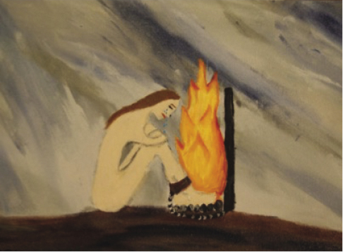
People with erythromelalgia suffer from periodic attacks of excruciating burning pain. To describe their pain, they use terms like “being on fire,” “being scalded,” or “feeling like hot lava has been poured into my body.” A picture entitled “Chained to Fire,” prepared by a fourteen-year-old girl to depict her erythromelalgia, is shown in figure 3.1.
Figure 3.1 A drawing depicting the pain of erythromelalgia, entitled “Chained to Fire,” prepared by Bailey Deacon when she was fourteen years old, and submitted to an art contest sponsored by The Erythromelalgia Association in 2012. As in many patients with erythromelalgia, the pain is most severe in the feet. Reproduced courtesy of Bailey Deacon, Todd Deacon, and The Erythromelalgia Association.
The burning pain of erythromelalgia is usually symmetrical—both sides of the body tend to be affected—most commonly the feet, sometimes the hands, and occasionally the tip of the nose or ears. Superimposed on a lower level of ongoing discomfort, the severe pain of erythromelalgia comes in bursts or attacks. These are triggered by mild warmth—such as the subtle warmth that comes from putting on shoes or socks, entering a warm room, or even mild exercise like walking. On a warm day, a walk across a parking lot can be enough to trigger severe pain. The pain attacks are accompanied by redness of the affected limbs. On a scale of 0 (“no pain”) to 10 (“worst pain I can imagine”), people with erythromelalgia describe their pain during attacks as a 7, 8, and too often a 9 or 10.
The pain in erythromelalgia is relieved by coolness. Characteristically, people with erythromelalgia will seek out cold places, walking barefoot in cool weather, and immersing their burning limbs in buckets of cold water or ice. This can lead to tissue injury, or worse, gangrene. Indeed, the literature contains reports of people with erythromelalgia who have sustained limb amputations or gone into septic shock as a result of infections due to skin breakdown from excessive cooling. Existing medications tend not to be helpful or are only partially helpful. Use of opiate medications is common, and death by overdose has occurred. A few patients have requested amputation of limbs because the pain is so severe, a maneuver that has in most cases not been helpful in the long run.
Erythromelalgia can occur in the context of other, more common disorders such as diabetes, multiple sclerosis, or disorders of the blood such as polycythemia vera in which the bone marrow produces too many blood cells. Erythromelalgia can also occur in isolation, where it is called “primary erythromelalgia,” or “primary erythermalgia.” About 5% of cases of erythromelalgia are now known to occur as an inherited disorder, as a result of mutations in a gene—these are called “inherited erythromelalgia.”
The human body is made up of cells—skin cells, muscle cells, blood cells, kidney cells, and many other types of cells, including nerve cells. Cells, in turn, contain protein molecules. Protein molecules are responsible for many of the activities of cells that keep them alive and allow them to perform properly in the body. Proteins are complex molecules assembled from smaller pieces called amino acids. There are twenty amino acids in humans. The amino acids line up in precise order, held together like links in a chain, to form a protein. One can also visualize the amino acids, in precise sequence, as being like a carefully designed string of multicolored beads. The identities of the amino acids—the colors of the beads, and the precise order in which they are strung together—are essential to the proper structure and function of the protein.
To form a functional protein molecule that can work properly within the body, the protein must contain the correct amino acids, in the correct order, and the string of amino acids must be folded into one particular conformation. Imagine scrunching up a string of colored beads in one’s hand, so that bead number 101 lies next to bead number 148, and bead 160 touches bead 194, and so on. Within the scrunched necklace, the correct sequence of beads, which come in twenty colors, and a very precise folding configuration are necessary for the overall, three-dimensional shape of the protein to be correct, so that the protein can work properly. One bead of the wrong color—one wrong amino acid—or a missing bead, and the string may not fold into the needed three-dimensional structure. And with that incorrect configuration, the protein may not work properly. That is what happens, for example, in sickle cell anemia, where one of the 146 amino acids is substituted by another, incorrect amino acid within the β-globin component of hemoglobin, an iron-containing protein that plays an essential role, transporting oxygen within the blood. One bead of the wrong color, and hemoglobin does not work properly.
The blueprint for proteins is contained in the human genome. Each of our cells contain twenty-three pairs of chromosomes, a total of forty-six. These contain the 20,000 genes in the human genome. The gene for a particular protein encodes the amino acids, in precise sequence, that make up that protein. There are two copies of each gene, one from an individual’s father, and the other from his or her mother. Genes are made of DNA (deoxyribonucleic acid) and consist of two strands, coiled around each other to form a double helix, that contain smaller molecules called nucleotides. For their discovery of the double helix configuration of DNA, James Watson, Francis Crick, and Maurice Wilkins were awarded the Nobel Prize in 1962.
There are four types of nucleotides within DNA: They are labeled A, T, G, C, and they can be considered as the letters within the alphabet of the genome. Since there are twenty amino acids which must be encoded with this alphabet, and only four letters to use, a series of three nucleotides is needed to encode each amino acid. It is as if each amino acid is identified by a code word of three letters, or three nucleotides. In 1961, at the age of 34, Marshall Nirenberg, working at the National Institutes of Health (NIH), made a stunning discovery that was a first step toward breaking the code. His experiments showed that the triplet TTT encodes the amino acid phenylalanine (Nirenberg and Matthaei 1961). He went on to a Nobel Prize, which he shared with Robert Holley and Har Gobind Khorana. Severo Ochoa (who had received a Nobel in 1959 for his work on the synthesis of RNA) went on to complete the identification of the DNA codes for all twenty amino acids.
By the mid-1960s it was clear, for example, that ATG codes for the amino acid methionine, that CCA codes for proline, and that GC, followed by any of the four nucleotides, codes for the amino acid alanine. And so forth for each amino acid. So, a stretch of a protein made up of a methionine, then a proline, and then an alanine would be encoded as follows:
ATGCCAGCT
This was the “Rosetta Stone” of genetics. By knowing the sequence of nucleotides within a gene, one could discern, amino acid by amino acid, the precise sequence of amino acids within a protein. Researchers in molecular biology laboratories were exhilarated. The genome, they hoped, would hold the key to understanding life.
Medical science moves forward in waves. Sometimes racing, sometimes crawling. And, not infrequently, multiple waves move medical research ahead on several fronts in parallel at the same time, like waves in the ocean, crashing simultaneously on different parts of a beach. This seems to have been true for the man on fire syndrome, because just as these advances in understanding DNA were occurring, physicians were beginning to recognize that this disorder had to be caused by an abnormality, hidden somewhere in the 20,000 genes that make up the human genome.
Every family has its heroes. Within the Alabama family, there was a person who recognized that, either then or sometime in the future, something might be done to help people with the man on fire syndrome.
In 1965, at the urging of a pediatrician, a mother in the Alabama family took her young daughter, with burning feet, to the Mayo Clinic in Rochester, Minnesota, a thousand miles away. It must have taken immense effort to arrange for a consultation so far away.
The visit was worth the effort.
The Mayo physicians recognized the girl’s disorder as Mitchell’s disease and appreciated that, in this case, it occurred as a familial or genetic disease. By the mid-1960s, the Alabama family knew that they had a very unusual disorder, and that it had a name, “familial erythromelalgia.” In a brief paper in the Journal of Laboratory and Clinical Medicine, Mayo physician Mahlon Burbank and two colleagues noted that “we have had the opportunity to study a family in which 19 out of 51 family members, comprising 5 generations, have typical erythromelalgia. Study of this sibship indicates that the disorder in this family is inherited as a dominant trait” (Burbank, Spittel, and Fairbairn 1966). Now it was clear that the pain in this family was related to genes. The pain and redness arose in the genes, not in the mind.
Birmingham, Alabama, is fortunate to be the home of a medical school with a strong tradition of research, at the University of Alabama Birmingham. UAB was fortunate, in turn, to have on its faculty a medical geneticist, Dr. Wayne Finley. Following a period of training at the Institute for Medical Genetics at the University of Uppsala, Sweden, Finley and his wife, Dr. Sara Crews Finley, established at UAB the first medical genetics program in the southeastern United States. Over the ensuing years, the Finleys, either individually or together, published more than 250 professional abstracts, articles, and chapters on various aspects of medical genetics.
In 1986 a young girl in the Alabama family was seen at a local clinic for a urinary tract infection. The pediatrician was struck by her unusual condition and suggested that the child see Dr. Finley, who was a friend. Recognizing the girl’s red, hot feet, Finley read the 1966 Burbank paper, contacted Burbank, and presciently decided to create a family tree. Finley did not have a grant to fund the work, but he persuaded the Departments of Dermatology and Pediatrics at UAB to partner with him in supporting it.
The first full-length article in the scientific literature on the Alabama family was published in 1992 by Finley and four other authors (Finley et al. 1992). Burbank was listed as the last author. The summary at the beginning of this pivotal paper notes that it “updates the family reported by Burbank 1966,” although the introduction states that the initial patient studied for this paper “was different than Burbank’s (and) we did not realize we were studying the same extended family.” The paper carefully described the pedigree of the Alabama family—twenty-nine affected persons in five generations—and outlined their clinical features. It correctly posited that the disease “may be an autosomal dominant trait,” in which a person must inherit one mutated copy of a gene from their affected parent to get the disease. Noting that “additional families must be studied,” Finley and his coworkers concluded that “the mechanism for initiation of pain is not yet known.” Indeed, Finley could not know—for the methods were not yet available—that mutations in one particular gene, one out of 20,000, were the cause of inherited erythromelalgia not just in this family, but in people with inherited erythromelalgia around the world.
A large family, containing multiple individuals with a rare disorder, presents an opportunity for medical researchers. Which one, out of the thousands of genes in the human genome, is responsible for their disorder? And what has gone wrong within that gene to cause disease? There was a lot of territory to cover, and a first step was to narrow the field using an assessment called “linkage analysis.”
Linkage analysis takes advantage of the fact that all genes contain “single nucleotide polymorphisms” or SNPs. Each SNP is a minor variation in a gene, the substitution of a nucleotide for the one present in the majority of the population. A SNP can be thought of as a relatively inconsequential “mis-weave.” SNPs do not necessarily cause disease. However, they provide markers that geneticists can use to study that gene. Polymorphisms tend to be inherited, like mutations, and polymorphisms located on the same gene as a mutation, especially polymorphisms located close to the mutation within the gene, tend to be inherited along with, or “linked” to, the mutation. It is relatively straightforward to map the pattern of polymorphisms within a family, and if the polymorphism is present in all of the affected family members and none of the nonaffected members, it suggests that that gene containing the SNP is the site of the mutation. Depending on the pattern of inheritance and the SNPs that are studied, this type of linkage analysis can point, not just to a candidate gene, but to a specific region within that gene.
Importantly, linkage analysis depends on probabilities: What is the probability that a particular gene, or a particular region within a gene, is related to a disease? If there is just a single affected family member, an apparent association of a polymorphism linked with the disease could be a random event and is not necessarily an indication of disease causation. It is only when multiple affected family members show the same pattern of linkage that the probability of a random association goes down, and the probability of having found the culprit gene goes up.
The Alabama family was nearly ideal for linkage analysis: a disease that is dramatic in its clinical presentation and thus easy to recognize, and a large number of affected family members in multiple generations. The Alabama family offered a good chance of pointing the way to a gene for pain. It is not surprising that medical researchers wanted to study that family.
Well before it was recognized as a genetic disorder, Joost Drenth had investigated erythromelalgia as a medical student in The Netherlands. Together with his mentor Professor Jan Michiels at the Erasmus Medical Center in Rotterdam, he had revised the diagnostic classification of various forms of erythromelalgia and described the development of erythromelalgia as a complication of treatment with certain medications (Drenth 1989; Drenth and Michiels 1990). By the mid-1990s Drenth, now trained in medical genetics in Paris, was searching for a family with erythromelalgia, the larger the better. As he worked to find a family, he discovered a pedigree of a family with erythromelalgia in a textbook on medical genetics. By 1995 he had written to American physicians, trying to find this family. His initial inquiries failed to elicit a response, and this closed the book for several years. In 1998 Drenth learned that Dr. Michiels had contacted Dr. Finley to ask for DNA from the Alabama family, and arranged for Dr. Peter Heutink at the Erasmus Medical Center, an expert on linkage studies, to attempt to do this type of analysis. The project had stalled, however, because there were inconsistencies in a few patients in the link between clinical status and genetic status.
Now working in Nijmegen, where he had been appointed professor of internal medicine, Drenth contacted Michiels in early 1999, offering to trace new families. Alternatively, he suggested, he might be able to resolve the incongruences. Later that year, Drenth’s detective work paid off as he resolved the inconsistencies in Dr. Finley’s family. Now the linkage analysis could move ahead. The resulting paper, which included Drenth, Finley, Michiels, and Heutink as authors, was entitled “The Primary Erythermalgia-Susceptibility Gene Is Located on Chromosome 2q31-32” (Drenth et al. 2001). The analysis showed that the gene was located on chromosome 2. Chromosome 2 is a large chromosome that contains nearly 1,500 genes. Drenth’s study pointed to the gene’s being located between two markers within a particular, small region of the chromosome, about 3% of its total size. The linkage analysis had narrowed the search to a specific portion of chromosome 2, termed q31–32. At that time, sodium channel genes had not yet been mapped to that region, so these investigators could not know that their results pointed toward a sodium channel gene. Nevertheless, their result was important: It had limited the search from 20,000 genes to about 50. The search for the pain gene no longer required finding a needle in a haystack. Now the search could focus on a small but still formidable tangle of hay.
The search now moved to Beijing where a young dermatologist, Yong Yang, was seeing patients within a major referral clinic at the Peking University First Hospital. Yong Yang was interested in genetic causes of dermatological disease, and he had become aware of a Chinese family containing three generations of patients with burning pain and redness in their hands and feet. The disorder began in each of the patients in early childhood. In each, reddening of the skin and severe pain were evoked by warmth and relieved by cooling.
As with the Alabama family, a first step in analyzing this Chinese family was to do a linkage analysis. And as with the Alabama family, this analysis pointed to a well-defined region within chromosome 2. By now, however, it was known that this region of chromosome 2 contained a cluster of sodium channel genes, including SCN9A, the gene encoding sodium channel NaV1.7. Every gene within this part of chromosome 2 was a candidate as a potential culprit, but there was something special about SCN9A: NaV1.7 is present at high levels within pain-signaling DRG neurons.
Together with colleagues at the Chinese National Human Genome Center, Yang began the task of sequencing the coding portions of the SCN9A gene from this family. The results, a sequence of letters—one for each nucleotide—revealed the solution to a puzzle. They showed that one nucleotide (one out of thousands making up the gene) had been changed:
AAC CTC ACC
had been changed to
AAC CAC ACC
This change, of nucleotide A for nucleotide T at position 2573 within exon 15 of the gene, indicated that there was a “missense mutation,” a mutation that substitutes a histadine for a leucine at position 858, within the mutant NaV1.7 channel. The same L858H change was found in other affected individuals within the family. Importantly, the L858H substitution was not present in unaffected family members. The family was not a huge one, and in the absence of information about the effect of the mutation on function of the NaV1.7 channel, the analysis suggested, but did not prove, that the mutation caused the disease. Yang and his colleagues had another advantage, however: a very large population available in China for them to study. Within this large population, in another patient with a similar but sporadic pain syndrome, they found another mutation, I848T, substituting a threonine for an isoleucine, at a nearby position in precisely the same gene. Again, the mutation was not present in unaffected relatives.
Yong Yang’s paper (Yang et al. 2004), entitled “Mutations in SCN9A, Encoding a Sodium Channel Alpha Subunit, in Patients with Primary Erythermalgia,” appeared in the March 2004 issue of the Journal of Medical Genetics. When I saw the title of the article, I initially told my team that it was a bad day and retreated to my office. We had hoped to find families with inherited pain so we could sequence their sodium channel genes, and it appeared that we had been scooped.
It was only after a cup of black coffee and reading the article that I realized what had happened. Yong Yang and his colleagues had indeed identified two mutations in the SCN9A gene encoding NaV1.7 in an inherited pain disorder, the man on fire syndrome. This was an important step forward. But finding the mutations did not prove that they caused these patients’ pain. Appropriately, Yang el al. ended their paper with the conclusion “Mutations in SCN9A may cause primary erythermalgia.” More work was needed to move from “may cause” to “do cause,” to show that the mutations actually caused the disorder.
Yang and his colleagues were dermatologists and medical geneticists, but they were not neuroscientists. They did not ask the questions that a neuroscientist would have asked, that would establish a causal role for the mutations. Now it was time for us to ask those questions. Some mutations produce amino acid substitutions that are detrimental to channel function and cause disease. But other mutations produce amino acid substitutions that are not detrimental to channel function and do not cause disease. When a mutation of an ion channel such as a sodium channel is encountered by a neuroscientist or channel biologist, it immediately triggers these questions: Does the mutation change the functional properties of the channel, that is, does it change the manner in which the channel works? If so, in what ways? If the mutation changes the functional characteristics of the channel, does that mean that nerve cells carrying the mutant channel will function in an abnormal way? And finally, if the mutation produces changes in the function of nerve cells, can these changes explain the disease? These questions—the “functional profiling” of the mutant channels which might link them to disease—had to be asked. And we were in a position to answer those questions. What had appeared to be a bad day was, in fact, a good day. My colleagues reassembled in my office. There was a lot of work to do. The ball was now in our court.
We rapidly planned the “must do” experiments. Each of the mutations substituted a single amino acid in a part of the channel called the “S4–S5 linker.” The linker acts as a hinge connecting the voltage sensor which controls the state of the channel with the channel pore, which must open to produce an electrical current. We needed, therefore, to assess the effects of the mutation on opening or gating of the channel. Serendipity now came into play. Five years earlier, we had carried out a detailed analysis of the “wild-type” or normal NaV1.7 channel (Cummins, Howe, and Waxman 1998). So, we already had a high-fidelity understanding of the behavior of the normal NaV1.7 channel, and we had the gene encoding it in our freezer. And we had a strong toolbox for studying sodium channels. We could insert the gene for normal NaV1.7 channels into immortalized cells like HEK (human embryonic kidney) cells which do not normally contain any sodium channels, and study the gating of the channel in this quiet background. And we could do the same thing with mutant NaV1.7 channels.
It took us only a few months to thaw the DNA for the wild-type NaV1.7 and, using that DNA as a starting point, to create the mutant gene for the L858H and I848T mutant NaV1.7 channels. The DNA was then inserted into cells in tissue culture so that electrophysiologist Ted Cummins could use a technique called “voltage clamp” to determine the effect of the mutations on the function of the NaV1.7 channel. The results were dramatic. Both mutations shifted activation, or opening, of the channel in a hyperpolarizing direction. The analysis was relatively straightforward because the shifts in activation were large, 13 mV and 14 mV. One millivolt (mV) is one one-thousandth of a volt; that may not seem large, but from the point of view of a neuron, it is huge. The shifted activation made it easier to activate the channel so that the mutant channels turned on too easily. Both mutations also slowed the channel deactivation process whereby the channel closes after stimulation ceases; slowed deactivation meant that, once they were turned on or activated, the mutant channels remained activated longer then they should. Finally, both mutations enhanced the amplifying effect of the channel on small depolarizing stimuli. This was like turning the volume up on a hearing aid so that small sounds were amplified, but too much.
My colleagues and I in New Haven excitedly discussed the findings as they came in from our recording rigs using terms like “trifecta.” Our experiments had established, at the channel level, the pro-excitatory effect of the L858H and I848T mutations, which made the channel hyperactive. Now we had some evidence for a causative role of the mutant channels in setting men on fire. Our experiments were beginning to show us how mutations in NaV1.7 produce pain. We published our paper on these findings in late 2004 (Cummins, Dib-Hajj, and Waxman 2004).
Our knowledge that the mutant NaV1.7 channels were overactive, a change that would be predicted to make pain-signaling neurons hyperactive, brought us close to proof that the mutations caused the man on fire syndrome. But to make the case conclusively, we needed more definitive evidence. We wanted to more directly answer the following question: What effect do the mutant channels have on pain-signaling DRG neurons? We knew that NaV1.7 channels were present in these cells, within the dorsal root ganglia (DRG) hanging just outside the spinal cord. These primary sensory neurons send peripheral axons, within peripheral nerves, to innervate the body surface; and they send a process centrally into the spinal cord, to synapse with second-order cells within the pain pathway. The functional role of DRG neurons is to carry pain messages from the periphery, the body surface, to spinal cord second-order neurons, which in turn send impulses upward toward the brain. To determine the effect of the mutant channels on the firing of DRG neurons, we needed to insert the mutant gene into these cells, then let the cells grow in tissue culture. After the cells had been in culture long enough for the mutant gene to produce mutant NaV1.7 channels, we could then record the electrical activity of these cells in response to precisely calibrated stimuli, using a technique called current clamp. This analysis would require a large number of very precise measurements. It was going to take a major effort. Thus, in planning for this study, we asked “which mutation is likely to teach us the most?” This brought us back to the Alabama family.
By now, we knew the identity of the family and where they lived. After obtaining approval from the human studies committee at Yale, we contacted them. I sent a team to Birmingham and obtained DNA from seventeen affected family members. We found a mutation in SCN9A, the gene for NaV1.7, in all of them. This mutation had not been previously described. We also obtained DNA from five unaffected family members; none of them carried the mutation. The large number of DNA samples, twenty-two for this family, was in itself important. Genetics depends on probabilities. The presence of a mutation in a family with disease can suggest that it causes disease, but there is always the possibility that the mutation is benign and appeared in particular patients by chance. But with twenty-two DNA samples the likelihood of a “false positive” was much smaller. The large number of people in the Alabama family and the observation that the mutation “segregated with disease” provided strong evidence that the mutation was disease-causing. To be sure, however, we needed to show that the mutant channels from the Alabama family made pain-signaling neurons hyperactive.
This mutation, F1449V, replaced a phenylalanine with a valine, in another functionally important part of the channel (Dib-Hajj et al. 2005). As with the L858H and I848T mutations, voltage-clamp experiments showed us that the F1449V mutation hyperpolarized activation. This pro-excitatory change at the channel level suggested that the mutation produced pain. To make an airtight case that the mutation was disease-causing, we needed to answer the question: Does the mutation change the firing properties of pain-signaling neurons? If so, do the mutations shift the activity of these nerve cells in the appropriate direction? Two electrophysiologists and two technicians worked in tandem as we moved toward an answer. Cell by cell, they assessed the effect of the mutant channels until, finally, they had enough data. Our laboratory buzzed with conversation as they announced their findings. Our observations on DRG neurons, described in Dib-Hajj et al. (2005), provided a striking parallel to the pain described by the people in the Alabama family. The presence of mutant F1449V channels lowered the threshold for firing of DRG neurons. In other words, a smaller stimulus was needed to trigger an action potential in DRG neurons containing the mutant channels. And, at a given stimulation level, the frequency of firing was much higher in DRG neurons containing the mutant channel. So, as a result of the mutation, pain-signaling DRG neurons were more likely to fire. And when they fired, these pain-signaling neurons fired at abnormally high frequencies. We now had a convincing link of NaV1.7 to pain.
From Burbank’s initial observations on members of the Alabama family in 1966 and the study by Finley et al. in 1992, both of which suggested that the man on fire syndrome is a genetic disorder, it had taken until 2002 for Drenth to capitalize on linkage analysis to point to a particular part of one chromosome, containing about fifty genes. It took two more years for the story to move to Beijing where the first erythromelalgia mutations were identified, and then to New Haven where we showed how these mutations change function of the NaV1.7 channel, making it hyperactive. One year later, focusing again on the Alabama family, we showed how these mutations cause DRG neurons to scream when they should be whispering, closing the loop to pain.
It had taken from 1966 to 2005, thirty-nine years, for the Alabama family’s genome to reveal its secret. We had crossed the threshold into Huxley’s science fiction. A gene for pain had been found and its role in disease had been uncovered. But, as exhilarating as discovery of the pain gene had been, there were even more exciting things to come.
References
Burbank MK, Spittell JA, Jr, Fairbairn JF. 1966. Familial erythromelalgia: Genetic and physiologic observations. Journal of Laboratory and Clinical Medicine 68(5): 861.
Cummins TR, Dib-Hajj SD, Waxman SG. 2004. Electrophysiological properties of mutant NaV1.7 sodium channels in a painful inherited neuropathy. J Neurosci 24(38): 8232–8236.
Cummins TR, Howe JR, Waxman SG. 1998. Slow closed-state inactivation: A novel mechanism underlying ramp currents in cells expressing the hNE/PN1 sodium channel. J Neurosci 18(23): 9607–9619.
Dib-Hajj SD, Rush AM, Cummins TR, Hisama FM, Novella S, Tyrrell L, et al. 2005. Gain-of-function mutation in NaV1.7 in familial erythromelalgia induces bursting of sensory neurons. Brain 128(Pt 8): 1847–1854.
Drenth JP. 1989. Erythromelalgia induced by nicardipine. BMJ 298(6687): 1582.
Drenth JP, Finley WH, Breedveld GJ, Testers L, Michiels JJ, Guillet G, et al. 2001. The primary erythermalgia-susceptibility gene is located on chromosome 2q31-32. Am J Hum Genet 68(5): 1277–1282.
Drenth JP, Michiels JJ. 1990. Three types of erythromelalgia. BMJ 301(6758): 985–986.
Finley WH, Lindsey JR, Jr, Fine JD, Dixon GA, Burbank MK. 1992. Autosomal dominant erythromelalgia. Am J Med Genet 42(3): 310–315.
Nirenberg MW, Matthaei JH. 1961. The dependence of cell-free protein synthesis in E. coli upon naturally occurring or synthetic polyribonucleotides. Proc Natl Acad Sci USA 47: 1588–1602.
Yang Y, Wang Y, Li S, Xu Z, Li H, Ma L, et al. 2004. Mutations in SCN9A, encoding a sodium channel alpha subunit, in patients with primary erythermalgia. J Med Genet 41(3): 171–174.