To understand the muscle involvement in each exercise you need at least a rudimentary knowledge of the names and functions of the main muscles of the human body, as outlined in this chapter. Most of the deep, hidden muscles have been excluded, however, because of the complexity of the entire system. For example, there are many deep muscles in the back, between and around the vertebrae.
But first you need to know the basics of the skeleton.
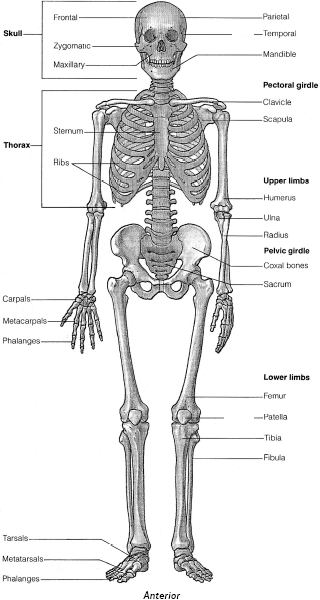
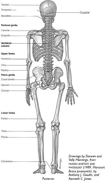
The bones comprise the framework to which the muscles are attached. There are 206 bones in the human body, including 28 in the skull. These are arranged into a cranial group (eight bones), facial group (14 bones), and the three tiny bones in each ear (the auditory ossicles).
The skeleton is divided into two regions: The axial skeleton consists of 80 bones and is comprised of the skull, spine, and thorax (ribs and sternum). The appendicular skeleton consists of 126 bones and is comprised of the shoulder girdle, pelvis, and limbs.
Most of the bones of the skeleton are joined to one another by movable joints, or articulations.
The toes (14 phalanges) articulate with the five metatarsals (the framework of the instep), which articulate with the tarsus (seven tarsal bones of the ankle, including the calcaneus, or heel bone). The talus—uppermost tarsal—is the primary bone of the ankle joint. The ankle joint articulates with the distal ends of the fibula and tibia (shinbone).
The proximal ends of the fibula and tibia articulate with the distal end of the femur (thighbone), at the knee.
The proximal end of the femur articulates with the pelvic girdle (hip bones), which in turn articulates solidly with the sacrum of the vertebral column. The pelvic girdle supports the weight of the upper body, and distributes it to the lower limbs.
The torso is connected to the vertebral column through the rib cage (12 pairs of ribs, and the sternum).
The vertebral column has 33 or 34 bones in a child, but because of fusions that occur later in the lower spine, there are usually 26 separate bones in the adult vertebral column. The skull is attached to the top of the vertebral column at the first vertebra, called the atlas.
Above and to the rear of the rib cage, are the pectoral or shoulder girdles (a clavicle or collar bone, and a scapula or shoulder blade, for each girdle). Each shoulder articulates with the proximal end of the humerus.
The distal end of the humerus articulates, via the elbow, with the proximal ends of the ulna and radius. The distal end of the radius articulates with the wrist.
The hand, or manus, is composed of the wrist, or carpus (eight small, oval-like bones called the carpals), the metacarpus (five metacarpals), and the phalanges (or fingers, comprised of 14 bones in each hand).


There are more than 600 muscles that move the skeleton and some soft tissues, such as the lips and eyelids. Movement is produced by contraction and relaxation of opposing muscle groups, at joints.
Calf muscles
Group of seven posterior muscles below the knee, divided into superficial and deep groups, whose functions include extending the ankle (pointing the toes). The two main muscles are the meaty two-headed gastrocnemius and, beneath it, the soleus. The gastrocnemius connects the heel to the femur, and the soleus connects the heel to the tibia and fibula—the gastrocnemius crosses the ankle and knee joints, while the soleus crosses the ankle joint only. The tendons of these two muscles, together with the plantaris, fuse to form the Achilles tendon.
Exercises that train the calves are calf raises.
Other muscles below the knee
There are four anterior muscles, which move the toes and foot—the largest is the tibialis anterior, which runs alongside the tibia. And two muscles extend along the lateral surface of the fibula—peroneus longus, and peroneus brevis, which lower and evert the foot.
Hamstrings
The three muscles of the rear thigh: biceps femoris (two-headed muscle), semitendinosus, and semimembranosus. They flex the knees, and contribute to hip extension (rearward movement of the femur). The hamstrings are abbreviated to hams.
The primary exercise that trains the hamstrings is the leg curl. Deadlifts, squats, leg press, and back extensions also work the hamstrings.
Quadriceps femoris
Group of four muscles of the frontal thigh: rectus femoris, vastus lateralis, vastus medialis, and vastus intermedius. The tendons of insertion of the four quadriceps muscles form the patella tendon.
The rectus femoris connects the tibia (through the patella) to the pelvis, whereas the other three connect the tibia (through the patella) to the femur. The rectus femoris flexes the femur (raises it) at the hip joint and extends the leg at the knee joint; the other three quadriceps muscles extend the leg only. The quadriceps are abbreviated to quads.
Exercises that train the quadriceps include squats, parallel-grip deadlift, and leg press.
Sartorius
The longest muscle in the body, which runs diagonally across the frontal thigh, from the proximal end of the tibia, to the outer edge of the pelvic girdle. The sartorius flexes the femur, and rotates the femur laterally.
Adductors, thigh
There are five major adductors of the femur, in the inner thigh: pectineus, adductor longus, adductor brevis, adductor magnus, and gracilis. They connect the pelvis to the femur, except for the gracilis that connects the pelvis to the tibia. They are responsible for adduction, flexion, and lateral rotation of the femur.
Squats, and the leg press, work the thigh adductors. A wider stance increases adductor involvement.
Buttocks
The muscular masses posterior to the pelvis formed by the three gluteal muscles (glutes): gluteus maximus, gluteus medius, and gluteus minimus. They extend (move rearward), rotate, and abduct the femur. (A group of six smaller muscles beneath the buttocks rotates the femur laterally.)
Exercises that train the buttocks include squats, deadlifts, and leg press.
Iliopsoas
The single name for three muscles—iliacus, psoas major, psoas minor— that fuse into a single tendon on the femur. These muscles originate on the pelvis or on some of the lower vertebrae, and are hidden from view. They flex the femur, and rotate it laterally. They are called the hip flexors. (Another hip flexor is the rectus femoris, of the quadriceps.)
The hip flexors are worked by most abdominal exercises.
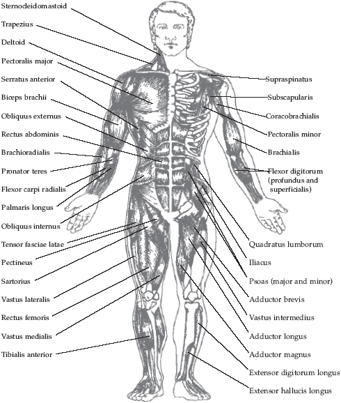
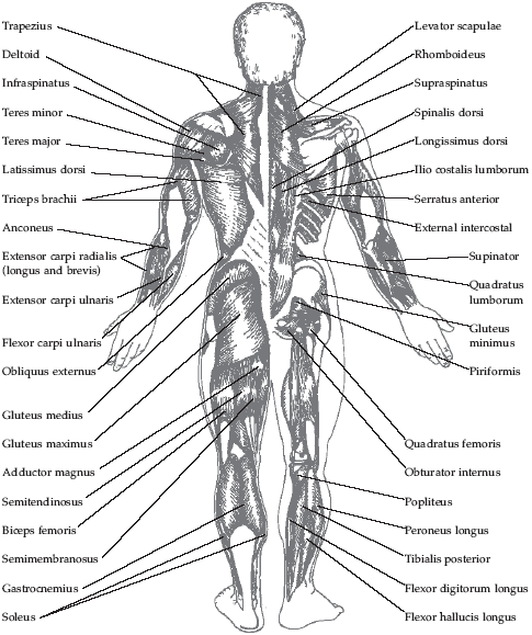
Some of the musculature shown on the right side of each anatomy chart is different from that shown on the left. This occurs where the outer layer of muscle has been omitted in order to show some of the deeper musculature.


Drawings by Eleni Lambrou, based on those of Chartex Products, England.
Erector spinae
Large muscles of the vertebral column—the iliocostalis, longissimus, and spinalis groups—that stabilize the spine, extend it (arch the back), and move the spine from side to side. Some of the muscles produce rotation, too. They are abbreviated to erectors, and are also called the sacrospinalis.
Squats, deadlifts, and back extensions train the erector spinae.
Multifidii
Large muscle group deep to the erector spinae, from the sacrum to the neck, which extends and rotates the vertebral column.
The multifidus group is worked by the rotary torso, twisting crunch, and the same exercises that train the erector spinae.
Rectus abdominis
The frontal, “six-pack” muscle of the abdominal wall, connecting the pelvis to the lower ribs. It compresses the abdomen, and flexes the trunk. The rectus abdominis is abbreviated to abs.
Exercises that train the rectus abdominis include variations of the crunch.
Obliques
The two muscles at the sides of the abdominal wall—external abdominal oblique, and internal abdominal oblique—connecting the ribs with the pelvis. They compress the abdomen, and flex and rotate the trunk.
Exercises that train this muscle include side bends, and crunches.
Transversus abdominis
Deep muscle of the abdominal wall, beneath the rectus abdominis, and obliques. It compresses the abdomen, and flexes the trunk.
Exercises that train this muscle include variations of the crunch.
Quadratus lumborum
Deep muscle either side of the lower spine that helps form the rear of the abdominal wall. Unlike the other muscles of the abdominal wall, the quadratus lumborum doesn’t compress the abdomen; instead, it depresses the ribs. When one side acts alone, it bends the spine to the side; when the two sides act together, they extend the spine.
Exercises that train this muscle include side bends, and back extensions.
Serratus anterior
The muscle on the rib cage underneath and slightly forward of the armpit, which gives a ridged appearance on a lean body. It protracts and rotates the scapula.
Pectoralis major
The large muscle of the chest connecting the chest and clavicle to the humerus. It adducts, flexes, and medially rotates the humerus. The pectorals are abbreviated to pecs.
Exercises that train the pecs include bench presses, and parallel bar dips.
Pectoralis minor
The muscle beneath the pectoralis major, connecting some ribs to the scapula. It protracts the scapula, and elevates the ribs.
Latissimus dorsi
The large, wing-like back muscle that connects the humerus to the lower vertebrae and pelvic girdle. It adducts, extends, and medially rotates the humerus. These muscles are abbreviated to lats.
Exercises that train the latissimus dorsi include the machine pullover, pulldown, and rows.
Rhomboids
The rhomboideus major and rhomboideus minor, which connect some of the upper vertebrae to the scapula. They retract and rotate the scapula.
Exercises that train the rhomboids include the pulldown, and rows.
Rotator cuff muscles
The rotator cuff is where the tendons of four small muscles in the upper back and shoulder area—supraspinatus, infraspinatus, teres minor, and subscapularis—fuse with the tissues of the shoulder joint. The rotator cuff muscles—or articular muscles of the shoulder—are involved in abduction, adduction, and rotation of the humerus.
The external rotators are usually neglected, and are trained by the L-fly.
Trapezius
The large, kite-shaped muscle that connects the skull, scapulae, clavicles, and some upper vertebrae. It retracts, elevates, depresses and rotates the scapula, and extends the head (moves it rearward). The trapezius is abbreviated to traps.
Exercises that train the trapezius include shrugs, rows, and the deadlift and its variations. The neck extension works the upper traps.
Sternocleidomastoid
The muscle at the sides of the neck, connecting the sternum and clavicles to the skull. Acting together, both sides of the sternocleidomastoid flex the head and neck; when acting separately, each muscle produces rotation and lateral flexion.
The four-way neck machine is the preferred exercise for this muscle.
Deltoid
The shoulder cap muscle, and a prime mover of the humerus—it abducts, flexes, extends, and rotates the humerus. It has three heads: anterior, medial, and posterior. The deltoids are abbreviated to delts.
Exercises that train the deltoids include the dumbbell press, barbell press, and lateral raise.
Biceps brachii
The two-headed muscle (long, and short heads) of the front or anterior surface of the arm, which connects the upper scapula to the radius and forearm muscle, and flexes the forearm and thus the elbow joint, and supinates the forearm. The biceps are abbreviated to bis.
Exercises that train the biceps include curls, pulldown, and rows.
Brachialis
The muscle of the front of the arm beneath the biceps, which connects the humerus to the ulna, and flexes the forearm and elbow joint.
Exercises that train the brachialis include curls, pulldown, and rows.
Triceps brachii
The three-headed muscle (long, medial, and lateral heads) on the rear or posterior surface of the arm, which connects the humerus and scapula to the ulna, and extends the forearm (and the elbow joint). Just the long head of the triceps adducts the arm. The triceps are abbreviated to tris.
Exercises that train the triceps include bench presses, presses, parallel bar dips, and the pushdown.
Forearms
The anterior surface (palm side) has eight muscles spread over three layers, most of which are involved in flexing the wrist and fingers. The posterior surface has ten muscles spread over two layers, involved in extending the wrist and moving the fingers.
Timed hold, deadlifts, shrugs, grippers, rows, pulldown, and finger extension train the forearms, along with all exercises that work the grip.