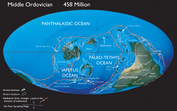
Plate 1. Ordovician continents and oceans. The reconstruction shown is for the Middle Ordovician, about 458 million years ago, about 5.5 million years older than the beginning of the Cincinnatian. Compare to Plate 2. The position of Cincinnati (*) on the paleo-continent Laurentia was south of the equator. Map courtesy of C. R. Scotese, PALEOMAP Project (www.scotese.com).
Plate 2. Late Ordovician continents and oceans. This reconstruction is for the youngest two stages of the Ordovician, the Ashgillian and Hirnantian. This timeslice includes the Cincinnatian interval. Dots indicate sites where reefs and related deposits are known for these stages, and colors indicate the dominant groups contributing to these deposits. Tan = land areas, light blue = epicontinental seas, and darker blue = deep oceans. Map from Kiessling et al. (2003), and courtesy of Wolfgang Kiessling.
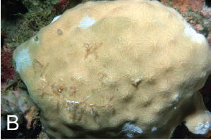
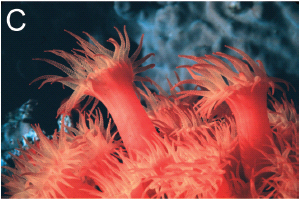
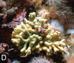
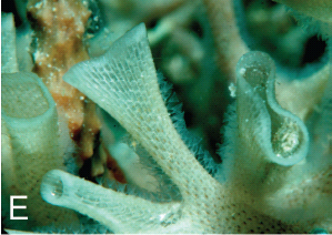
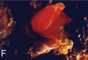
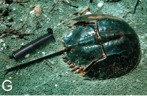
Plate 3. Living invertebrates related to type-Cincinnatian fossil groups. All photos by D. L. Meyer except as noted. A. Sponge, Pericharax sp., Lizard Island, Great Barrier Reef, Australia. Green fluorescent dye was released at base of sponge, taken into sponge’s filtration system, and ejected as stream from osculum at top, as a demonstration of the sponge’s filter-feeding capability. B. Coralline sponge, Acanthochaetetes wellsi Hartman and Goreau, submarine cave at 18 m depth, Palau Islands. Diameter 8 cm. Sponge tissue is a thin veneer over a calcareous skeleton. Coralline sponges are similar to Ordovician stromatoporoids. C. Scleractinian coral, Tubastrea coccinea Lesson, showing tentacle-bearing polyps, Curaçao, Netherlands Antilles. Photo by William K. Sacco. D. Bryozoan, Heteropora pacifica Borg, San Juan Islands, Washington, depth 24 m. Colony form very similar to some Ordovician bryozoans. E. Bryozoan, unidentified cheilostome, Lizard Island, Great Barrier Reef. Australia, depth about 18 m. Note extended tentacles along funnel-shaped section at center. Funnel-shaped section will act as a “chimney” to direct water away from the colony after it has been filtered by the tentacles. F. Brachiopod, Thecidellina, Curaçao, Netherlands Antilles. These brachiopods are cemented by the pedicle valve to the undersides of coral colonies. Note 90 degree gape of valves and extended tentacles of the lophophore. Width about 4 mm. Photo by William K. Sacco. G. Horseshoe crab, Family Limulidae, Subfamily Tachypleinae, Mersing, Malaysia.
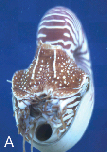
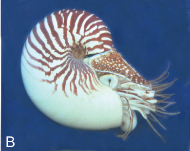
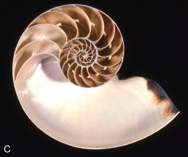
Plate 4. Nautilus, the only living cephalopod with an external shell. Photographs by Richard Arnold Davis. A., B. Nautilus macromphalus, specimen trapped at Lifou and photographed in aquarium at Nouméa, New Caledonia. A. Note the hyponome, the circular opening below the head. B. Note countershading pattern in which stripes are confined to upper side of shell. C. Nautilus pompilius, cross-section of shell, showing chambers and siphuncular tube.
Plate 5. A. Scolecodont, one element of an annelid worm jaw apparatus, Nereigenys alata Eller, CMC IP 1952, FairviewFormation, Dearborn Co., Indiana, x 70. B. Conodont, one element of an apparatus, Phragmodus undatus Branson and Mehl, CMC IP 50705, Kope Formation, Campbell Co., Kentucky, x 227.
Plate 6. Allolichas halli (Foerste), Dan Cooper collection, Richmondian, Waynesville Formation, Franklin Co., Indiana. This exceptionally rare, complete specimen shows a darker coloration around the margin of the carapace that may be a remnant of original coloration. This species was assigned to Amphilichas until 2002 when Holloway and Thomas placed it in Allolichas. Scale in mm.
Plate 7. Isotelus maximus Locke, CMC IP 50168, Richmondian, Adams Co., Ohio. This exceptional complete specimen is 37.5 cm in length and was collected and prepared by Thomas T. Johnson. Isotelus is the State Invertebrate Fossil of Ohio.
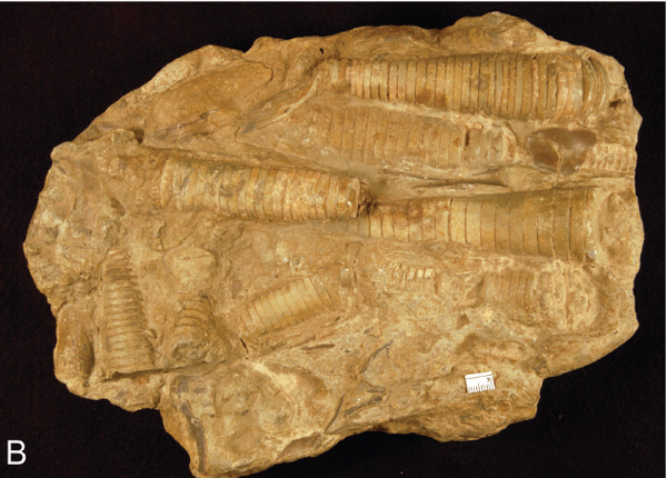
Plate 8. A. Flexicalymene retrorsa on internal mold of nautiloid, CMC IP 50844, Arnheim Formation, Thomas Weaver Collection. Trilobite is located on phragmocone (chambered) section of the nautiloid mold, with head toward body chamber. Scale in mm. B. Parallel alignment of orthoconic nautiloids, CMC IP, Cincinnatian. Scale in mm. Note that not all specimens on the slab are aligned.
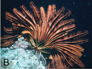
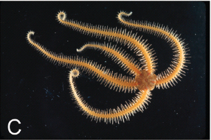
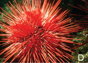
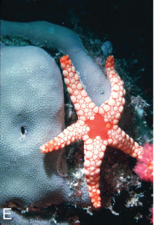
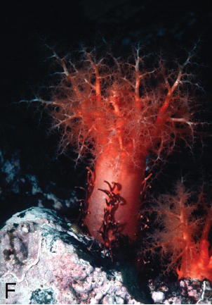
Plate 9. Living echinoderms. A. Stalked crinoids (sea lilies), Neocrinus decorus Thomson, northeastern Straits of Florida, height about 1 m, 420 m depth. B. Unstalked crinoid (feather star), Pontiometra andersoni (P. H. Carpenter), Palau Islands, 4 m depth, arm length about 12 cm. C. Ophiuroid (brittle star), Ophiothrix sp., Caribbean Panama, disk diameter 5–10 mm. D. Echinoid, Strongylocentrotus franciscanus, San Juan Islands, Washington, diameter about 15 cm. E. Asteroid (sea star), Fromia nodosa Clark, Seychelles, Indian Ocean, arm length about 40 mm. F. Holothuroid (sea cucumber), Cucumaria miniata (Brandt), San Juan Islands, Washington, height about 15 cm. A. by Charles G. Messing, all others by D. L. Meyer.
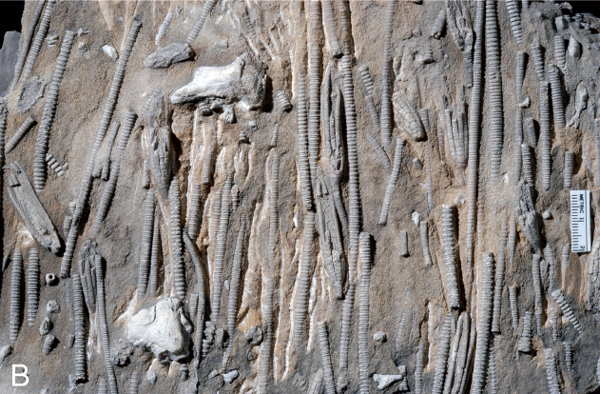
Plate 10. Cincinnatian disparid crinoids. A. Iocrinus subcrassus (Meek and Worthen), CMC IP 46012, Waynesville Formation, Franklin Co., Indiana; note large crown size and dense packing of crinoidal material. B. “Logjam” of Ectenocrinus simplex (Hall), CMC IP, Fairview Formation, Hamilton Co., Ohio; note parallel alignment of crinoids.
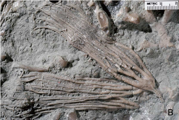
Plate 11. Cincinnatian cladid crinoids. A. Cupulocrinus polydactylus (Shumard), MUGM 28344, Oxford area, Butler Co., Ohio, six crowns, X1. B. Plicodendrocrinus casei (Meek), MUGM 28353, Liberty Formation, southwestern Ohio, two crowns, one overlaps the cup of the other, X1.3.
Plate 12. Late Ordovician paleogeography of the eastern United States. Light blue = shallowest water over Lexington Platform, medium blue = shallow subtidal depths, dark blue = deeper subtidal depths, buff = Queenston Delta, brown = Taconic highlands, the eventual location of the Appalachian Mountains. Courtesy of Steven M. Holland.
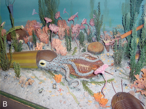
Plate 13. Dioramas of the Cincinnatian sea. A. Cincinnati Museum of Natural History, by Paul Marchand, about 1959, under direction of Kenneth E. Caster. B. Museum of Comparative Zoology, Harvard University.
Plate 14. The Cincinnatian, by John Agnew, 2007.