Chapter 6
The Operating Room, Instruments and the Surgical Team
- The operating room
- Equipment
- Instruments
- Sterilisation
- The surgical team
- Sterile technique
- The operation
- The close of the operation
This chapter discusses the dental surgery or theatre, the instruments for oral surgical operations and the preparation of the surgical team.
The Operating Room
The operating room in both hospital and general dental practice should be of simple design, the walls and furniture should be made of easy-to-clean materials, and the equipment normally required should be accommodated without overcrowding.
It should be well ventilated and kept at an even temperature of 18–21°C, without undue humidity. In hospital theatres this is best done by positive pressure air-conditioning, which also prevents contamination from the outside atmosphere. There should be a recovery room with experienced nursing staff where the patient may recover on a bed or trolley within easy reach of surgeon and anaesthetist until the patient is fit to return to the ward or go home.
Equipment
Light
The light source should provide adequate illumination without undue production of heat, and be easily adjusted to shine into the mouth. A headlamp or fibre-optic light attached to a handpiece is recommended for operations involving the palate or deep cavities such as cysts or the antrum.
Suction Apparatus
No procedure, however minor, particularly under general anaesthesia, should be attempted without suction apparatus. This must be tested before the operation starts and whenever possible an alternative form of suction should be available in case of breakdown. Electrical apparatus is very powerful but does occasionally fail. Compressed air can also provide powerful suction in a similar way. Whichever method is used, a catchment bottle must be included in the circuit so that if roots or any foreign objects are lost the bottle may be searched.
Radiographic Viewing Box
This should be placed such that the surgeon can see it without moving from the dental chair or operating table. It should incorporate a spotlight. Digital radiographs will require a suitable computer terminal.
The Dental Motor (Drill)
Though the conventional dental handpiece can be sterilised, its attachment to the dental electric engine or air motor presents a problem. The surgeon may be contaminated from the cable drive unless this is covered with a sterile sleeve. Alternatively, sterilisable surgical motors and handpieces are available, but due to their high cost these are usually only found in hospital practice. For the clean and rapid cutting of the bone without overheating it is necessary that the bur be washed by a continuous stream of sterile water. Handpieces with an integral irrigation system are available and provide automatic irrigation of the bur. The air-rota is not advocated for oral surgery due to the risk of contamination of the wound with oil and the introduction of air into the tissues, causing surgical emphysema. The modern electrical motor gives very adequate speed and torque.
The Dental Chair
This should be of a design in which the patient can lie flat and the operator may work seated, as this is the position of choice. The light, drill and suction should be sufficiently adjustable that they can be used with a supine patient from either right or left side.
Electrical Equipment
Where this is to be used in the presence of anaesthetic vapours which may form explosive mixtures of gases, the equipment, particularly the drill, must be adequately sealed and earthed to prevent sparking or a build-up of static electricity which might cause an explosion.
Diathermy
Diathermy or electric cautery can be very useful in the control of haemorrhage encountered during surgery of the soft tissues. Monopolar diathermy uses a negative electrode on the body of the patient and is usually employed under general anaesthesia in situations requiring the greater current available. This is less localised than bipolar diathermy which uses current between the beaks of the diathermy forceps to achieve ablation in areas where it is important to reduce the local damage, e.g. around nerves.
Lasers
Modern lasers give excellent control for dissection of soft tissues. Cells in the path of the cut are vapourised with little damage on either side. Their principal advantage in excisions in the mouth is the relatively small amount of postoperative pain, and a reduction in tissue swelling. Stringent safety measures must be taken during the use of the equipment to avoid damage to the patient and the operators. Laserproof glasses should be worn by all personnel in theatre at all times when the laser is in use to protect the eyes. Under general anaesthesia the endotracheal tube should also be protected to avoid inadvertent puncture, and metal instruments should be avoided to decrease possible reflection of the beam.
Cryotherapy
Cryosurgery using liquid nitrous oxide, carbon dioxide or nitrogen destroys cells by intracellular ice crystal formation which ruptures the cell membrane. Healing of the tissue damage is by regeneration of normal tissue. It is of particular benefit in the treatment of benign soft tissue lesions and fluid-filled lesions such as haemangiomas.
Operating Microscope
This is essential equipment for microvascular surgery and nerve repair and is increasingly used for apical surgery. Up to 40x magnification is possible.
Instruments
The selection of hand instruments depends on the surgeon’s preference. In the succeeding chapters instruments suitable for the various procedures are suggested. It is the surgeon’s responsibility to check that all those needed are readily available. They should be laid up on sterile towels on a trolley.
Care and Maintenance of Surgical Instruments
The principles of care and maintenance are:
- to clean the instruments thoroughly
- to examine them for defects
- to repair or discard those that are defective.
Mechanical Cleaning
Mucus and clotted blood may harbour and protect bacteria and make it impossible for steam to reach them. The first step in the process of sterilisation is to scrub all instruments until clean with a brush under a running cold water tap. A bath agitated by ultrasonic vibrations produces a very high standard of cleanliness, especially for hinged instruments and for suction tubes and heads. The latter should have cold water sucked through them immediately the operation is finished.
Cleaning also includes stripping down, cleaning and oiling all working machinery such as handpieces.
Examination for Defects
Broken or bent instruments should never be used as their breakage during surgery can result in serious damage to the patient, either directly from the fractured ends or during the retrieval of the fragment (see Chapter 10). Disposable items, burs, injection needles and scalpel blades are discarded after use.
Sterilisation
- Kills all microorganisms
- Chemical or physical methods are used
- Steam at high pressure (autoclave)
- Boiling water only disinfects.
Physical and chemical methods are both in use today. Physical methods include wet and dry heat, and gamma radiation (used commercially for sterilising packed instruments such as scalpel blades). Boiling water is no longer regarded as a safe method of sterilisation as it only disinfects and does not kill sporing organisms.
Autoclaves use steam under pressure. Some are high vacuum but all depend on downward displacement of air by steam. Steam at 2 kg/cm2 pressure gives a temperature of 134°C, which will destroy all organisms and spores within 3 minutes. It is the method of choice for dressing and towels, but they must be packed loosely to allow the steam to circulate. Blunting of instruments is due to oxidation, which should not occur in a properly functioning autoclave, so it can be safely used for sharp instruments. Vapour phase inhibitor (VPI) paper can be used to wrap instruments such as burs, which tend to rust if autoclaved. Handpieces must be cleaned and oiled before being placed in the autoclave. The oil must not become oxidised or lose its oily properties at high temperatures.
Dry heat is effective in ovens that have fans to ensure even heat distribution and a door-lock that prevents opening during the sterilising period. The cycle is much slower and it takes half an hour at 160°C to destroy organisms and spores. It may be used for sharp instruments and handpieces. Both autoclaves and hot-air sterilisers are made with controlled cycles that cannot be interrupted once started, so that sterility of the instruments is ensured. The efficiency of the cycle must be checked periodically by the use of Brownes’ tubes.
Chemical sterilising is not regarded favourably by bacteriologists as most of the solutions available are not considered reliable. Glutaraldehyde is effective against vegetative organisms and spores if the instruments are immersed in it for 10 hours, after which it must be washed off with sterile water as it is irritant to tissues. Because of the length of the sterilising time and the irritant properties, the use of glutaraldehyde is confined to those instruments that cannot be heat sterilised. Glutaraldehyde or hypochlorite solutions may be used to disinfect instruments potentially heavily contaminated with viruses (such as hepatitis) before these are cleaned and sterilised.
Both gamma radiation and ethylene oxide are used commercially for the sterilisation of disposables such as scalpel blades and sutures, but their toxicity precludes their use in the surgery.
To sterilise with certainty, autoclaving is the method of choice. Dry-heat sterilising is the next best and only where both of these are impossible should chemical methods be considered.
Instruments Packs
In hospital practice instruments are sterilised wrapped in paper containers. These are permeable to the steam in the autoclave and, providing the latter is of an evacuating type, are dry at the end of the cycle. Packs may be made up of one instrument or a complete layout for an operation, including towels. They can be stored for up to 6 months ready for immediate use. The packs are duplicated according to the frequency with which they are used and may be prepared for local anaesthetic injections, flap preparation, suturing and so forth. This system is seldom available in general dental practice, making it necessary to sterilise and lay up instruments for each operation separately.
The Surgical Team
- Surgeon
- Anaesthetist
- Assistant
- Nursing staff.
The Surgeon
The surgeon is responsible for checking the identity of the patient and the nature of the operation to be performed, the operation and the surgical safety of the patient. Time out procedures are now performed routinely, in an effort to eliminate wrong site operations and ensure patient safety, when the patients ID is checked against the consent form and preoperative checklist. This task is paramount and the surgeon should devote every attention and effort to it, informing and encouraging the rest of the team to assist in the endeavour. This responsibility applies to any procedure, planned or accidental, inflicted on the patient, including those carried out by assistants. The surgeon must be informed and satisfied that all instruments, swabs and packs are accounted for at the end of the operation.
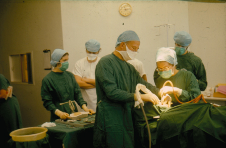
The others in the team should support the surgeon and always relate information that will either alter or facilitate the outcome (Figure 6.1). In a surgical emergency, where speed and efficiency are needed, the surgeon should take the lead in managing this.
Figure 6.1 The surgical team. The surgeon is only one part of this, and cohesive working within the team promotes better care for the patient.

The Anaesthetist
The anaesthetist assesses the patient’s fitness for general anaesthesia, chooses the anaesthetic agent, prescribes suitable premedication and administers the general anaesthetic. He will supervise the moving of the patient to and from the ward, and on and off the operating table, as well as their recovery from the anaesthetic and such post-anaesthetic complications as may arise.
During the operation the anaesthetist is responsible for the patient’s airway, which should include packing of the throat and the removal of the pack after operation. They should continually assess the patient’s general condition and report any deterioration to the surgeon, so that a mutual assessment of the situation can be made. The anaesthetist’s opinion about the safety of the patient is accepted and surgical procedures planned or modified accordingly.
The Surgeon’s Assistant
The oral surgeon’s assistant in the hospital theatre is either a qualified colleague or a member of the nursing staff. In the dental surgery assistance will usually be extremely effectively delivered by a dental nurse with appropriate training. The assistant will produce the patient’s notes and radiographs, and help with the preparation of the patient, by cleaning and towelling up the operation area and assembling the suction and other equipment needed.
The assistant will retract tissues to give the surgeon the best possible access, clear fluid from the field of the operation with suction or swabs and remove solid debris with forceps. Assistance with haemostasis by applying pressure, artery forceps or diathermy, and cutting the ends of the sutures for instance all contribute to the smooth flow of the operation.
The longer two people work together the greater will be the degree of teamwork possible, with mutual benefit to all concerned. The surgeon may ask for further help in various ways. Involving qualified colleagues, using their previous experience, can extend their field of interest and responsibility.
The Nursing Staff
Before the operation begins the nursing staff will select and lay up those instruments, drugs and dressings they know to be necessary. They check the working of the dental engine and suction apparatus, and of all electrical appliances.
In the operating room they share the responsibility of ensuring that the correct patient is present and that the correct operation is being performed prior to its start.
They follow the progress of the operation closely to anticipate the surgeon’s needs and inspect and clean instruments, drawing attention to any that become broken or damaged. At the beginning the end of the operation they count all swabs, packs, dressings, instruments and needles, and tell the surgeon if any are missing before the patient leaves the theatre or the outpatient surgery.
Preparation of the Surgical Team
Those taking part in a surgical operation must be free from infection, especially of the respiratory tract or skin, which could be transmitted to the wound. All are responsible for their own safety and must develop a sensible routine that avoids skin or conjunctiva from being contaminated by blood or saliva from patients. The use of personal protection equipment (PPE), at minimum gloves, masks and eye protection, are now mandatory.
Sterile Technique
- Minimising contamination of the wound
- Wearing ‘scrubs’ and clogs
- Scrubbing up
- Sterile gowns and gloves
- Sterile instruments
- Preparing operation site with antiseptic solution
- Avoiding contamination during surgery.
Dress
All those entering a hospital operating theatre must change from their normal clothes into freshly laundered uniform trousers and shirt (‘scrubs’). This not only reduces the risk of contamination but is cooler and more comfortable. The hair is covered with a paper cap and a mask is worn over the mouth and nose. Safety glasses should be worn to avoid splashing of blood or irrigation fluid into the eyes. Shoes are changed for rubber boots or clogs used only in the theatre.
In the dental surgery a sterile gown worn over everyday clothes is put on for each patient. A mask and protective glasses should be worn. The use of a cap is optional.
Scrubbing Up
Those who have to handle sterile instruments or dressings must undertake the ritual of ‘scrubbing up’. The arms are bared to the elbow and all rings and watches removed. The hands and forearms are washed under a running tap, using an antiseptic detergent solution (chlorhexidine or iodine based). They are well soaped and washed from the tips of the fingers up towards the elbow in a ritual that covers every area thoroughly. The fingernails must be kept trimmed short for satisfactory cleansing, which is done with a sterile scrubbing brush or nail scraper. The suds are rinsed away under the tap, being made to flow off at the elbow, not at the hands. This is continued for 4 minutes by the clock. The hands are then dried on a sterile towel by wiping from the hands up to the elbows. For minor procedures carried out in the dental surgery sterile gloves are put on and the operation commenced. For more extensive operations or where there is a risk of infection of staff and in theatre to conform with normal practice, a sterile gown and gloves are worn.
Gowning Up
The sterile gown is lifted from its container folded in such a way that if held correctly with both hands at its neck, it will unroll and fall with the sleeves hanging away from the operator. The arms are then placed in the sleeves and a second person standing behind draws the gown over the shoulders and fastens the tapes and belt at the back. The belt avoids billowing and rubbing of the gown and is an antistatic precaution.
Surgical gloves are then put on. There is now a large selection on the market to cater for all aspects of surgery. Powder-free gloves are now the norm and alternatives for those who are latex sensitive or patients who are latex allergic are available. The gloves are taken from their envelope by the cuffs, which are folded down over the palms. The cuff of the right glove is held in the left hand and the right hand thrust in. The right hand then lifts the left glove by placing the fingers under the rolled cuff and the left hand is thrust in. The cuffs are turned back over the wrist to cover the sleeve. In this way at no time will the hands have been in contact with outside of either gown or gloves. From this moment if any unsterilised object is touched, the operator must gown up again.
Closed gloving further enhances the sterile technique. After gowning, both hands remain covered by the sleeves. The left glove, with its fingers directed towards the elbow, is placed with the palm surface against the left sleeve. The cuff of the glove is grasped through the material by the thumb and fingers of the left hand. The right hand (through the gown material) grasps the outer part of the glove cuff and turns it over the sleeve of the gown. The glove is drawn onto the left hand by pulling glove and sleeve up the forearm (Figure 6.2).
Figure 6.2 Donning gloves (closed glove method): (a) glove is placed over hand still within gown, fingers pointing up arm; (b) cuff of glove is grasped through gown by left and right hand, glove is pulled over left hand; (c) left hand is then thrust into glove; (d) left hand then positions glove on right hand; (e) and pulls glove over hand while; (f) hand emerges from gown into glove.

Preparation of the Patient
In hospitals, a label carrying the patient’s name, address and hospital number is attached to the wrist. The details should be checked against the patient’s notes. A valid consent form must be available before the general anaesthetic is started. When local anaesthetic is being used mistaken identity may be avoided by direct questioning of the patient.
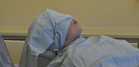
The patient’s position on the table is adjusted if unsatisfactory, following which the operator then cleanses the site of operation, usually the mouth and surrounding skin, with a swab held in forceps and dipped in a detergent (chlorhexidine), care being taken to protect the eyes. The patient’s body and head are then covered with sterile towels in such a way as to leave only the operation site exposed (Figure 6.3). For extraoral procedures the mouth may also be covered.
Figure 6.3 Patient is protected by sterile towels to reduce contamination.

In the outpatient surgery the patient should be asked to wash the mouth thoroughly with a mouthwash and in the case of female patients all cosmetics should be removed. A sterile towel may then be pinned round the patient’s neck and a sterile cap placed over the hair, to prevent contamination of the instruments or of the operator’s hands. The patient’s eyes are protected from the light and instruments by dark glasses.
Preoperative Check
In theatre, with the patient lying on the operating table, intubated and with the throat packed off, the surgeon and nursing team should repeat the identity checks, consulting the identity bracelet, consent form and notes as necessary to avoid wrong operations. Similar checks should take place in the dental surgery with the awake patient, particularly when sedation is to be given.
The Operation
All members of the team must work in a comfortable position to avoid fatigue. It is traditional to stand for most procedures in operating theatres; however, modern equipment makes it possible for the surgeon and the assistant to work seated, which is much less tiring. With this in view the position and height of the table and chair, instrument trolleys and other apparatus are adjusted before the operation is commenced. In the dental surgery this should be done before the surgeon and their assistant scrub up.
The mouth can be held open with a rubber prop placed between the molar teeth. For operations under local anaesthesia a prop often helps the patient by resting the muscles and joints. Under general anaesthesia the mouth must not be opened by force because of the danger of fracturing teeth or damaging the temporomandibular joint.
Where the patient is given a general anaesthetic, teeth can be damaged or dislodged during induction. Special care must be taken by anaesthetists and surgeons where loose teeth are present.
The Close of the Operation
The surgeon refers to the notes to confirm that all the surgery planned has been completed and tells the anaesthetist of the completion of the procedure. It must be ascertained that all bleeding is controlled and that wound closure is satisfactory. Packs or drains to be left in the mouth or wound are securely sutured in place. The mouth is searched for any clot, debris or swabs and a final count is made of the teeth extracted, of swabs, needles and small instruments. With the anaesthetist’s agreement the throat pack may be removed and the nursing staff should be informed also. Any debris is removed from the mouth and oropharynx, after which the patient is handed over to the anaesthetic team for the recovery phase.
The surgeon will then write up the notes of the operation. This is done immediately for the information of the ward staff and other colleagues. The value of this is particularly pertinent in an emergency.
Operation notes may be written in the following form:
Date
Name and number of patient
Time of commencement of operation
Surgeon and assistant named
Anaesthetist named
1. Anaesthetic. Local anaesthetic, general anaesthetic, type, pack used.
2. Operation described logically. Incision – reflection – bone removed – teeth extracted or fracture reduced and fixed, etc. – debridement of wound – closure of wound – sutures – dressings applied – removal of throat pack – condition of patient on finishing – time of completion of operation.
The assistant will remain with the anaesthetist to man the suction apparatus and advise about the operation site. The patient must remain under the supervision of the anaesthetist, preferably in a special recovery area, until sufficiently recovered to control their own airway. Initially the patient may be nursed on their left side in the recovery position to allow drainage of blood or saliva forwards out of the mouth, but should be fully conscious before returning to the ward.
It is usual in hospital practice for a nurse from the ward to fetch the patient from the operating theatre. The recovery nurse should hand over the patient, describing which procedures have taken place, showing the site of the operation, and giving instructions verbally and in writing for the immediate postoperative nursing. Both anaesthetist and the surgeon may be involved in this process if there are any special circumstances of which the ward should be aware.
The patient will be moved on a trolley equipped with an oxygen cylinder and mask and will be accompanied back to the ward by two people. One of these should be a trained nurse who gives undivided attention to the patient and in an emergency stays with them while assistance is summoned.
Further reading
Cottone JA, Terezhalmy GT, Molinari JA (1995) Practical Infection Control in Dentistry, 2nd edn. Williams and Wilkins Media, Philadelphia.
Experience of wrong site surgery and surgical marking practices among clinicians in the UK. http://qshc.bmj.com/content/15/5/363.full.pdf
Mulcahy L, Rosenthal MM, Lloyd-Bostock SM (1998) Medical Mishaps: pieces of the puzzle. Open University Press, Buckingham.
Sentinel events: approaches to error reduction and prevention (1998) Joint Commission Journal on Quality Improvement 24(4): 175–86.