FOR A 3.3-POUND (1.5-KG) FATTY BLOB WITH THE approximate consistency of tofu, the human brain has to be admired. After all, it’s managed to formulate the theories of relativity and quantum mechanics, written all the great novels, and, perhaps most astoundingly of all, has evolved sentience, self-awareness, and the ability to contemplate its own origin and existence.
That said, it’s also given us John Travolta’s Battlefield Earth and the truly execrable Dirty Grandpa.
For the most part, though, the human brain is a veritable powerhouse. It generates around 23 watts of heat while its owner is awake, and consumes around 20 percent of the oxygen that’s funneled into the bloodstream from the lungs. The brain of an adult human is made up of around 100 billion nerve cells, or neurons. These communicate with each other through electrical impulses and by the exchange of messenger chemicals, known as neurotransmitters. Each neuron can be wired like this to as many as 10,000 others, forming a network of up to 1,000 trillion connections. And the structure of these connections is responsible for storing our knowledge and memories.
Perhaps the biggest outstanding mystery of the brain is consciousness. How did this tangled web of synaptic connections develop the power not just to store information and perform computations, but to be aware and to have conscious experiences? These experiences—the redness of the color red, the flavor of a burrito, the smell of a sea breeze—are known as “qualia” to neuroscientists, and they are subjective to each individual, defying description except perhaps by analogy.
Qualia must correspond to certain physical processes in the brain, and identifying these so-called “neural correlates of consciousness”—the firing of a certain group of neurons to signify the aroma of a rose, for example—has been a major focus for scientists.
In 2014, a breakthrough was made when researchers found that electrically stimulating a brain region called the claustrum induced a loss of consciousness, which was immediately reversed the moment the current was switched off again. Later, in 2018, scientists were able to identify two parts of the brain that contribute to our experience of free will—our sense of being in control of our actions. You can read more about that discovery later on in this chapter. Other researchers have even developed theories positing that free will and our experience of qualia all derive from applying the weird laws of quantum mechanics to the functioning of the brain. However, these claims have been greeted with a good deal of skepticism.
In separate research, some astonishing insights into the nature of the brain have been revealed using medical scanners. Scientists have used functional magnetic resonance imaging (fMRI) to identify patterns of activity that they say can reveal whether someone’s telling lies or being truthful, and can give away personal prejudices, political leanings, and even brand preferences (Coke or Pepsi?). FMRI works by coaxing radiation from hydrogen atoms residing within water molecules in the body by stimulating them with radio waves and magnetic fields. Measuring the strength of the radio waves emitted in response reveals regions of increased blood flow. And this acts as a tracer for which brain regions are most active.
More recently, fMRI scanning has been combined with a field called “machine learning.” This is a branch of data science that enables computers to learn and make discoveries from raw data, doing so with minimal human intervention. Researchers at Kyoto University in Japan gathered fMRI scans of patients’ brains, taken while they looked at various images. The set of scans and the input images were fed to a kind of machine-learning agent called a “deep neural network,” which was then able to look at new fMRI scans and deduce with reasonable accuracy what pictures the subjects in each case were thinking about. Pretty cool, huh?
More on that later. Brains scans can even yield insights into the decision-making process. This was demonstrated by scientists who were able to peer inside the brains of rats. In an experiment that you’ll soon read about, the researchers placed rats in a maze and, based on the observed patterns of brain activity, they were able to predict which path through the maze each animal was about to take.
Also coming up, find out how magic mushrooms can boost your creativity. Researchers from Leiden University, in the Netherlands (surprise, surprise), have found that so-called microdosing (taking tiny amounts of psilocybin, the active ingredient in ’shrooms) led test subjects to come up with more original solutions to problems. (And, just to be clear, no, none of these involved enlisting the help of ninja unicorns on mopeds.)
Plus, we discover the facial feature that’s a dead giveaway of a narcissist. Find out just why it is that one or two (or was it 10?) drinks too many can leave you a little hazy on the details of what went on last night. And get the lowdown on why conscientiousness is, apparently, the most attractive characteristic in a sexual partner (according to heterosexual couples, anyway).
As Miles Monroe put it in the 1973 movie Sleeper: “My brain? It’s my second favorite organ!”
84
WE JUST FOUND THE PART OF THE BRAIN RESPONSIBLE FOR FREE WILL
by Rosie McCall
THE PHILOSOPHER THOMAS HOBBES DESCRIBED FREE will (or “liberty”) as “the absence of all the impediments to action that are not contained in the nature and intrinsical quality of the agent”—which, in plain English, is the ability to act without any outside constraint, be that an overbearing boyfriend/girlfriend or something altogether more whimsical, like fate.
The scientific definition, however, is far more specific. Essentially, it comes down to two cognitive processes: volition and agency. Volition is “the desire to act,” whereas agency is “the sense of responsibility for our actions.”
Thanks to a study published in the Proceedings of the National Academy of Sciences in 2018, scientists have identified the exact locations in the brain responsible for these processes and, therefore, our perception of free will.

Researchers from Beth Israel Deaconess Medical Center, in Boston, studied 28 cases where brain injury had affected patients’ volition and left them with no desire to move or speak (a condition known as “akinetic mutism”). They also examined a further 50 cases where agency had been impaired, so that the patients were left feeling as though their movements weren’t their own (“alien limb syndrome”). The team looked at scans to find the lesions in each patient’s brain responsible.
This revealed a diverse range of injury locations but, interestingly, all lesions appeared within one of two networks. The injuries related to akinetic mutism were connected to the brain’s anterior cingulate cortex (responsible for motivation and planning), whereas almost all the injuries (90 percent) related to alien limb syndrome were connected to the brain’s precuneus cortex.
To confirm that these two networks are indeed the two areas responsible for free will (as defined by science, not Hobbes), the researchers examined the effect of brain stimulation on free-will perception in healthy volunteers and looked at images of the brains of psychiatric patients with abnormal free-will perception. Both revealed alterations in the same networks.
While the information is not going to stop moral philosophers and sociologists from debating the meaning (and the very existence) of free will, the researchers hope it will prove useful when it comes to helping patients with volition- and agency-inhibiting injuries.
85
ARTIFICIAL INTELLIGENCE RE-CREATES IMAGES FROM INSIDE THE HUMAN BRAIN
by Jonathan O’Callaghan
A TEAM OF RESEARCHERS SAY THEY HAVE USED MACHINE-learning to re-create images from “thoughts” inside patients’ brains.
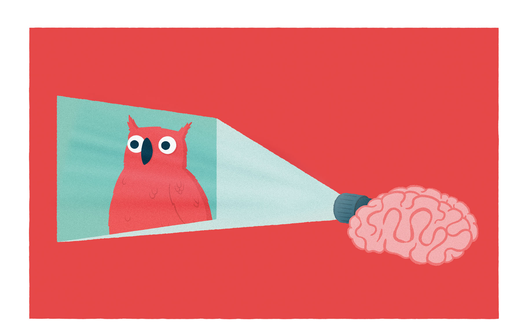
The research, reported in January 2018 by Science magazine, was conducted by scientists from Kyoto University in Japan and led by Yukiyasu Kamitani. Using functional magnetic resonance imaging (fMRI), the team said they were able to reconstruct images by analyzing scans of brain activity. An artificial intelligence agent, known as a deep neural network (DNN), was able to process the scans to re-create pixel-by-pixel images that resembled the originals.
“The results suggest that hierarchical visual information in the brain can be effectively combined to reconstruct perceptual and subjective images,” the team wrote in their paper.
The research builds on earlier work by the same team that found that brain activity could be decoded to reveal the sensory inputs producing it. Other researchers have reported similar work in this field.
“This is a significant improvement on their earlier work,” Professor Geraint Rees, a neuroimaging expert from University College London, told The Times.
In this latest paper, the researchers used three subjects (two males, aged 33 and 23, and one female, aged 23). They were shown images of things like a mailbox and a lion, as well as geometric shapes and alphabetical letters.
The subjects viewed the images while inside an fMRI scanner, with their heads held securely in place. They took part in multiple scanning sessions, each lasting a maximum of 2 hours, spread over a period of 10 months.
The participants first stared at each image for a number of seconds, while their brain activity was recorded. This data, alongside the input image, was then used to train the DNN—to teach it the patterns of brain activity corresponding to different features in a visual image.
Later, the subjects were asked to remember one of the images they had seen and picture it in their mind. Using the DNN, the researchers then attempted to decode the signals recorded by the fMRI scanner and produce a computer-generated reconstruction of the original image.
Some of the results were rather remarkable, with the DNN able to reproduce images of a DVD player, feet with socks on, a fly, and more. However, it wasn’t too hot on other images—like a person with a cowboy hat, or a snowmobile.
“Our approach could provide a unique window into our internal world by translating brain activity into images,” the team noted.
87
GROWING UP POOR PHYSICALLY CHANGES THE STRUCTURE OF A CHILD’S BRAIN
by Madison Dapcevich
A LONG-TERM ANALYSIS OF HUNDREDS OF ADOLESCENT brains suggests that the socioeconomic status (SES) of a child’s family may play a role in the early development of brain areas responsible for learning, language, and emotion.
To study the connection between parents’ income and education levels and their children’s cognitive development, researchers from the US National Institute of Mental Health scanned the brains of more than 600 individuals over the course of their lives between the ages of 5 and 25. Then they compared these neuroimages against data on their parents’ education and occupation, as well as each participant’s IQ.
When it comes to the relationship between brain anatomy and SES, not much changes from childhood to early adulthood. This led researchers to suspect that preschool life is a pivotal time in which associations between socioeconomic status and brain organization first begin to develop.
The authors, writing in the Journal of Neuroscience, found correlations between SES and total gray matter volume, and between volume levels in the prefrontal cortex, which is the area of the brain associated with personality development, and the emotion-regulating hippocampus. The areas of the brain responsible for emotional development, learning, and language skills were found to be more complex in youngsters whose parents were better educated and worked in professional careers.
“Early brain development occurs within the context of each child’s experiences and environment, which vary significantly as a function of socioeconomic status,” wrote the authors. A child’s early life experiences and environment depend on her family background, through factors such as her parents’ income, education, and occupation. And these impact a child’s mental health, cognitive development, and her academic achievements. Understanding how such things physically change the brain could help researchers understand how SES is associated with different life outcomes.
The researchers note that the link they have found between SES and cognitive development represents only one possible set of interactions between childhood environment, anatomy, and cognition.
89
A TECHNIQUE TO CONTROL YOUR DREAMS HAS BEEN VERIFIED FOR THE FIRST TIME
by Stephen Luntz
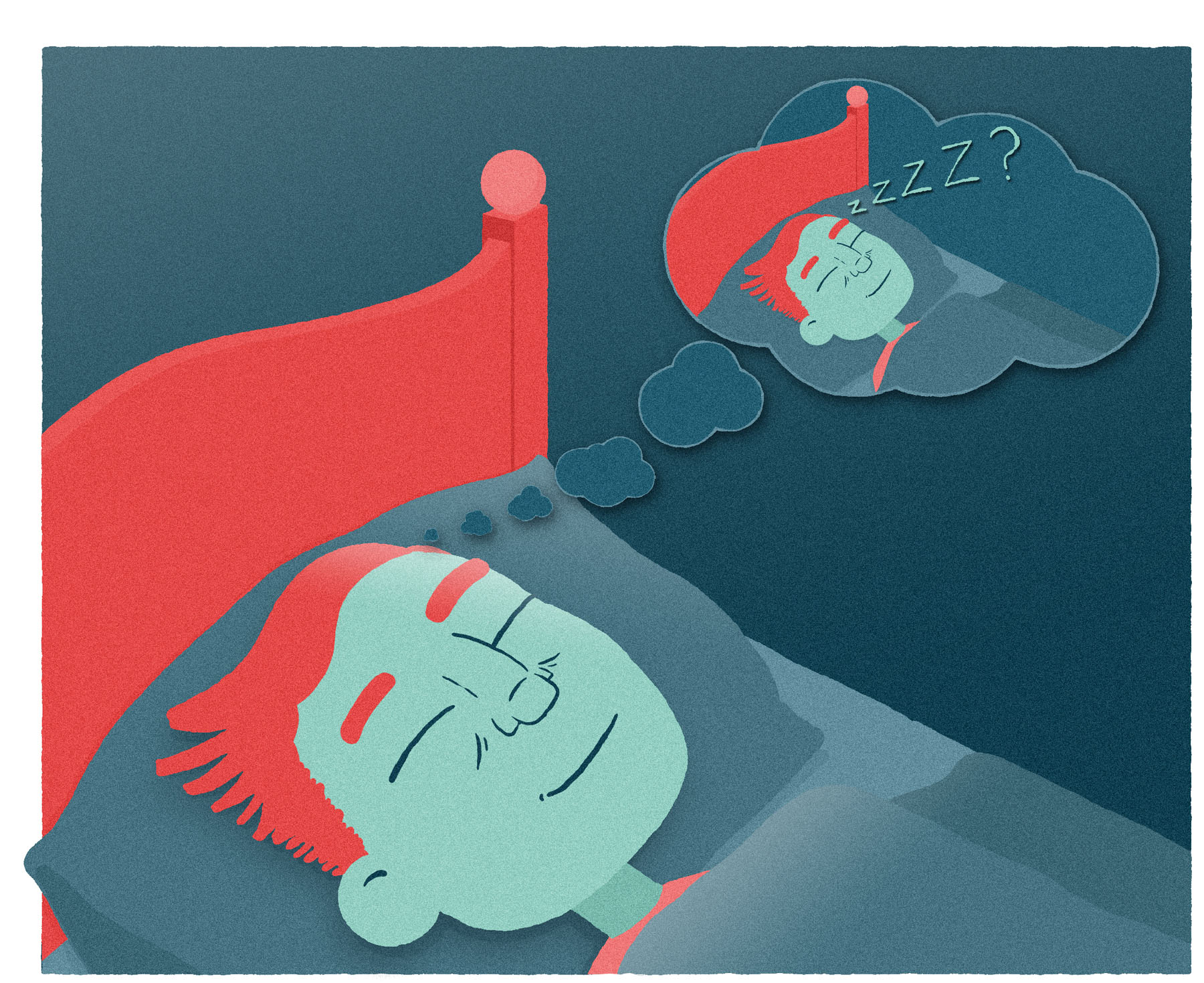
GET READY TO HAVE THE DREAMS OF… WELL, OF YOUR dreams. A technique to induce lucid dreaming—a state of dreaming that can be consciously experienced and controlled—was independently verified for the first time in 2017. More than half the participants in the study lucidly dreamed during the trial, a record-breaking success rate.
Lucid dreaming is the term given to the state where dreamers are aware that they are dreaming, and have some control over how the dream progresses. Once considered a myth, science has confirmed that lucid dreams exist, and has found techniques to help induce them. Nevertheless, some of these require advanced equipment, and others are far from reliable.
This is unfortunate, as they are considered a potential tool for healing traumas and for controlling unhealthy behavior. So Dr. Denholm Aspy, of the University of Adelaide, decided to investigate.
Aspy instructed 169 participants in techniques developed to induce lucid dreaming. One of these, called “reality testing,” gets people into the habit of regularly checking to make sure they really are awake. The other, called “mnemonic induction of lucid dreams” (or MILD), has participants set alarms to wake them after five hours and recite the line “The next time I am dreaming, I will remember that I’m dreaming,” before going back to sleep.
Writing in the journal Dreaming in 2017, Aspy reports that reality testing on its own produced no benefit, but of those who tried the combination of reality testing and MILD, 53 percent had a lucid dream during the trial period, while 17 percent were successful each night. This exceeds the results of any previous study conducted without physical interventions, such as trying to awaken participants once they were in dream sleep, he told IFLScience.
Approximately 55 percent of us experience a lucid dream at some point in our life. Aspy himself became interested in lucid dreaming after having one as a child, and says he made it the topic of his psychology PhD research after having a lucid dream the night before he was due to begin his doctoral studies.
Most lucid dreamers initially wake up quickly, Aspy told IFLScience, but with practice they can learn to extend their lucid dreams for up to an hour.
90
HERE’S WHAT HAPPENS TO ALCOHOLICS’ BRAINS WHEN THEY QUIT DRINKING
by Ben Taub
SCIENTISTS HAVE COME UP WITH SURPRISING EVIDENCE that may explain why recovering alcoholics find it so hard to stay off the booze.
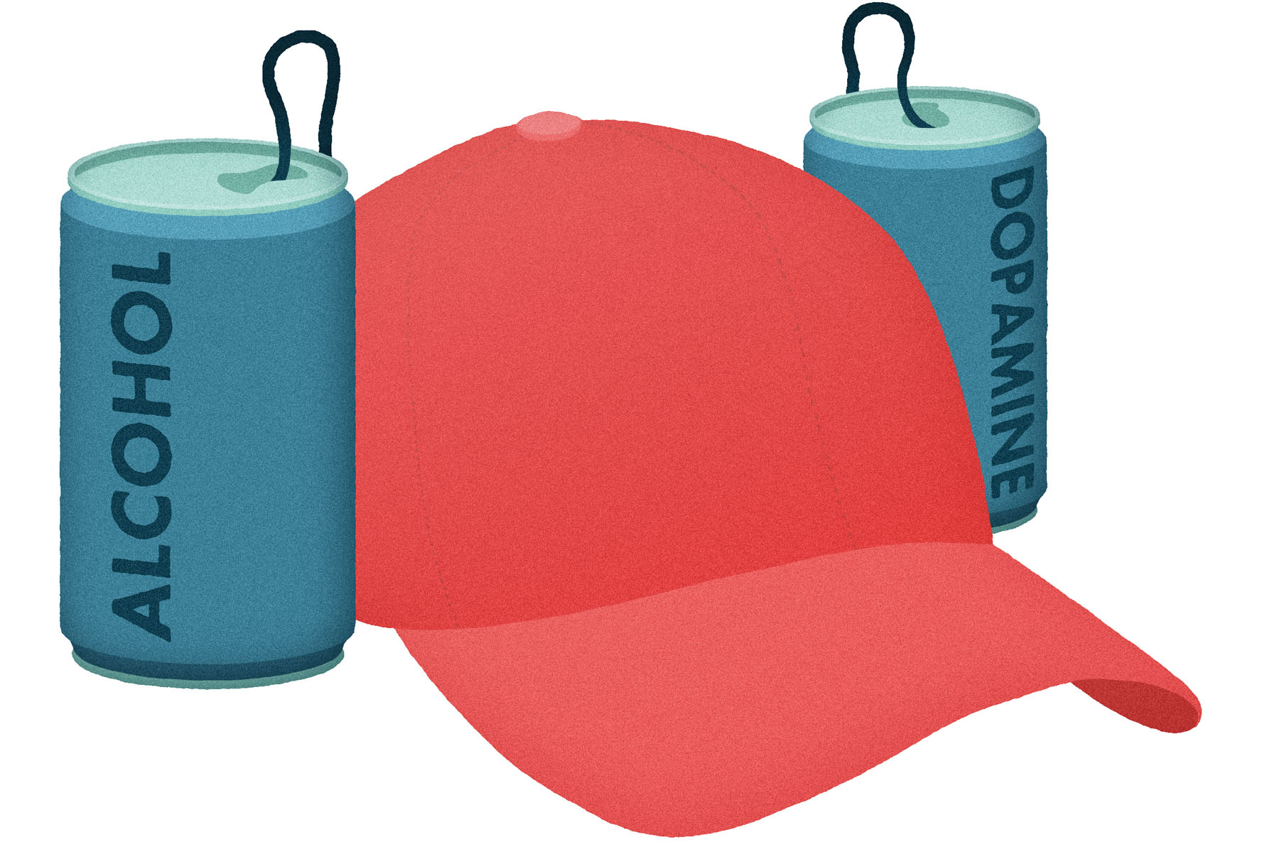
Publishing their findings in 2016 in the Proceedings of the National Academy of Sciences, the researchers suggest that when an alcoholic stops drinking, the brain’s ability to use the neurotransmitter dopamine changes, altering the wiring of the brain’s reward system.
Like many drugs, alcohol is known to stimulate the production of the chemical messenger dopamine, which activates the so-called reward center of the brain. Previous studies into the nature of addiction have revealed that the dopamine response is significantly reduced in alcoholics, leading to a need to drink more in order to feel a buzz. But why?
To investigate, researchers began by examining brain tissue from deceased alcoholics. They found that these brains had fewer D1 dopamine receptors than normal brains. D1 receptors are the sites on the membranes of neuronal cells to which dopamine binds, causing these neurons to become excited. Any reduction in these receptor sites would therefore be expected to decrease the brain’s responsiveness to dopamine, explaining why alcohol fails to satisfy.
The brains were also found to have fewer dopamine transporter sites, which allow for any unused dopamine to be sucked back up and recycled. As with D1 receptors, the disappearance of these sites is likely to hinder the brain’s ability to use dopamine.
Next, the study authors used radiography techniques to track dopamine levels in the brains of alcohol-dependent rats that were denied alcohol for several weeks.
They discovered that dopamine levels dropped during the first six days. However, after three weeks, the researchers noted that dopamine levels were in fact elevated, as the number of available receptor and transporter sites plummeted, so that the rats’ brains resembled those of the deceased alcoholic humans. Significantly, at the three-week mark, the rats displayed continued behavioral effects associated with alcohol cravings.
The study authors concluded that, while acute alcohol withdrawal may be associated with lowered dopamine levels, prolonged abstinence actually leads to dopamine levels in the brain being higher than normal. Crucially, they say that both of these states are representative of a dysfunctional reward system, increasing an addict’s vulnerability to relapse.
92
THE KEY TO A HAPPY SEX LIFE SOUNDS PRETTY UNSEXY, ACCORDING TO THIS STUDY
by Tom Hale
HOLLYWOOD MOVIES AND OTHER ROMANTICIZED DEPICTIONS of love like to imagine “good sex” as a spontaneous splurge of impulsive animal desire. However, according to a new German study, the key to a healthy and fulfilling sex life could actually be… conscientiousness. Wild, right?
A study published in July 2018 in the Journal of Sex Research looked at what personality traits and partner types tend to create a happy sex life. They came to the unlikely conclusion that those who are big on planning and organization tend to report fewer problems and higher levels of satisfaction in the bedroom.
Psychotherapists from Ruhr University in Germany reached this conclusion by quizzing 964 German couples—98 percent of whom, it should be added, were in heterosexual relationships—about their sex life and satisfaction. They were asked intimate details, such as how easily they were aroused, how inhibited they were, and how they thought they performed sexually.
The researchers also asked the respondents to complete a questionnaire to assess how they scored on the Big Five personality traits: Conscientiousness, Agreeableness, Extroversion, Neuroticism, and Openness to Experience.
“Studies have shown that most of these personality traits and sexuality-related traits are relevant,” Julia Velten, a post-doctoral fellow in clinical psychology and psychotherapy, told the news website Quartz.
However, it came as a surprise how strongly conscientiousness was correlated with sexual satisfaction. The researchers speculate that people who display this trait might actually make more thoughtful lovers, as they are more likely to carefully engage with their partners, make sure they are fulfilled, and remain focused on resolving any hiccups in the relationship.
“Individuals low on emotional stability or agreeableness may be more likely to behave in a way (i.e., express criticism, avoid communication) that triggers a negative response from a partner, which in turn may lead to inadequate sexual communication and result in lower sexual functioning,” the study authors write.
“High conscientiousness can be especially beneficial when it comes to putting effort into a satisfying sexual life or to postponing one’s own needs and interests to focus on resolving a sexual problem within the context of committed, long-term relationships.”
So, if you’re looking to spice things up in the bedroom, you just need to remember your pen and your day planner.
93
MICRODOSING MAGIC MUSHROOMS COULD SPARK CREATIVITY AND BOOST COGNITIVE SKILLS
by Tom Hale
ADVOCATES OF MICRODOSING CLAIM THAT TAKING TEENY doses of magic mushrooms and other psychedelic substances can inspire creative thought, boost your mood, and even enhance your cognitive function, all without the risk of a so-called “bad trip.”
But aside from loose anecdotal evidence from Silicon Valley bros, what does the science say? A team of researchers from Leiden University in the Netherlands decided to find out. Their admittedly small-scale study, published in October 2018, is the first of its kind to experimentally investigate microdosing of magic mushrooms and its cognitive-enhancing effects within a natural setting.
Reporting in the journal Psychopharmacology, the researchers looked into how magic mushrooms, aka psilocybin or truffles, affected the brain function of 36 people at an event organized by the Psychedelic Society of the Netherlands. The participants were given a one-off dose of 0.01 ounces (0.37 grams) of dried truffles and asked to solve three puzzles.

It’s worth noting that microdosing usually involves taking regular small doses in the hopes of obtaining a cumulative effect. Nevertheless, the researchers claim that they observed some subtle yet profound changes. The test subjects appeared to be drifting through the puzzle-solving tasks with great ease while creating solutions that were notably more original and flexible than those before they microdosed. This is what the study authors called “changes in fluid intelligence.”
“Our results suggest that consuming a microdose of truffles allowed participants to create more out-of-the-box alternative solutions for a problem, thus providing preliminary support for the assumption that microdosing improves divergent thinking,” lead author Luisa Prochazkova of Leiden University in the Netherlands explained in a statement.
“Moreover, we also observed an improvement in convergent thinking, that is, increased performance on a task that requires the convergence on one single correct or best solution.” In sum, the findings of the study are pretty much what the anecdotal evidence has been hinting at for years.
The doors of scientific research into psychedelics have only recently opened, but there’s been a wealth of research looking into their potential benefits. Some of the most promising findings so far have come from studies of their potential to ease depression and other mental health conditions. While the pros and cons are not yet crystal clear, many researchers are welcoming the fact that this intriguing subject is now open for critique and investigation.
“Apart from its benefits as a potential cognitive enhancement technique, microdosing could be further investigated for its therapeutic efficacy to help individuals who suffer from rigid thought patterns or behavior, such as individuals with depression or obsessive-compulsive disorder,” Prochazkova explained.

95
HOW AND WHY ORGASM FACES DIFFER AROUND THE WORLD
by Tom Hale
ARE FACIAL EXPRESSIONS UNIVERSAL ACROSS THE GLOBE? You certainly might assume so. If you’re hit on the thumb with a hammer, you’ll make a pretty similar face to someone who was raised in a totally different culture on the other side of the planet.
Only, when it comes to orgasms, that’s not true at all.
Research published in 2018 looked at how two different cultures, Western and East Asian, interpret facial expressions associated with pain and orgasms. Pain was pretty much universal. But they discovered some deep cross-cultural differences between orgasm faces. While Westerners expect to see their partner with a wide-eyed expression and a gaping mouth, East Asian people anticipate a smiling face with tight lips.
“This finding is counterintuitive, because facial expressions are widely considered to be a powerful tool for human social communication and interaction,” the study authors wrote.
As reported in the Proceedings of the Natural Academy of Sciences, where the research was published, psychologists led by the University of Glasgow, in Scotland, reached these findings by asking 80 people (half male and half female, half white European and half East Asian) to observe a number of computer-generated faces and label them as showing either “pain,” “orgasm,” or “other.” The results were used to create a series of better facial animations that more than 100 people then scored and rated.
The findings indicate that people from both cultures associated the same expression—lowered eyebrows, gritted teeth—with the sensation of pain. Why, then, do they appear to associate different expressions with a universal sensation of sexual pleasure?
“It is likely that Westerners and East Asians display different facial expressions in line with the expectations of their culture,” the study authors speculated.
“These cultural differences correspond to current theories of ideal effect that propose that Westerners value high arousal-positive states, such as excitement and enthusiasm, which are often associated with wide-open eye and mouth movements, whereas East Asians tend to value low arousal-positive states, which are often associated with closed-mouth smiles.
“That is, Westerners are expected to display positive states as high arousal, e.g., excited, whereas East Asians are expected to display positive states as low arousal, e.g., calm.”
The study authors hope that their work will pave the way for more research looking into the role of cultural and perceptual factors in facial expressions, something that is likely to become even easier with the accelerating development of facial recognition technologies.
96
SCIENTISTS CAN READ RATS’ MINDS AND PREDICT WHERE THEY WILL GO NEXT
by Alfredo Carpineti
SCIENTISTS HAVE SUCCESSFULLY DEMONSTRATED MIND-reading. They were only able to read the minds of rats, but, hey, it’s a start.
In an area of the brain known as the hippocampus, there are special neurons known as “place cells.” These fire up whenever an animal enters a particular place in its environment to construct a “cognitive map” in the brain. In earlier research, this has allowed scientists to figure out where a rat is, based on which neurons are active.
In 2018, researchers also managed to decipher the rat’s intentions. As reported in the journal Neuron, the team noticed that the activation of a particular place cell could be linked not just to where the rat was, but also to where it was going next.
They placed rats at the center of an eight-way junction, three arms of which contained hidden food, and recorded brain activity in each animal’s hippocampus. The team were interested in testing both reference memory, which allows the rat to remember which arms contain food and which don’t, and working memory, which allows the rat to remember which arms it has visited and which it still needs to explore.
In reference memory tests, the sequence of place cells firing gave the researchers an idea of the rat’s next move. “This gives us an insight into what the animal is thinking about space,” senior author Jozsef Csicsvari, from the Institute of Science and Technology Austria, said in a statement. “We used this concept to understand how rats think during tasks that test their spatial memory. The animal is thinking about a different place than the one it is in. In fact, we can predict which arm the rat will enter next.”
The team could even predict more than just where the rat was going. They could also tell when a rat was about to go down the wrong path. “When the rat makes a mistake, it replays a random route,” Csicsvari added. “Based on the place cells, we can predict that the rat will make a mistake before it commits it.”
98
WHY DO YOU LOSE YOUR MEMORY WHEN YOU GET REALLY DRUNK?
by Aliyah Kovner
HAVE YOU EVER HAD TO DEAL WITH SOMEONE WHO IS blackout drunk? Or maybe it’s you who’s ended up in this state? Either way, the experience may leave you wondering why heavy drinking tends to erase all memory of the experience.
Since we here at IFLScience love answering life’s strange questions, let’s dive on in.
According to insights from the latest research on the subject, alcohol-induced amnesia is theorized to occur because alcohol, which can cross the blood-brain barrier, interferes with a receptor found on neurons involved in the formation of memories. The inhibitory process—first identified in the early 1990s but still not fully understood—unfolds when ethanol (pure alcohol of the sort found in our drinks) finds its way to the pyramidal neurons within a region of the hippocampus known to be crucial to memory formation. Pyramidal neurons are a type of nerve cell that receives information from other cells before sending one integrated signal onward.
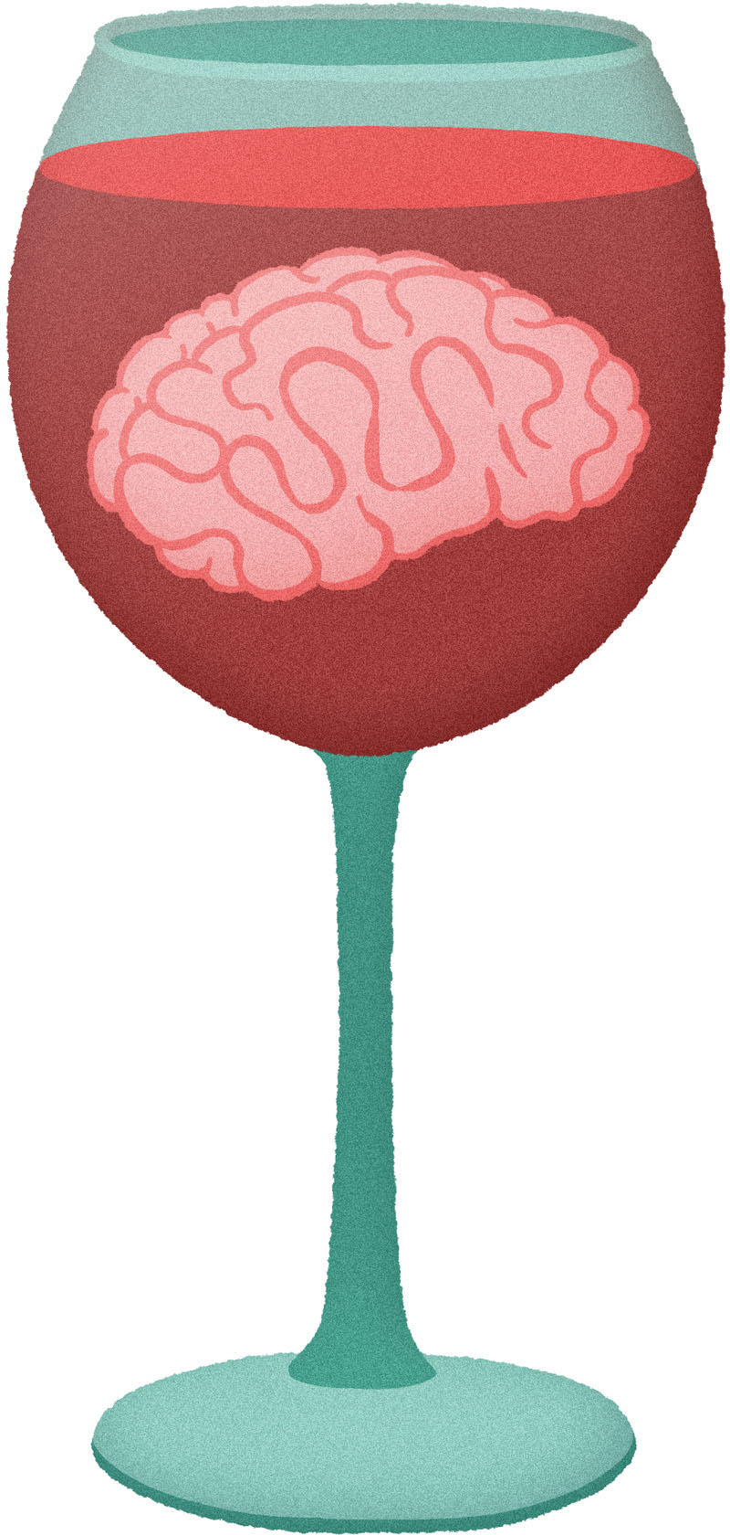
In a 2011 study, published in the Journal of Neuroscience, lead investigator Dr. Charles Zorumski and his colleagues used experiments in rats to tease out more details about ethanol’s effects. They determined that alcohol messes with a receptor on the pyramidal neurons, causing them to release steroids that inhibit neuron plasticity. And no neural plasticity means no changing synaptic connections, and therefore no memory formation.
“The mechanism isn’t straightforward. The alcohol triggers these receptors to behave in seemingly contradictory ways, and that’s what actually blocks the neural signals that create memories,” Dr. Zorumski said in a statement. “[This finding] may explain why individuals who get highly intoxicated don’t remember what they did the night before.”
He explained that consumption of copious amounts of alcohol appears to block some receptors while activating others. His group’s insights counter the long-held notion that alcohol impairs memory simply by killing brain cells.
“Alcohol isn’t damaging the cells in any way that we can detect,” Zorumski added. “As a matter of fact, even at the high levels we used here, we don’t see any changes in how the brain cells communicate. You still process information. You’re not anesthetized. You haven’t passed out. But you’re not forming new memories.”
Past research has shown that acute stress can also impair memory formation, though the process is actually boosted immediately following the threatening event, when corticosteroid hormones involved with the physiological stress response are still high (which makes sense from an evolutionary perspective because animals need to remember and learn from threats they encounter). Some evidence also suggests that long-term stress interferes with memory as well.
Though most of this work has been done in animals, it is predicted to work the same way in humans, meaning that there is a decent probability that if you consume copious amounts of booze, and/or get anxious, then your brain’s ability to store new information will be compromised.
