1
Easier than you think
Professor Kirk makes genetics as easy as A C G T.
SEAMUS KIRK1
[1 It’s possible that my son is not an entirely impartial critic.]
My friend and colleague Steve Withers, a geneticist himself, often refers to others as having ‘a brain the size of a planet’. Many people think that you need a bulging cranium to understand genetics. There’s an aura of difficulty around the subject … which turns out to be a complete con. Genetics is remarkably straightforward. If, by the end of high school, you could manage primary-school mathematics with reasonable confidence, you will have no difficulty with the essentials of genetics.
Why do people think it’s hard? Perhaps it’s just that there is a great deal of detail — thousands of different conditions, all of which vary in their severity, many of which overlap with each other. To fully understand genetic disease, you need to know a bit about how cells work, and there is an awful lot of detail there, too. It’s all just information piled on information, though — anyone can understand any individual part of it.
To prove the point: perhaps the most important piece of information in genetics is the relationship between DNA and proteins. This relationship is similar to, but much simpler than, the relationship between letters and words. Here are the facts:
Proteins form a lot of the ‘stuff’ of the body — they are the building blocks of cells, and of the padding between the cells. Anytime your body has a job to do, it gives it to a protein. If your cells wanted to make a car, every single mechanical and electrical component would be made from proteins … and so would the garage you parked the car in; it’s not just for moving parts. Proteins themselves are made up of amino acids.
DNA is a chemical that contains information. This information is written in an alphabet with only four letters: A, C, G, T. They stand for four nucleobases,2 the chemical building blocks of DNA.
[2 You may be more familiar with the term ‘nucleotide’, which is a nucleobase attached to the other structural elements of DNA.]
Unlike English, the language of DNA has only 21 words. The spelling for those words always involves three nucleobases — it’s a code of threes. In English, CAT means a furry parasite, but, in this language, it means the amino acid histidine. There are 20 amino acids represented in this language, and the 21st word is ‘stop’. A gene is a stretch of DNA that codes for a particular protein — so it’s a string of groups of three that say, ‘Put a histidine in. Then put a glycine in. Then a proline. Okay, now stop.’
You can think of nucleobases as the letters, amino acid names as the words they spell, and genes as sentences. Each sentence explains how to build a particular protein, and each molecule of DNA contains many of these sentences. It’s a manual for building parts of the body.
That’s it. The fundamental basis of genetics. Far simpler than learning to read, and we ask six-year-old children to do that. Even better, there’s no need to actually learn the language — you just need to understand that there is a language, and how it works. After more than 20 years in genetics, I only know the spelling of three or four of the words in the code. The rest I look up when I need to.
There are no concepts in genetics more complex than the one you just learned, if you didn’t know it already. The rest is just detail.
Fortunately, although genetics is simple, it is also fascinating. Take chromosomes, for instance.
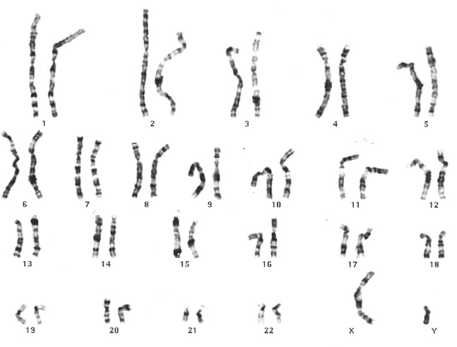
Chromosomes, the physical form our DNA takes within cells, are wholly remarkable structures. You’ve probably seen pictures before, but, just in case, here’s an example.
This is a particularly good set of chromosomes — they’re mine. One of the lesser known perks of training in genetics used to be the chance to prepare and examine your own chromosomes, and who could resist an opportunity like that? Today’s trainees don’t get the chance, for fear they might find out something they don’t want to know. It’s a pity — there is something immensely satisfying about staring at your own genome down a microscope. I imagine it’s a bit like seeing video of your own heart after an operation, but without the inconvenience of having your chest cracked open to get the pictures.
A genome is the totality of an organism’s genetic information, and every living thing has one — you, me, a slug, a blue whale, the kale in the salad you had for lunch, the microbes living under your waiter’s fingernails.3 Bacteria have genomes; protozoa and fungi have them; viruses have them, too. And in everything from bacteria on up, the genome is organised into chromosomes. The number of chromosomes varies enormously between species, and there is no clear link between how complex an organism is and how many chromosomes it has. Bacteria, to be sure, have only one or two, compact and circular. Male jack jumper ants — far more complex than a bacterium — also have only a single chromosome. But Atlas blue butterflies have 450.
[3 Eww.]
The chromosomes you see in the picture were captured at a very particular moment in their existence. It’s easiest to look at chromosomes when they are like this, at a point part way through cell division. They are compressed, and easily recognised as separate structures. Humans (mostly) have 23 pairs of chromosomes. They are 46 long, thin threads of DNA, totalling about two metres, in each of the trillions of cells in your body. Two metres may not sound like much, until you remember that a typical cell nucleus — which holds almost all of the cell’s DNA — is only six millionths of a metre across. If the nucleus were the size of your lounge room, and DNA were made of string, there would be 1,000 kilometres of string in the room with you — enough to stretch from London to Berlin, or from San Francisco to Portland.
Most of the time, that string is not bunched up into the tight bundles that you’re seeing in the picture. It’s a delicate gossamer, stretched and twirled through the nucleus, not completely on the loose but organised, coiled around proteins called histones. This DNA-protein combination is called chromatin, and it is the stuff of life.
DNA, famously, carries information. It carries it through generations, and through deep time. Your DNA is the result of a continuous, unbroken chain of events that has lasted for billions of years. It has been copied, over and over, subtly changing as it went, starting with the first, simple living things that emerged in some warm, shallow, long-forgotten sea. It has endured through many different forms, through mammals, through proto-humans, through the whole of humanity’s existence, until your conception. It carries the memory of that long journey with it. Though we may forget, our genes do not.
Work in genetics for a while, and each chromosome develops its own flavour — not a personality exactly; it’s more that there are things that spring to mind when someone mentions each of them. Chromosome 1 has a pale section near the top — you can see it easily on mine. Remove that section on one of the two copies at conception, and the child who results will have intellectual disability, and a distinctive facial appearance, with deep-set eyes and low-set ears. Chromosome 7 is home to the cystic fibrosis gene, the goal of a race to discovery and a rich scientific prize (that race was won by Lap-Chee Tsui, a dual Hong Kong/Canadian citizen who was working in Toronto at the time). 17 is where BRCA1, one of the breast cancer genes, can be found. The story of the race to find BRCA1 is a darker one, and its consequences are still playing out in the patent courts and in people’s lives today. Chromosome 15 is associated with Prader-Willi syndrome and Angelman syndrome, two very different disorders forever locked together, strange dance partners. There are a few places in the human genome where genes remember which parent they came from and are switched on or off accordingly, and chromosome 15 contains one such region. Chromosomes 13, 14, 15, 21, and 22 are the acrocentric chromosomes: their waists are where their heads should be. Sometimes, they actually fuse together, head to head (a Robertsonian translocation). The Y is a wasteland, a dying chromosome littered with the corpses of broken genes. It hardly has any reason left to exist, and yet it struggles on.
Chromosome analysis, also known as karyotyping, was the original genetic test. There were other medical tests before the karyotype that could detect genetic disorders — examination of a film of blood under a microscope to diagnose sickle cell disease, for example. But this was a test that was purely genetic. More than that, it was the first, and, for a long time, the only, genomic test: it examines the whole of a person’s genome for abnormalities in one go. It’s a bird’s-eye view, lacking in detail by today’s standards, but it has stood the test of time, and we are still using it today.
It has always intrigued me the way human experience builds up around a new technology. Take flying, for instance. Hardly any time had passed after the invention of powered flight before aviation developed its own received wisdom. Aviate, navigate, communicate.4 There are old pilots, and there are bold pilots, but there are no old, bold pilots. Nothing is less use to a pilot than altitude above you and runway behind you.
[4 For when a pilot is in difficulty: aviate — first, do what you need to in order to keep the aircraft flying; navigate — next priority is to figure out where you are and where you might be able to land; communicate — once the first two are under control, you need to talk to the ground, and to other aircraft.]
The same thing has happened with cytogenetics (the study of chromosomes), and even with the newer genetic technologies. There are known traps for young players. There’s the way we’ve always done things (it’s always worked, so why change it?). And, already, young though the field is, there is tradition.
Part of that tradition has to do with the naming of parts. Look at the chromosomes in the picture and you’ll see that some have a waist part way along their length. This is the centromere, the structure that anchors and guides the chromosome during cell division. It’s never exactly in the middle of the chromosome, which means that there is a short arm and a long arm on either side of it: these are named the p and q arms.
Why p and q? In 1966, early in the story of chromosome analysis, a meeting was convened in Chicago5 to discuss standardisation in the description of chromosomes. It was decided that the short arm would be the p arm — for ‘petit’, French for ‘small’. There had been discussion of calling it s for ‘short’, but the French cytogeneticist Jérôme Lejeune was evidently a persuasive man. And perhaps this was a tactical concession by those who wanted to claim the long arm for themselves.
[5 Perhaps because of the outcome, there’s a myth among geneticists that this happened at the Paris nomenclature meeting of 1971. When you read the records of that meeting, however, it’s obvious that the p/q question had long been settled by then. The same story alleges that q was chosen because it’s the next letter in the alphabet. I have been telling medical students this tale for many years, and never bothered to check until I was writing this. My apologies to all those I’ve misled.]
By the time p was agreed upon, it was late in the night. English speakers pushed for the long arm to be l, but it was pointed out that this could easily be confused with the numeral 1. Nobody wanted to let the French have both arms, so there was something of a stalemate. This was broken by the English geneticist Lionel Penrose, who suggested q, because it favoured no language, and because, in another branch of genetics, there was a famous equation, p+q=1, which suggested that with the p arm and the q arm, you have the whole of the chromosome. At this point, it seems, everyone was sick of the dispute, and welcomed the chance to settle the matter and get to bed.
Looking along the arms of a chromosome, cytogeneticists learned to recognise patterns of light and dark staining, due to the interaction of the chromosome material with the dyes used in preparing the slides. You can see these bands in my chromosomes. We’ve already looked at the top (the end of the p arm) of chromosome 1; combine that with the fact that 1 is the largest chromosome and you won’t have trouble finding it. Now look at chromosome 7 — it’s a medium-sized chromosome with a prominent dark band near the end of the p arm. You’ll never mistake a 1 for a 7 now, and you should be able to pick out either in a crowd. Congratulations! You’re on your way to becoming a cytogeneticist.
Sorting the chromosomes by size, from 1 to 22 plus the X and Y (although it turned out that 21 is actually a little smaller than 22), and by their bands, with ever finer divisions in those bands, led to a system of addresses. Chromosome 1 was divided into 1p and 1q. 1p was divided into 1p1, 1p2, 1p3 … and so on, until today we have addresses like 1p36.33 — chromosome number, chromosome arm, band (3), sub-band (6), sub-sub-band (3), and even sub-sub-sub-band (3). Each of these is only visible at finer and finer resolutions, and needs more and more skill to distinguish. When I started in genetics, this was one of the main ways to make a genetic diagnosis: a skilled scientist would look down a microscope and see a subtle change — something missing, something extra, something rearranged. The subtlety of the abnormalities a good cytogeneticist can pick is extraordinary.
When I tried to do this, I struggled even to tell the chromosomes apart, because they don’t come all lined up in pairs in the cell — they lie higgledy-piggledy on a glass slide, at all angles, often crossing over one another. It takes at least a year of staring down microscopes at chromosomes under supervision to become a skilled cytogeneticist, and many more years to become an expert. And sometime, perhaps quite soon, technology will make the job, and these skills, obsolete.
It matters a great deal how much chromosome material a person has. Having too much or too little can have serious consequences. Apart from the remnant that is the Y chromosome, 21 is the smallest of the chromosomes, with the fewest genes. Even so, having three copies of chromosome 21 instead of two causes Down syndrome, a complex condition that affects almost every system in the body. Having only a single copy of chromosome 21 instead of two is fatal, incompatible with surviving even the early stages of pregnancy. There are several other syndromes that relate to whole chromosomes. A child with an extra copy of chromosome 18 has Edwards syndrome,6 for instance. And we’ve already seen what an extra copy of 13 can do.7
[6 Named for the British geneticist John Hilton Edwards, who described the condition in 1960. This was probably the first example of a genetic condition that was first defined by finding its underlying cause — rather than there being a recognised pattern of features first, with a cause identified later.]
[7 The X and Y are special in this regard — see chapter 4 for more on this.]
The pictures of chromosomes we see have a long history. Early studies of the cell led to the discovery that grasshoppers have huge germ cells (the cells that become eggs or sperm) and proportionally huge chromosomes, making them easy to study at a time when microscopes were weak and difficult to use. By the beginning of the 20th century, a link had been established between chromosomes and heredity. But it was decades before this promising start was fulfilled and the first firm connection between chromosomes and a human disease was made. For much of the 20th century, we didn’t even know how many chromosomes humans have. The number was thought to be 48, not 46, and so everyone who looked counted 48.
Sir Alexander Fleming’s discovery of penicillin is the most famous example of lab error gone wonderfully right. Fleming, already a well-known researcher, was studying the bacterium Staphylococcus aureus. He went away for a holiday and came back to find that a culture plate that had been left lying around in his (notoriously untidy) lab had been invaded by a mould, and that around the growths of mould were areas cleared of bacteria. Fleming made some progress in understanding the properties of penicillin, including limited attempts to isolate the substance and develop medical applications, but he concluded that this was probably not going to work, and abandoned the line of research. It was left to others — particularly Howard Florey (an Australian) and Ernst Chain (British, but born in Germany), working together in Oxford — to develop the discovery into a usable drug. Despite this, although Florey and Chain shared the Nobel prize with Fleming, they are far less remembered than Fleming.
So you would think that Tao-Chiuh Hsu, who made a similarly serendipitous discovery that had a profound effect on a whole field of medicine, but who then also successfully developed and applied it, would be at least as famous as Fleming. It’s one of the vagaries of science history that pioneers are not equally remembered for their contributions — which is why you know who Fleming was, but have never heard of Hsu.
It’s a pity, really, because Hsu was a man of remarkable character as well as a great pioneer of science; he should by now have been the subject of at least one biopic. There’s a picture of him on the wonderfully named website of the American Cytogenetics Conference, chromophile.org. The photograph was taken in 2000. Hsu is clasping the inaugural ACC Distinguished Cytogeneticist Award, an ambiguously shaped glass object. In this image, Hsu looks like a kindly old uncle. But half a century before that photograph was taken, he was a young man possessed of an adventurous spirit. In the early 1950s, he left China, a very different China from today, to do research on fruit flies (the famous Drosophila melanogaster, beloved of geneticists if not of orchardists) at the Texas Drosophila Laboratory in Austin. Texas is famous for many things — the Space Center in Houston, the Cotton Bowl, the Alamo. In a more rational world, the Texas Drosophila Laboratory would be better known than any of them.
Many years later, in a ‘mini-autobiography’ published in the American Journal of Medical Genetics, Hsu praised the openness of the America he had come to, and the willingness of everyone he met along the way to help him. He tells the story of the only incident of racism he ever encountered. En route from the South to a meeting in New Hampshire, Hsu committed an act of heinous driving, reversing on a busy highway after missing a turn. An angry taxi driver paused to deliver his verdict. ‘Watch where you are going, you damn Confederate!’
Hsu had come from a technologically undeveloped nation, where cars were rare. He launched himself from a scientific backwater into the forefront of genetic science, a place where he was to stand tall for the decades to come.
The laboratory error in this case happened in 1956, and involved making up solutions that were needed to examine chromosomes. An assistant misread the instructions and mixed one chemical with too much water, making a hypotonic (overly dilute) solution. This caused the cells to swell, allowing the chromosomes to separate so that, instead of being a tangled snarl on the slide, they were much easier to see and distinguish from one another. Hsu seized on this chance opportunity, worked out exactly what had gone so unexpectedly right,8 and how to consistently achieve the effect, and published his results.
[8 It took him three months of painstaking effort, changing one step at a time in the process, over the course of countless repetitions, to work out what the difference had been.]
Almost immediately, Joe Hin Tjio (who is remembered, by geneticists at least) and Albert Levan (who mostly is not) were able to use the method to show that in humans, the chromosome number is 46, not 48. If you can’t even count chromosomes correctly, you can’t hope to find chromosomal abnormalities. Now this was possible, and soon afterwards, in 1959, a French team (Lejeune the p persuader, with Marthe Gautier and Raymond Turpin) reported the extra copy of chromosome 21 that is found in the cells of children with Down syndrome. This opened the door to the discovery of many other chromosomal conditions. Even more importantly, better cytogenetics meant the ability to correctly identify individual chromosomes and to create accurate genetic maps. The Human Genome Project, and most of modern genetics, rests on that mistake in Hsu’s lab.
As it happened, in the same period, the study of DNA was making real progress at last — Watson and Crick’s paper (based on experimental data from Rosalind Franklin) describing the double helix structure of DNA was published in 1953. Their work led to the understanding of the relationship between DNA and proteins described at the start of this chapter.
Between Hsu, Watson, Crick, and the many others on whose work theirs rested, genetics was on its way.