Anthony C. Chu1 and Fernanda Teixeira2
1Hammersmith Hospital, Imperial College Healthcare NHS Trust, UK
2Imperial College Healthcare NHS Trust, UK
Acquired ichthyosis is a condition that arises in adulthood but is clinically and histopathologically similar to hereditary ichthyosis vulgaris. It is rare, and should raise the suspicion of an associated internal disease, particularly malignancy, endocrinopathy, some infections, autoimmunity or a reaction to medication [1].
Acquired ichthyosis usually arises in adult life and clinically resembles hereditary ichthyosis. It is not, however, inherited but is associated with a systemic disorder.
Acquired ichthyosis is a rare condition. There are no available data on prevalence.
Acquired ichthyosis occurs mainly in adulthood but acquired ichthyosis in children with systemic disease has been reported [2].
See Box 87.1
The pathogenesis of acquired ichthyosis is not fully understood; it differs according to the entity with which it is associated.
In cases associated with diabetes, it is supposed that the changes in the skin are due to structural abnormalities in proteins resulting from non-enzymatic glycosylation [23, 24]. However, well-controlled diabetics can also show ichthyotic changes, and for these, an abnormal host immune response has been proposed, probably against components of the granular cell layer [10]. The same hypothesis has also been advanced to explain acquired ichthyosis associated with autoimmune disorders [17], whereas those cases associated with tumours seem to be due to secretion by neoplastic cells of transforming growth factor α (TGF-α), which may exert a mitogenic action on keratinocytes [25].
Histologically, the epidermis shows compact hyperkeratosis with a thinned or absent granular cell layer.
Living in a hot and humid climate may hide the clinical manifestations of filaggrin deficiency [26]. Severe xerosis mimicking acquired ichthyosis can be observed in atopic individuals who immigrate from very humid atmospheres such as South-East Asia to Europe. In their home country, xerosis is not evident but the low humidity in Europe may precipitate an ichthyotic change in the skin.
The onset of acquired ichthyosis is typically sudden with initial involvement of the lower limbs but it may then generalize.
Symmetrical dark thick scaling appears on the legs in a pattern likened to the skin of lizards. The arms and trunk can also be involved, especially the back (Figure 87.1), but flexures are spared, due to the higher humidity in these areas. The face is unaffected in most cases, probably due to the size and number of its sebaceous glands, but the scalp shows abundant fine scales. Pruritus can be pronounced. There may be palmoplantar hyperkeratosis, with fissures that can become infected [1].
Acquired ichthyosis may improve with successful treatment of the underlying disease or cessation of the responsible drug.
The diagnosis of acquired ichthyosis is made clinically and confirmatory tests are unnecessary. Once the diagnosis has been made, a careful search for an underlying cause should be undertaken, with particular care not to overlook the possibility of occult malignancy, especially lymphoma. In addition to a full history, clinical examination, chest radiography and a detailed drug history should be obtained. Appropriate investigations should be performed to identify other potential causes.
The primary aim in the management of acquired ichthyosis is to identify the underlying cause of the disorder. Its treatment can lead to improvement of the dermatosis.
Treatment of the acquired ichthyosis is symptomatic and involves the use of retinoids and keratolytic agents.
Topical retinoids, particularly tretinoin and tazarotene, reduce the cohesiveness of keratinocytes [27].
Beta-hydroxyacids (salicylic acid) help to disaggregate the corneocytes.
Alpha-hydroxyacids (lactic or glycolic acids) produce loosening and desquamation of corneocytes, when applied twice daily. Urea 10–20% is an excellent humectant. Propylene glycol as a 20% preparation in aqueous cream hydrates the stratum corneum.
Acanthosis nigricans (AN) is a dermatosis that manifests as asymptomatic and symmetrical darkening affecting the skin of intertriginous areas, in particular the axillae, groins, submammary folds and neck. The skin in those regions is thickened, has a velvety texture, and may be studded by skin tags. It is particularly associated with obesity and insulin resistance.
AN may present as an isolated skin condition but may be associated with a large range of conditions ranging from obesity to endocrinopathies to internal neoplasms.
Benign AN is very common, and affects up to 20% of adults and 7% of children; this increases threefold if only overweight children are considered [1, 2, 3]. Malignant AN is rare.
AN can occur at any age. The benign form is most common in adults but may be present at birth and is not uncommon in obese children. The malignant form, which is rare, usually arises in older age groups but has been observed in children with Wilms tumours and osteogenic sarcomas [3].
AN has an equal sex ratio.
AN is more common in patients with darker skins. In one study AN was observed in 1% of white people, 5.5% in Latino populations and 13.3% in African American populations [4].
Obesity is the most common association with AN (previously called pseudo-AN), and can regress with weight loss [5].
Many syndromes (Box 87.2) have been associated with AN; they usually involve the endocrine system or accompany autoimmune disorders.
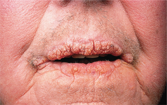
Malignant AN (described in more detail in Chapter 147) has been associated with an extensive range of internal cancers, but over 90% have been seen in patients with gastrointestinal cancer, of which two-thirds are gastric [6, 7, 8]. Other tumours associated with AN are listed in Box 87.3. Malignant AN may be accompanied by other cutaneous paraneoplastic phenomena, particularly florid cutaneous papillomatosis in which there is a rapid development of numerous warty papules on the trunk and the extremities that are clinically indistinguishable from viral warts (see Chapter 147).
The most common associations with benign AN are obesity [2, 9] and insulin resistance [1, 10, 11, 12]. Insulin-derived growth factor (IGF-1) receptors are overexpressed in obese patients with hyperinsulinaemia and insulin resistance [13]. IGF-1 can stimulate the proliferation of keratinocytes and dermal fibroblasts. Epidermal growth factor receptors and fibroblast growth factor receptors (FGFR) are also implicated. In Beare–Stevenson syndrome, activating mutations of FGFR2 have been identified, and in Crouzon syndrome and thanatophoric dwarfism, mutations of FGFR3 have been found [14]. FGFR3 mutations have been identified in the familial form of AN [15].
Some drugs may contribute to AN development: FGFR activation can be produced by certain medications used in stem cell transplantation, such as palifermin [16], and insulin can provoke the development of AN at injection sites by activation of IGF receptors.
In malignant AN, tumour-derived stimulating factors are produced, especially TGF-α, which is recognized by epidermal growth factor receptors. The levels of TGF decrease with tumour debulking, which may be followed by regression of the paraneoplastic phenomena [17].
AN is associated with a number of benign and malignant conditions with a common pathway of keratinocyte and fibroblast proliferation by circulating factors. Perspiration and/or friction are mechanical contributing factors which may be important in determining the characteristic distribution of AN in flexural areas.
Despite its name, AN shows no or minimal acanthosis or hyperpigmentation microscopically. Histology shows hyperkeratosis and papillomatosis with finger-like upward projections of dermal papillae. Pigmentation is due to the hyperkeratosis; there is no increase in melanocyte numbers or in melanin production. Pseudo-horn cysts may be present. In mucosal lesions, parakeratosis may be observed [3].
The familial form of AN is inherited in an autosomal dominant fashion. In familial AN, FGFR3 mutations have been identified [15].
AN usually starts as asymptomatic darkening of the skin of the neck, axillae and groins. With time, the patches become thicker and may develop skin tags in the affected areas. Pruritus is not common.
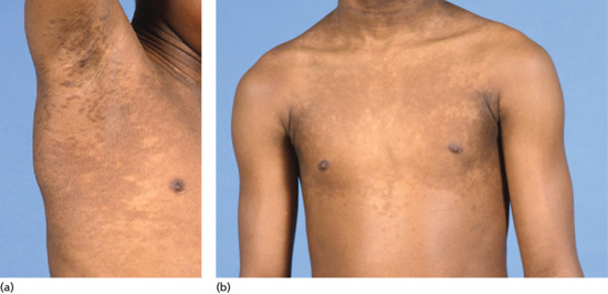
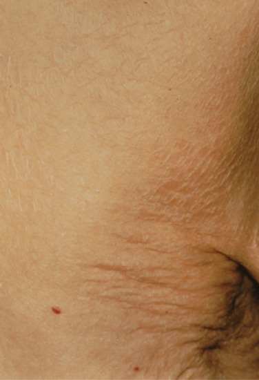
AN presents as symmetrical velvety dark patches which are most commonly seen in the axillae, groins and on the back and sides of the neck (Figure 87.2). The back of the neck is the most common site in children. Skin tags (acrochordons) may be present in affected areas. AN may become widespread with delicate velvety furrowing of mucosal surfaces and involvement of the eyelids and conjunctivae. Associated nail changes include leukonychia and subungual hyperkeratosis.
HAIR-AN is a familial syndrome, that manifests as hyperandrogenaemia, insulin resistance and acanthosis nigricans. It typically affects young black girls, who develop polycystic ovaries, hirsutism, clitoral hypertrophy and frequently have high plasma testosterone levels. This condition, which is also known as type A insulin resistance syndrome, is described in more detail in Chapters 90 and 149.
Type B insulin resistance syndrome is characterized by the association of AN with diabetes and hyperandrogenism, or with an autoimmune disease (including systemic lupus erythematosus, systemic sclerosis, Hashimoto thyroiditis and Sjögren syndrome).
Familial AN is rare and transmitted as an autosomal dominant trait with variable penetrance. It manifests early in life and tends to stabilize in the teenage years. In some patients, it can improve with age.
Drug-induced AN has been associated with many different medications, particularly hormones, insulin, systemic corticosteroids, testosterone and exogenous oestrogens, including oral contraceptives [18, 19]. One of the most common associations is with nicotinic acid. The dermatosis tends to resolve after discontinuation of the offending agent.
Generalized AN is very rare, and seen only in children. There is generalized hyperpigmenation and velvety thickening of the skin, and extensive investigation fails to show any associated systemic abnormality [20, 21, 22].
Acral AN is more common in skin phototypes 5 and 6. It is not associated with systemic disease, and manifests as a velvety thickening and hyperpigmentation of the skin on the dorsa of the hands and feet, especially the knuckles [23].
Unilateral AN (or naevoid AN) is very rare, and is assumed to arise from a somatic mutation during embryogenesis. It can appear in infancy, but not always, and cases have been reported with onset in childhood or adulthood. Clinically, it appears as a pigmented plaque, solitary or along a line of Blaschko, and resembles an epidermal naevus. Histopathologically, the typical changes of AN are seen. It has been described on the face and scalp, chest and abdomen, back and thighs [24].
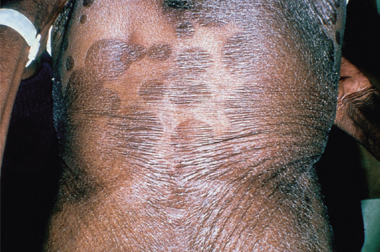
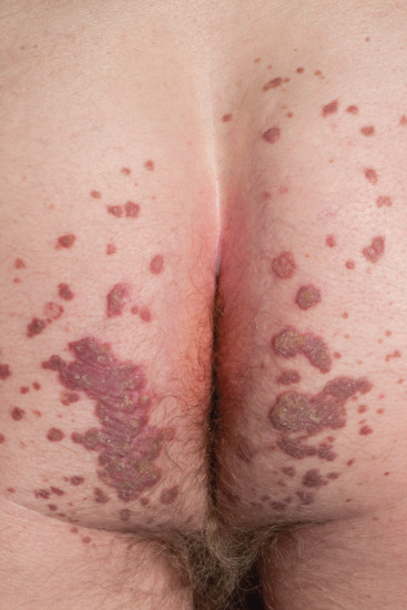
Malignant or paraneoplastic AN (Figure 87.3) can be associated with an extensive range of internal cancers (see above and Box 87.3).
The major differential diagnoses are as follows:
Fairly simple investigations will exclude these diagnoses.
Benign AN is not associated with systemic disease but generally persists and may be a significant cosmetic problem.
AN associated with metabolic abnormalities and insulin resistance may improve with treatment of the underlying condition. AN associated with obesity may improve with dietary restriction and weight loss.
The prognosis of patients with malignant AN is poor, with an average survival of only 2 years from diagnosis.
In adult-onset AN, patients should be screened for underlying endocrinopathy or malignancy. A sensitive test for insulin resistance is serum insulin, the levels of which may be elevated before the onset of diabetes or elevation of glycosylated haemoglobin levels.
Management of AN is the management of the underlying condition. In familial AN or AN not associated with an underlying condition, treatment is aimed at improving the cosmetic appearance of the condition.
Confluent and reticulated papillomatosis (CARP) is an uncommon disorder of epidermal keratinization characterized by the development of hyperkeratotic papules which coalesce into confluent and, in places, reticulated plaques on truncal skin. An abnormal reaction to commensal microorganisms has been postulated to play an aetiological role.
Gougerot and Carteaud first described this entity in 1927 [1]. It is a controversial disease and many had considered it to be a specific form of AN [2]. Diagnostic criteria have been established and it is now considered a specific form of cutaneous papillomatosis [3, 4].
CARP is a rare disease and there are no data on the prevalence of the disease.
CARP is predominantly a disease of young adults. In one series of patients, the mean age at onset was 19 years [2].
It is likely that the sex incidence is equal but different reports have shown a predominance of women in white populations but a predominance of men in Japan. A study from the Mayo Clinic [4] and a recent study from Lebanon shows an equal sex incidence [5].
CARP has been reported in all ethnic groups.
The pathogenesis of CARP is poorly understood. It is thought that an abnormal host reaction to Malassezia organisms may be relevant in some patients [6, 7] and it has also been suggested that actinomycete bacteria may play a role [8]. These hypotheses are discussed in more detail later.8 However, the anti-inflammatory properties of the antibiotics might be the true reason for the response.
Abnormal keratinocyte differentiation has been found on transmission electron microscopy and by immunohistochemical studies,which show an increased expression of involucrin, keratin 16 and Ki-67 [9]. These changes might explain the clustering of the condition in some families [10, 11]. The number of Odland bodies is increased and this is associated with a higher turnover of epidermal keratinocytes as seen in psoriasis [12]. The role of metabolic abnormalities in the development of CARP is gaining supporters [2]. CARP has been associated with obesity and insulin resistance [13], as well as thyroid dysfunction and Cushing disease [14].
There is hyperkeratosis and papillomatosis, with decrease in the thickness or disappearance of the granular cell layer. There may be increased melanin in the basal cell layer and in the stratum corneum, and this is reflected in the colour of the plaques. The dermis shows at most a mild non-specific inflammatory infiltrate [15].
Fungal stains frequently show Malassezia yeasts on the surface of the epidermis.
A number of reports have demonstrated the presence of Malassezia organisms in CARP. This has led to speculation that CARP is due to an abnormal host reaction to Malassezia. Several studies have shown, however, that only about half of patients with CARP have significant yeast populations on affected skin [2, 6, 7]. Although some patients with CARP respond to topical imidazoles or other antifungal agents, about half of cases fail to respond [5, 6, 7] and in those cases only a few spores can be retrieved from the skin surface.
Some patients with CARP appear to respond to tetracycline and macrolide antibiotics, which has led to the suggestion that skin bacteria may be responsible for this disease. In 2005, a previously unknown actinomycete bacterium of the genus Dietzia was isolated from the skin of a patient with CARP. Sensitivity studies showed sensitivity to tetracycline and erythromycin and the patient cleared with tetracycline. The organism, which has subsequently been named Dietzia papillomatosis, has been implicated in a case of septicaemia but, to date, there have been no further reports linking it with CARP [8]. It has been suggested, however, that the response to antibiotics may be due to their anti-inflammatory rather than their antibacterial properties.
Although familial forms of CARP have been reported [10, 11] they are too rare to postulate a genetic inheritance.
Clinical features (Figure 87.4)
The patient is usually a young postpubertal individual. Plaques of CARP are located mainly on the trunk, especially on the presternal, interscapular and epigastric areas. They are generally asymptomatic.
Patients first notice pigmented, 1–2 mm diameter hyperkeratotic papules on the trunk. These coalesce to form greyish-blue plaques which are confluent at the centre but become reticular towards the periphery of the plaques. Over weeks or months, the plaques spread to involve the lower abdomen and pubic areas [2]. The face and limbs may be affected. Localized forms affecting only the face or pubic area have been reported [16, 17]. Mucous membranes are not involved. A number of different patterns of skin involvement have been described [18].
The most common differential diagnoses proposed in cases of CARP include the following:
CARP is a chronic disease with remissions and exacerbations. If CARP responds to treatment, it often relapses when treatment is withdrawn.
CARP is a chronic disease and is purely cosmetic, so no treatment is an option if the condition does not bother the patient. In overweight patients, weight reduction may result in improvement. In some women, CARP may improve during pregnancy or with the use of the oral contraceptive.
A wide range of oral antibiotics have been found to be effective in CARP [21]: the most effective appears to be minocycline [4, 19, 20]. Topical mupirocin ointment has been of benefit in some cases [21].
Topical and systemic antifungal agents, including selenium sulphide shampoo, have been used with success in some patients [2, 22] but the results have been very variable. Topical retinoids and vitamin D analogues have been used with mixed results [23, 24, 25].
Both high- and low-dose isotretinoin have been used in the treatment of CARP with varying results [26, 27].
Pityriasis rotunda (PR) [1] is a dermatosis of unknown cause that presents as perfectly round, slightly erythematous or hyperpigmented plaques, with fine scaling, usually located on the trunk or buttocks, arms and legs.
PR is a rare condition with very few reports in the literature. The most common form of PR is type 1 PR which is typically seen in older Asian or African individuals and is usually associated with underlying systemic disease or malignancy [2]: it may resolve with treatment of the underlying condition [2, 3]. Type 2 PR is usually familial and presents in younger white patients: it is not associated with underlying disease.
PR is rare with most reports coming from South Africa, Sardinia or Japan.
Type 1 PR is usually seen in patients in their 60s. Type 2 PR is usually seen in patients in their forties.
PR has an equal sex incidence.
Most case reports of PR have come from South Africa in black populations and the few reports from the UK have been in patients of African descent [4]. Type 1 disease is also seen in East Asian patients with most reports from Japan; it is rare in white populations [5]: type 2 PR has been reported principally from Sardinia.
Type 1 PR has been associated with hepatocellular carcinoma. In a series of 10 black patients from South Africa with PR, 70% were associated with hepatocellular carcinoma [6]. In a further study from South Africa examining hepatocellular carcinoma, 15.9% of 63 patients had PR [7]. In the South African black population, PR has been associated with a number of systemic diseases listed in Box 87.4.
In Asian populations, PR is associated with underlying malignancy but not specifically with hepatocellular carcinoma [8].
The aetiopathogenesis of PR is unknown. Histologically, it shares some characteristics with ichthyosis vulgaris and some authors feel it is a variant of this disease [9]. In South African patients it has been associated with malnutrition, but this is not generally accepted as a cause of the disease.
Histological changes are restricted to the epidermis with hyperkeratosis and loss of the granular cell layer.
Type 2 PR is inherited as an autosomal dominant trait [10].
PR is an asymptomatic condition and is often diagnosed incidentally in patients being investigated for other disease.
PR presents as asymptomatic thin finely scaling plaques ranging from 0.5 to more than 20 cm in diameter located on the trunk, buttocks, arms and legs. The plaques, ranging in number from a few to more than 100, are sharply demarcated and can coalesce, forming polycyclic plaques. They are pink in lightly pigmented skin to dark brown in skin of phototypes 5 and 6 (Figure 87.5).
PR types 1 and 2 are sporadic and familial, respectively (see earlier).
Differential diagnosis includes post-inflammatory hyperpigmentation following fixed drug eruption, erythrasma, tinea corporis, pityriasis versicolor and psoriasis.
Once type 2 PR develops it usually persists lifelong. In type 1 PR associated with an underlying disease, treatment of the disease may result in resolution of the PR.
A skin biopsy might be helpful to exclude other conditions, as the diagnosis of PR is one of exclusion.
Skin scraping and mycological examination will exclude dermatophytosis and pityriasis versicolor.
Lesions of PR are notoriously resistant to therapy, but some improvement has been achieved with topical treatment.
Keratosis pilaris (KP) is an inherited abnormality of keratinization affecting the follicular orifices with varying degrees of keratotic follicular plugging, perifollicular erythema and follicular atrophy.
KP [1, 2] is a common skin condition, characterized by follicular keratotic papules and perifollicular erythema. Because up to half of the population present with the condition to some degree, it can be considered a variant of normal rather than a disease.
KP is a very common condition affecting 50–80% of adolescents and about 40% of adults.
KP often presents in the first decade of life and may worsen around puberty. In some patients, the disorder improves with age but any age group can be affected from childhood to old age.
Females appear to be more frequently affected than males.
KP is not more prevalent in any racial group.
KP may be associated with ichthyosis vulgaris and atopic eczema [3]. These conditions are, however, all common and the association may be coincidental. Some other reported associations are listed in Box 87.5. More recently, patients with BRAF-positive tumours under treatment with vemurafenib and sorafenib have been noted to develop KP as a side effect [4].
A plug of excess keratin is formed, possibly due to a defect of corneocyte adhesion at the follicular orifice, and this impedes the hair from emerging. The hair can become ingrown and result in an inflammatory response [18].
Histology of KP shows hyperkeratosis, hypergranulosis and plugging of hair follicles. In the dermis, there is a mild perivascular lymphocytic infiltrate in the upper dermis.
KP is inherited as an autosomal dominant trait with variable penetrance [18]. Reports have identified a partial monosomy in the short arm of chromosome 18 in patients with severe forms of KP and ulerythema ophryogenes [19].
KP may show seasonal variation, improving in the summer [20].
KP generally starts in children, most commonly on the extensor surfaces of the upper arms, and can worsen around puberty.
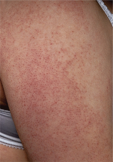
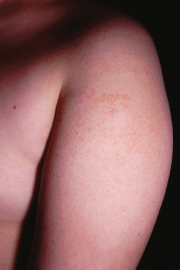
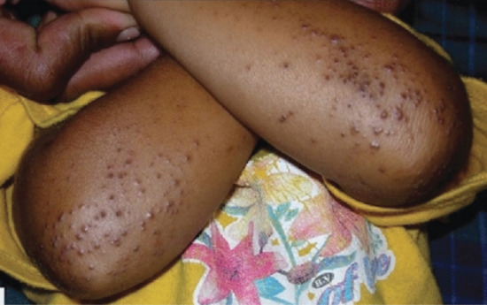
There are small, keratotic papules on the extensor aspects of the limbs, particularly the arms (Figure 87.6) and thighs [20]. The buttocks and the lumbar areas are also frequently affected. These areas acquire a ‘goose-bump’ appearance and rough texture. Lesions can become pustular with superficial pustules developing in affected follicles, precipitated by rubbing on clothing. On the buttock, deeper inflammatory lesions and nodules may develop.
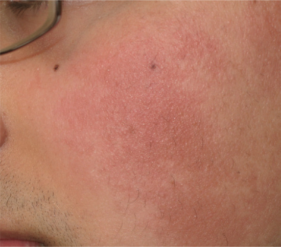
Erythromelanosis follicularis faciei et colli [21–23] is a condition that has been described as a subtype of KP and seen in India and other countries in the Far and Middle East. It manifests as follicular hyperkeratosis accompanied by erythema and hyperpigmentation, and affects, as the name indicates, the face, particularly the cheeks and neck (Figure 87.7).
KP atrophicans is a more inflammatory form of KP which results in follicular fibrosis and atrophy progressing to scarring alopecia. Three variants have been recognized as follows:
KP may be confused with a large number of dermatoses including the following:
Eruptive vellous hair cysts.
If follicular lesions become inflamed KP may be confused with the following:
Folliculitis.
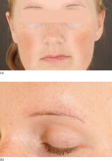
KP atrophicans faciei may be confused with rosacea.
In the majority of patients KP is a mild cosmetic disorder which improves with age. Hypopigmentation, hyperpigmentation and scarring may occur.
KP is a clinical diagnosis for which no tests are normally required.
KP is principally a cosmetic problem and many people do not know they have it. If it does not bother the patient, treatment is unnecessary. Benefit from topical therapy is often limited and many patients will not persist with it.
Keratolytics, particularly salicylic acid. Lactic and glycolic acid preparations also reduce roughness of the skin. Treatment may need to be continued for many years.
Topical retinoids may reduce hyperkeratosis and can be very successful when combined with 10% urea containing moisturisers. Topical retinoids are particularly useful for KP atrophicans faciei [28].
In severe KP, oral isotretinoin has been successfully used but relapse occurs on cessation [29].
Patients with severe fixed erythema from KP atrophicans faciei have been successfully treated with pulsed dye laser [30, 31].
Lichen spinulosus (LS) [1, 2, 3] is a rare idiopathic condition, characterized by the appearance of hyperkeratotic follicular papules arranged into large plaques.
LS was first described by Adamson in 1908 [4] as an acute eruption of grouped keratotic papules which form into plaques. They appear suddenly and are distributed symmetrically on the extensor surfaces of the limbs or on the trunk and neck [1, 2, 3]. They are coarse to the touch and typically measure 2–5 cm in diameter.
LS is a rare disorder. From the first description [4], no further reports of the disease appeared in the literature until 1990, when Friedman described 35 patients in a survey of 7435 patients attending a dermatology clinic in the Philippines, accounting for 0.5% of patients surveyed [3].
LS is a disease of children and young adults with an average age of onset of 16.2 + 10.1 years [3].
Case reports suggest an equal distribution of LS in males and females.
There is no predilection of LS in any ethnic group.
LS is not usually associated with any underlying systemic disease. There have been case reports of LS in HIV infection, Crohn disease and alcoholism, Hodgkin disease and syphilis [5, 6, 7]. Previously, LS has been associated with certain drugs including thallium, gold and diphtheria toxin.
The cause of LS is unknown. Some authors feel that LS is a variant of KP: the conditions share the same features on histology.
Hair follicles are dilated by a thick keratinous plug, and surrounded by a mild to moderate chronic inflammatory infiltrate.
LS generally erupts acutely and is asymptomatic.
Individual papules are follicular, measuring 2–3 mm in diameter and raised 1 mm above the surface of the skin with a pointed keratotic spine. Papules coalesce into plaques ranging from 2 to 5 cm in diameter. The patches are symmetrical and distributed on the trunk, buttocks (Figure 87.10), neck, knees and elbows. The face, hands and feet are usually spared. Plaques erupt in crops, grow rapidly and then remain stationary.
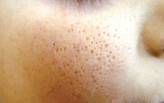
Spinulosis of the face, presenting with tiny keratotic spicules on the cheeks, may be a variant of LS.
All other causes of follicular papules should be considered in the differential diagnosis, including KP, lichen nitidus, phrynoderma, and pityriasis rubra pilaris.
LS is a chronic but purely cosmetic disease. LS can persist for many years, but in most patients, it resolves spontaneously within 1–2 years.
Diagnosis of LS is made clinically and can be supported by histology.
Management of LS is aimed at improving the cosmetic appearance. The mainstays of treatment are topical retinoids and keratolytics [8].
Keratosis circumscripta (KC) is a rare condition characterized by circumscribed areas of follicular hyperkeratosis.
KC was first described by Shrank in 1966 in 10 members of the Yoruba tribe in Nigeria [1]. It is rare and there remains controversy over its status as an individual dermatosis. In one report and review of the literature, the authors suggested that KC was in fact a form of psoriasis modified by environmental factors [2]. It has also been suggested that it is the same as type IV circumscribed juvenile-onset pityriasis rubra pilaris (see Chapter 36). Shrank's findings were, however, supported by a report of 10 patients from Kenya [3] and other authors have supported its recognition as an individual entity [4, 5].
KC is rare.
KC first develops in childhood and is persistent thereafter.
KC was first described in an African tribe and is more common in phototype 6 skin.
The aetiopathogenesis of KC is unknown.
Histology shows follicular plugging and moderate hyperkeratosis. There is no involvement of the dermis.
The condition appears to be sporadic but clustering of cases within a particular ethnic group would support a genetic predisposition.
KC normally starts at the age to 3–5 years with lesions developing quickly over a period of 2–3 weeks.
The lesions of KC are well-defined areas of diffuse and follicular hyperkeratosis affecting the extensor surfaces of the arms and legs and the trunk (Figure 87.11). In his original report, Shrank described elbow and knee involvement in all his patients and 4–5 cm round discs of follicular hyperkeratosis on each hip. The dorsa of the hands and feet were occasionally involved but the palms and soles were rarely affected. Lesions may become thickened and hyperpigmented or violaceous in colour.
A number of dermatoses can be confused with KC but can be clinically differentiated from this disease. These include the following:
Once established, the condition persists. Patients tend not to develop new lesions but existing lesions may slowly increase in size and become more keratotic.
KC is poorly responsive to treatment and generally does not improve with topical corticosteroids, topical retinoids or conventional keratolytics. 40% urea in white soft paraffin has been used successfully to improve the appearance of KC in one patient [3].
Phrynoderma, which literally means toad skin, is one of the cutaneous manifestations of vitamin A deficiency but may also be associated with other nutritional deficiencies. It manifests as follicular hyperkeratosis. It is described in more detail in Chapter 63.
Phrynoderma was first described by Nicholls in 1933 in African labourers and was recognized as a manifestation of vitamin A deficiency [1]. It is usually seen in children living in economically deprived countries.
Phrynoderma is more common in countries where malnutrition is prevalent, but can also be seen in Europe in individuals with liver cirrhosis, malabsorption syndromes or anorexia nervosa and in those who abuse alcohol or are homeless [2]. In economically deprived countries, it is seen in <5% of children and adolescents [3]. Recently, it has been recognized as a complication after bariatric surgery [4, 5].
Phrynoderma is commonest in children between the ages of 5 and 15 in economically deprived countries.
Sexes are equally affected.
No data available.
Phrynoderma was initially thought to be due purely to vitamin A deficiency. Vitamin A is essential for normal cellular growth and division, and maintenance of the immune response. Lack of this vitamin results in abnormal epidermal keratinization as well as immunosuppression [6, 7, 8]. Phrynoderma has now been described in patients with normal vitamin A levels and has been associated with other nutritional deficiencies including including B complex, riboflavin, vitamin C, vitamin E, essential fatty acids and malnutrition [1].
Follicles are dilated with compact keratin and patchy parakeratosis with no dermal reaction.
Clinical features (Figure 87.12)
Phrynoderma starts gradually with mostly non-pruritic lesions on the elbows.
Phrynoderma manifests as groups of papules, each one around 3–4 mm in diameter, with a central keratotic plug [9, 10, 11]. The papules have a follicular distribution and give the skin a rough texture. The elbows and knees are the most commonly affected areas but the buttocks and extensor surfaces of the limbs may be affected. In generalized disease, the trunk and face may be affected. Papules are skin coloured or hyperpigmented. The condition is usually associated with ocular signs, such as night blindness, conjunctival xerosis and ulcerations [12].
The differential diagnosis of phrynoderma includes the following:
Ocular involvement including the development of Bitot spots, conjunctival xerosis and blindness may be associated with phrynoderma.
All patients with phrynoderma should have their vitamin A levels measured. Serum vitamin A levels lower than 35 μmol/dL indicate hypovitaminosis.
In some patients with nutritional deficiency, there will be concomitant hypoproteinaemia. In this circumstance, values of vitamin A can appear reduced despite adequate vitamin A stores.
Treatment involves the correction of poor diet and administration of a multivitamin preparation containing vitamin A, since poor diet often results in other concurrent deficiencies. Oral administration is preferred over parenteral therapy. The dosage, in children aged 8 years or above and adults is 100 000 units daily for 3 days, followed by 50 000 units daily for 2 weeks, and then 10 000 units daily for 2 months, until liver storage is adequate [13].
Trichodysplasia spinulosa is a rare and disfiguring condition caused by infection of the hair inner root sheath by a polyoma virus in immunocompromised patients.
This is a rare condition first described in 1999 in immunocompromised patients [1]. It is due to a novel polyoma virus that appears to infect keratinocytes of the inner root sheath of the hair follicle.
Trichodysplasia spinulosa is rare with only a small number of reported cases in the world literature.
Trichodysplasia spinulosa has been reported in immunosuppressed patients of all age groups [2].
All ethnic groups can be affected.
Most patients with trichodysplasia spinulosa are immunosuppressed following organ transplantation [3]. The disease has been reported in a patient with lymphocytic leukaemia [4]. There have been rare reports of trichodysplasia spinulosa associated with systemic lupus erythematosus [5].
The trichodysplasia spinulosa polyoma virus (TSPyV) is an opportunistic virus: immunosuppression, whether caused by disease or medication, is a prerequisite for the development of trichodysplasia spinulosa.
In affected areas, the hair follicles show abnormal maturation with dilation of the follicular infundibulum, which is plugged by cornified eosinophilic keratinocytes containing large trichohyalin granules (see Chapter 89). Electromicroscopy shows 28 nm intracellular viral particles consistent with polyoma virus. Immunofluorescence studies using antibodies to trichohyalin and TSPyV VP1 protein have shown that the virus is restricted to the nuclei of inner root sheath cells [6]. However, virus has been identified using molecular techniques in the renal allograft of one affected patient [7].
Trichodysplasia spinulosa is caused by TSPyV. TSPyV is a group I double-stranded DNA virus of the family Polyomaviridae and genus Orthopolyomavirus and is related to the Merkel cell polyoma virus. A study in the Netherlands [8] has shown that the virus is common in the general population with a seroprevalence of 70%; it only becomes symptomatic if a carrier is immunocompromised. In contrast to Merkel cell polyoma virus, TSPyV causes dysplasia rather than neoplasia.
Clinical features (Figure 87.13)
The original patient described by Haycox et al. [1] was a heart transplant recipient who developed alopecia of the eyebrows followed by the appearance of multiple painful follicular papules with spiny excrescences which then rapidly coalesced to form a leonine facies.
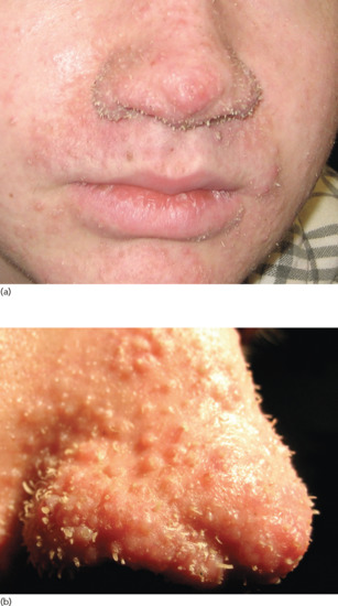
Shiny follicular papules with central spiny keratotic spikes form on the facial skin. The condition progresses rapidly with multiple disfiguring lesions. Alopecia of the eyebrows and scalp may occur. The condition is usually asymptomatic but may be mildly pruritic.
Trichodysplasia spinulosa is a very distinctive disease but in the early stages may be confused with other follicular keratotic dermatoses including the following:
All patients are immunocompromised.
Trichodysplasia spinulosa progesses unless specifically treated or unless immunosuppression is withdrawn. There have been no long-term reports of this disease so long-term prognosis is speculative.
Skin biopsy shows characteristic changes in the hair follicle. The virus can be identified using molecular or immunofluorescence techniques.
Trichodysplasia spinulosa is a persistent disease. In organ transplant recipients, reduction of immunosuppression has resulted in some improvement. Antiviral treatment has been successful in some patients [9, 10] and one patient responded to surgery followed by topical tazarotene [11]. Firm guidance on management cannot be given due to the paucity of reports but oral antiviral therapy would appear to be the treatment of choice.
Flegel disease (FD) is a rare dermatosis, first described by Flegel in 1958 [1]. It is characterized by the presence of flat keratotic papules on the lower legs and dorsa of the feet. It is a disease of the older adult, but it can be seen occasionally in younger persons.
FD is a rare dermatosis characterized by flat keratotic papules on the lower legs and dorsa of the feet. It is a disease of the older adult, but can appear occasionally in younger persons. Each papule measures 1–5 mm in diameter and is topped by a horny keratotic scale, the removal of which causes bleeding. Lesions are commonest on the dorsa of the feet and the lower legs, typically in older patients.
FD is a rare disease and there are no prevalence data in the literature.
FD is most commonly seen in mid to late adult life but cases in children have been reported [2].
FD is seen in all racial groups but appears to be more common in white populations.
A number of reports have suggested an association between endocrinopathies and FD [3], particularly diabetes.
The cause of FD is unknown. A number of hypotheses have been put forward. Some authors have suggested that UV light may play a role [4]. Electron microscopic studies have demonstrated a lack or paucity of Odland bodies in the stratum granulosum, suggesting a possible mechanism for localized hyperkeratosis [5, 6]. Immunological studies have shown that the dermal infiltrate in FD is predominantly T cell and that these cells are activated, suggesting that the disease could be the result of a cell-mediated immune response to keratinocytes [7].
Microscopic examination of a well-developed papule shows characteristic histological features. The stratum corneum is markedly thickened, eosinophilic and compact, and the underlying stratum spinosum is compressed. Patchy parakeratosis may be present. The intervening granular layer is thinned or absent. The surrounding epidermis can show papillomatosis. There is a lymphocytic dermal infiltrate in a band-like distribution beneath the affected epidermis [5].
Both a familial and a non-familial variant have been recognized. At least in some cases, the disease is inherited as an autosomal dominant trait [8].
Some authors suggest that exposure to the sun may be involved in the pathogenesis of FD.
Clinical features (Figure 87.14)
Small keratotic papules develop on the lower legs in middle-aged to elderly patients and slowly spread up the legs. The lesions are asymptomatic.
The lesions of FD are red/brown non-follicular keratotic papules measuring 1–5 mm in diameter. Rarely, the disease may affect the outer ear lobes, arms, palms, soles and oral mucosa [9]. If the scale is removed, the site is red and may have bleeding points. Involvement of the trunk is unusual but a generalized form of FD has been described [10].
Diseases with localized areas of hyperkeratosis are considered in the differential diagnosis, as listed in Box 87.6.
Skin biopsy will show the characteristic changes and will confirm a clinical diagnosis.
FD is difficult to treat and medical treatment needs to continue for prolonged periods.
The most consistent treatment results have been achieved with 5% fluorouracil, which needs to be continued for several months [11, 12, 13], dermabrasion and local excision [14].
Various systemic retinoids have been used with variable responses. Lesions tend to reform on cessation of treatment [15].
Treatment with topical vitamin D analogues have been reported with variable and inconsistent results [16, 17]. Topical retinoids and keratolytics have been used with disappointing results [12].
The term multiple minute digitate keratoses (MMDK) describes a rare clinically distinctive but aetiologically heterogeneous disorder of epidermal keratinization occurring in adults.
Multiple minute digitate hyperkeratoses is a term introduced by Goldstein in 1967 [1] and more fully characterized by Ramselaar and Toonstra in 1999 [2] for a rare disorder of keratinization in which multiple tiny spiky cutaneous keratoses appear in adult life. The more appropriate label MMDK is preferred to define this disorder [3].
MMDK is a rare disorder.
The genetic form generally presents between the ages of 20 and 30 years. MMDK associated with malignancy or other systemic disease presents later in life, typically in the sixth decade of life [4].
MMDK shows a slight male preponderance [1].
No racial predilection has been identified.
Post-inflammatory/reactive cases of MMDK may be associated with solar damage, radiotherapy or may be related to drugs including acitretin, simvastatin and ciclosporin [5].
Sporadic cases of MMDK may be associated with systemic disease or may be paraneoplastic [1, 6–8].
Three forms of MMDK have been proposed, although these divisions may not be real.
The genetic form appears to be inherited as an autosomal dominant trait and the disease becomes clinically apparent in the second and third decades of life.
MMDK has been associated with various drugs as well as following radiotherapy or chronic sun exposure.
Sporadic MMDK may be a paraneoplastic phenomenon but due to its rarity it is difficult to determine whether the associations with underlying neoplasms which have been noted are simply coincidental [6].
Histologically, MMDK shows discrete columns of compact hyperkeratosis arising from acanthotic interfollicular epidermis. The granular cell layer is intact. The dermis is uninvolved. Under the electron microscope keratohyalin bodies are smaller than normal but Odland bodies are present. The pattern of keratin is otherwise normal [9].
The early-onset familial form is inherited in an autosomal dominant fashion.
Patients report the sudden appearance of asymptomatic tiny spiny keratoses on the skin.
MMDK are typically located on the trunk and proximal limbs and may number in the hundreds (Figure 87.15). They are tiny flesh-coloured spikes that measure up to 2 mm in length and are non-follicular. Case reports have described patients with lesions restricted to the palms and soles [7, 8].
Other digitate keratoses including LS, phrynoderma, multiple filiform viral warts, trichodysplasia spinulosa and arsenical keratoses [10].
In sporadic cases of MMDK, investigations for age-related underlying malignancy should be performed.
There is no evidence for the treatment of MMDK. Reports have shown response to a number of treatments.
A porokeratosis is a clonal expansion of keratinocytes which differentiate abnormally but are not truly neoplastic. All forms of porokeratosis show a thin column of parakeratosis, the cornoid lamella, representing the active border. Squamous cell carcinomas may develop within lesions.
A porokeratosis is a clonal expansion of keratinocytes which differentiate abnormally but are not hyperproliferative. Porokeratoses may present as single or multiple lesions and may be localized or disseminated. All forms show a thin column of parakeratosis, the cornoid lamella, representing the active border [1, 2].
Mibelli described the classical form which bears his name in 1893. This was followed by descriptions of superficial and disseminated forms of porokeratosis by Respighi in 1893, linear porokeratosis in 1918, DSAP by Chernosky in 1966, disseminated palmoplantar porokeratosis by Guss in 1971 and punctate porokeratosis by Rahbari in 1977.
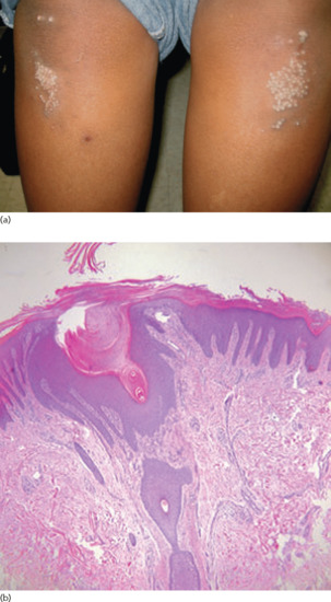
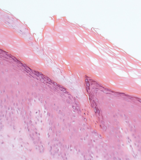
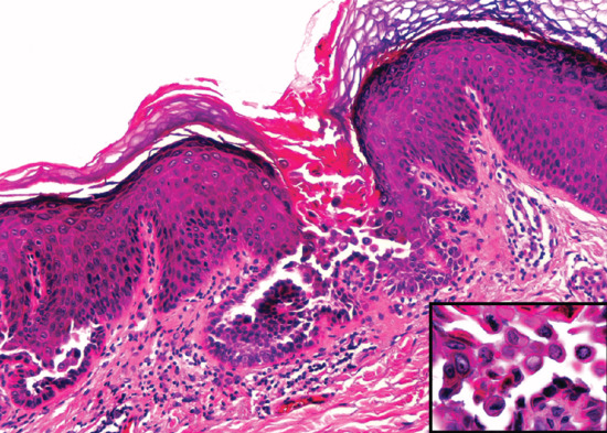
A working clinical classification of porokeratoses is shown in Box 87.7 [1, 2]. Lesions start as papules or plaques which develop into annular lesions with a thin, often thread-like elevated rim. Diagnosis is confirmed by finding the pathognomonic cornoid lamella on histological examination (Figure 87.16). It is likely that the different variants are related, as more than one type of porokeratosis has been reported in the same patient [3] and different types have been reported in different members of the same family [4]. Malignant change with development of squamous cell carcinoma may occur.
Porokeratosis is listed as a rare disease and classified as such by Orphanet and the Office of Rare Diseases (ORD) of the National Institutes of Health. DSAP is the most common form [5].
Classical porokeratosis of Mibelli and linear porokeratosis typically appear during infancy or childhood. Punctate palmoplantar porokeratosis and disseminated palmoplantar porokeratosis usually appear in adolescence while DSAP generally first manifest in adult life.
Porokeratosis of Mibelli, genital porokeratosis and punctate porokeratosis are more common in males whereas DSAP is more common in women. Linear porokeratosis has an equal sex ratio.
All forms of porokeratosis are seen predominantly in pale fair-skinned ethnic groups.
Porokeratosis represents a clonal expansion of keratinocytes [12] which show abnormal differentiation but are not hyperproliferative [13]. The cornoid lamella, which is the hallmark of porokeratosis, is composed of parakeratotic keratinocytes which result from either faulty maturation or an acceleration of epidermopoiesis. It has been shown that there is reduced keratinocyte loricrin and filaggrin expression and abnormal premature keratinocyte apoptosis underlying the cornoid lamella, indicating dysregulation of terminal differentiation [1]. Furthermore, abnormal DNA ploidy in keratinocytes has been demonstrated [14]. In DSAP mutations in the SART3 and MeValonate Kinase (MVK) genes have been found [5]: MVK has a role in keratinocyte differentiation and may protect keratinocytes from apoptosis in response to damage from UV radiation [15].
Drug-induced immunosuppression in various diseases including organ transplantation may predispose to porokeratosis [16]. Natural or therapeutic exposure to UV radiation are recognized trigger factors for DSAP and porokeratosis of Mibelli.
Identification of the cornoid lamella is essential for the diagnosis of porokeratosis. As this represents the peripheral thread-like border of the lesion it is essential that biopsy includes this border. Histologically, the cornoid lamella is a thin column of tightly packed parakeratotic keratinocytes within a keratin-filled invagination of the epidermis through the stratum corneum (see Figure 87.16) [1]. The underlying stratum granulosum may be absent or attenuated but is normal is other parts of the lesion. There is a perivascular or lichenoid lymphocytic infiltrate. Amyloid deposits have been reported in DSAP and in the intertriginous portion of perianal porokeratosis (porokeratosis ptychotropica). The central portion of a porokeratosis may show epidermal atrophy and areas of liquefaction degeneration.
All forms of porokeratosis have been reported to have familial clusters with autosomal dominant patterns of inheritance but with variable penetration. A number of very different chromosomal loci have been identified in DSAP, including 12q23.2-24.1, 1p31.3-p31, 16q24.1-24.3 and 18p11.3 [17]. Linear porokeratosis follows the lines of Blaschko and may be systematized, indicating genetic mosaicism.
Exposure to UV radiation is a factor in the induction of superficial actinic porokeratosis and porokeratosis of Mibelli.
Porokeratoses present with single or multiple papules or plaques which develop into annular lesions with a thin raised border. They are usually asymptomatic but may be pruritic and, if verrucous, may cause discomfort from pressure.
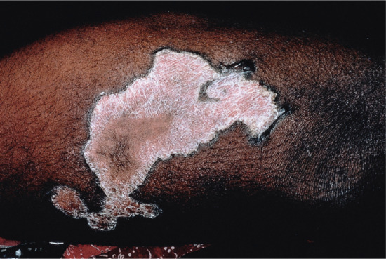
Porokeratosis of Mibelli starts as a single or small group of keratotic papules which may be pigmented. These gradually grow over years to form one or more irregular plaques with a thin, keratotic and well-demarcated border. The central area may be atrophic, either hyper- or hypopigmented, hairless and anhidrotic (Figure 87.17). Lesions are generally distributed on the extremities but can occur anywhere on the body. Occasionally, giant and verrucous forms of the disease may occur [18].
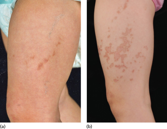
Linear porokeratosis generally occurs in infancy as unilateral streaks or plaques of reddish-brown papules along limbs or the side of the trunk, head or neck following Blaschko lines, indicating underlying somatic mosaicism (Figure 87.18). There is a higher risk of malignant change in linear porokeratosis than in other forms of porokeratosis [19].
Punctate palmoplantar porokeratosis is a rare type of porokeratosis in which seed-like punctate keratoses form on the palms and soles during adulthood [1].
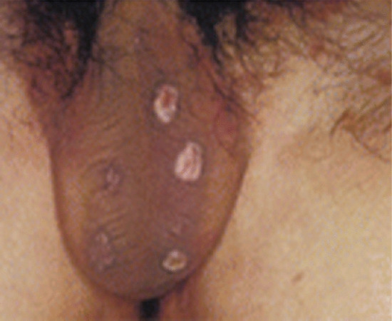
Genital porokeratosis is a rare localized type which it is important to be aware of as it is frequently misdiagnosed clinically. It occurs almost exclusively in men, more often affecting the scrotum than the penis (Figure 87.19) [20]. Vulval porokeratosis is much rarer [21].
Perianal porokeratosis which has been termed porokeratosis ptychotropica [22], is another very rare type which may be very inflammatory and thus elude diagnosis until appropriate biopsies have been taken (Figure 87.20).
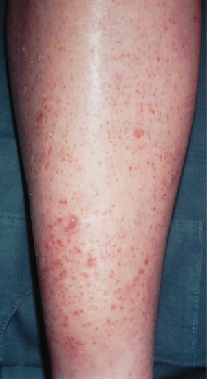
DSAP is the most common form of porokeratosis [1, 5], representing more than half of all cases. The condition is often overlooked as the lesions may be quite inconspicuous to the casual observer. Few or multiple flesh-coloured, pink or reddish-brown finely scaling macules with a thin but well-defined raised border start to appear in early adult life, predominantly on the lower legs and arms (Figure 87.21). Palms and soles are not affected. Lesions are generally small, <1 cm diameter and usually asymptomatic. Patients will often give a history of worsening of the condition following sun exposure.
Disseminated superficial porokeratosis is not necessarily related to sun exposure and will then present in both sun-exposed and sun-protected sites, including sometimes oral mucosa and genitalia. It may be associated with immunodeficiency (e.g. organ transplantation, malignancy, HIV infection) or may develop sporadically during childhood [1].
Systematized linear porokeratosis is a disseminated variant of linear porokeratosis which may be unilateral or generalized and follows the lines of Blaschko [1, 23].
Disseminated palmoplantar porokeratosis (porokeratosis palmaris et plantaris disseminata) is a rare generalized form of punctate palmoplantar porokeratosis in which palmoplantar lesions which first appear in the third decade of life are succeeded by multiple widely disseminated wart-like keratoses in both sun-exposed and sun-protected areas including oral mucosa and genitalia and often following Blaschko lines [1, 24].
CDAGS syndrome is a rare autosomal recessive disorder defined as craniostenosis, delayed closure of the fontanelles, cranial defects or deafness, anal anomalies, genitourinary abnormalities and skin eruption, which is often porokeratosis [25].
The main differential diagnosis of porokeratosis of Mibelli and disseminated palmoplantar porokeratosis is psoriasis. Disseminated superficial actinic porokeratosis may be confused with actinic keratosis or stucco keratoses. Differential diagnosis of linear porokeratosis includes linear verrucous epidermal naevus, lichen striatus and incontinentia pigmenti. Punctate palmoplantar porokeratosis may be mistaken for viral warts.
Cutaneous malignancies, particularly squamous cell carcinoma may occur. This is most often seen in linear porokeratosis [19]. Regular monitoring of patients is needed to identify and treat skin malignancies, especially in immunosuppressed patients.
All forms of porokeratosis are chronic with no tendency for spontaneous resolution.
Patients presenting with sudden onset of porokeratosis should be investigated for causes of immunosuppression including HIV and haematological malignancies.
In many patients, regular monitoring for evolving skin cancer may be all that is needed. In patients with immunosuppression or in linear porokeratosis where the malignancy rate is increased or in those requesting treatment, there are principally two approaches as follows:
Cryotherapy, 5-fluorouracil [26], imiquimod [27], curettage and cautery, photodynamic therapy, carbon dioxide laser [28], topical diclofenac [29] and topical vitamin D analogues such as calcipotriol [30] have all been used to treat porokeratosis with varying degrees of success.
Oral retinoids including isotretinoin and acitretin have been advocated in patients with porokeratosis who are immunosuppressed or have the linear form of the disease in order to reduce the risk of malignant change [31].
Transient acantholytic dermatosis (TAD) is a relatively common transient or persistent monomorphous, papulovesicular eruption mainly affecting the trunk which may be pruritic or asymptomatic It was first described by Grover in 1970 [1].
The term TAD is possibly misleading [2], for although it is self-limiting in some patients it may be very persistent in others. The physician needs a high index of suspicion to diagnose this disease, as clinically it resembles a number of inflammatory dermatoses and histologically is similar to several other dermatoses.
TAD is a relatively common inflammatory dermatosis but there are no incidence data available. In a Swiss study [2], only 24 cases of TAD were found amongst 30 000 biopsies taken. Hospital studies of incidence in hospital referrals in the USA and France have shown an incidence of about 0.1% [3].
TAD is a disease of older patients with a mean age at diagnosis of 61 years but may manifest throughout adult life [3].
The disease has a male predominance with a male to female ratio of 2.4 : 1 [3].
TAD is commonest in white populations.
TAD may be seen in conjunction with a number of other inflammatory dermatoses including psoriasis, asteatotic eczema, contact allergic dermatitis and contact irritant dermatitis. In one study, 11% of patients with TAD were found to have a concurrent inflammatory dermatosis [4].
TAD may also be associated with malignant disease including skin cancer and haematological malignancies [4].
The cause of TAD is unknown. A recent study has demonstrated autoantibodies against a number of proteins involved in keratinocyte development, activation, growth, adhesion and motility using proteomic microarrays to analyse immunoglobulin A (IgA) and IgG autoantibodies [5]. It is still unclear whether these autoantibodies are causative or are a reaction to the damage to keratinocytes seen in this disease.
TAD has been associated with exposure to natural UV radiation, heat and sweating, which has led to the hypotheses that the eccrine sweat glands are aetiologically involved. The disease has not, however been associated with artificial UV exposure and in some reports is more common in the winter [6]. A number of case series have reported the association of TAD with hospitalized and bedridden patients [7].
The primary histological feature of TAD is the presence of small foci of acantholysis with dyskeratosis, intraepidermal clefting and sometimes vesicle formation (Figure 87.22). Five patterns of acantholysis have been described, present either singly or in combination. The incidence of these patterns observed in the three largest studies on TAD are: pemphigus vulgaris-like (47%), Darier-like (18%), spongiotic (9%), pemphigus foliaceus-like (9%) and Hailey–Hailey-like (8%) [2] (see Table 87.1).
Table 87.1 Histological patterns in transient acantholytic dermatosis [2].
Darier like | Suprabasal acantholysis with scattered dyskeratosis and apoptotic cells (corps ronds and grains) throughout the epidermis |
Hailey–Hailey like | Acantholyisis throughout the stratum spinosum with more hyperplastic epidermis and no significant dyskeratosis |
Pemphigus vulgaris like | Suprabasal acantholyisis often with large numbers of eosinophils present |
Spongiotic | Epidermal oedema causing separation of keratinocytes and prominent desmosomes |
Pemphigus foliaceus like | Superficial clefting in the superficial epidermis |
Within the dermis, there is usually a sparse lymphohistiocytic, perivascular infiltrate. Lichenoid change with basal vacuolization has been reported; eosinophils and neutrophils may be present [8].
TAD is reported to be more common in the winter but may be exacerbated by UV exposure and sweating.
Patients usually give a history of the sudden onset of itchy papules on the trunk.
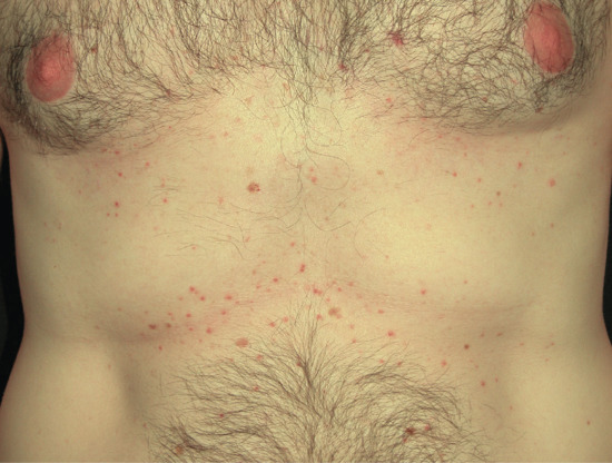
The normal presentation is of a papulovesicular erythematous eruption on the trunk of a middle-aged or elderly white male. It starts with small papules and vesicles that quickly crust and develop keratotic erosions (Figure 87.23). The eruption is usually very itchy and the patient presents with multiple excoriations, although in some patients there is no pruritus. In others, the distribution extends to cover the proximal limbs. In many patients, it may live up to its name and be transient, lasting 2–4 weeks; in some it may, however, persist for months or years or follow a chronic relapsing course.
Some authors use the term ‘persistent and recurrent acantholytic dermatosis’ to encompass those cases which do not rapidly resolve.
TAD has very characteristic clinical features and usually presents little problem in diagnosis. It may be confused with folliculitis, papular urticaria, scabies and herpes zoster. Galli–Galli disease, a rare acantholytic variant of Dowling–Degos disease occurs in a similar age group and may clinically resemble TAD but is usually more widespread, affecting the hands and groins and the reticulate pattern seen in Dowling–Degos disease may be present [9].
TAD has been shown to be associated with skin cancer and in one study 8% of patients had a haematological malignancy [3].
TAD may resolve spontaneously after weeks or months or persist for years. There are no clinical or histological prognostic signs in this disease.
Skin biopsy will confirm the clinical diagnosis. In view of the raised incidence of haematological malignancies seen in this disease, haematological work-up is advised.
Patients should be advised to avoid sunlight exposure, strenuous exercise and heat, all of which may exacerbate the disease.
In many patients, the disease resolves spontaneously after a few weeks and all that is needed is a potent topical corticosteroid for symptomatic relief. Topical vitamin D analogues and calcineurin inhibitors have been used. Systemic antihistamines can be used to control pruritus.
In more severe cases, short courses of systemic corticosteroids have been shown to give sustained improvement but rebound may occur. Systemic retinoids [10], phototherapy [11] and methotrexate have been used in more severe and refractory cases.
Recent case reports have claimed benefit from etanercept [12] and, in one patient with recalcitrant TAD, from photodynamic therapy [13].
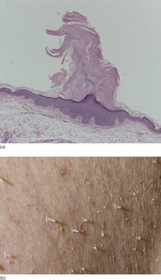
Keratolysis exfoliativa is a common disease of young adults in which discrete areas of superficial skin peeling occur on the palms, starting as air-filled blisters leading to a circinate or irregular annular pattern of scaling. This may be associated with localized hyperhidrosis.
Keratolysis exfoliativa represents an acquired form of non-inflammatory skin peeling. The condition was first described by Wende in 1919 [1].
Keratolysis exfoliativa is a common condition but there are no epidemiological data available on it.
Keratolysis exfoliativa typically affects young adults.
No sex predilection described.
No data.
Keratolysis exfoliativa may be associated with local hyperhidrosis.
The cause is unknown but a recent study suggests that premature corneodesmolysis is the main pathological mechanism [2]. This study showed no association with atopy and filaggrin mutations were not found.
Histology and electron microscopy show cleavage and partially degraded corneodesmosomes within the stratum corneum [2]. There is no inflammatory infiltrate.
Keratolysis exfoliativa often presents in the summer with warmer weather. It can be aggravated by detergents, solvents and irritants.
Clinical features (Figure 87.24)
Keratolysis exfoliativa starts as a sudden onset of discrete scaling on the palms of the hands.
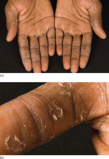
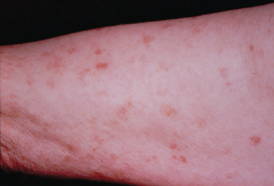
Keratolysis exfoliativa presents initially as small superficial blister-like air-filled pockets on the palms and palmar aspects of the fingers or occasionally the feet. These are formed as the result of focal separation of superficial layers of corneocytes from the stratum corneum. The roofs of the pockets rupture centrally as they expand centrifugally, leaving a ragged rim of residual scale surrounding an irregular superficial dry erosion. There is no irritation and vesicles are not present. Peeled areas of skin lack normal barrier function and may become dry and fissured, particularly on the fingertips.
Keratolysis exfoliativa is a very distinctive disorder but may be confused with pompholyx, psoriasis, tinea manuum, epidermolysis bullosa simplex or acral skin peeling syndrome (see Chapter 65).
Keratolysis exfoliativa may be associated with local hyperhidrosis.
Keratolysis exfoliativa is generally self-limiting but may recur each summer.
No investigations are needed.
Keratolysis exfoliativa is usually a self-limiting condition and treatment may not be needed. Patients should be advised to avoid contact with detergents and other irritants.
Emollients and moisturisers, particularly moisturisers containing agents such as urea or salicylic acid. Topical steroids may exacerbate the condition by further drying the skin.
Xerosis cutis (dry skin) and asteatosis (lacking in fat) are alternative terms used to describe an acquired abnormality of the skin which has lost its normal soft smooth surface and feels dry and rough to the touch. This may be the result of a range of endogenous and exogenous factors, especially ageing and low ambient humidity, and is associated with impaired epidermal barrier function. Xerosis cutis may be accompanied by pruritus (winter itch) and predisposes to eczematous inflammation (eczéma craquelé or asteatotic eczema).
Xerosis cutis is very common and almost physiological in old age. Xerosis cutis (dry skin) and asteatosis (lacking in fat) are alternative terms used to describe an acquired abnormality of skin which has lost its normal soft smooth surface and feels dry and rough to the touch. The condition is associated with an impaired barrier function of the stratum corneum, as reflected by increased transepidermal water loss (TEWL) and reduced water content. Xerosis may however be more accurately assessed by clinical examination [1].
Xerosis is also a common component of eczematous skin diseases, e.g. atopic eczema (atopic xeroderma). This is discussed in detail in Chapter 41.
Xerosis cutis is very common and almost universal in old age.
It generally first manifests in the seventh decade of life and progresses with age.
It appears to be slightly more common in men.
Xerosis cutis is universal in older age groups. In children, it is said to be more prevalent in sub-Saharan Africa, possibly due to frequent use of soap and high local temperatures: TEWL has been shown to be higher in phototype 6 skin compared to other skin types [2].
It may be observed in association with marasmus, malnutrition, diabetes, renal failure and renal dialysis.
Xerosis cutis was initially thought to be due to reduced sebum production with age. The major factors in the development of dry skin, however, appear to be changes in stratum corneum function and lipids [3]. There is a deficiency of all stratum corneum lipids as well as premature expression of involucrin and persistence of corneodesmosomes [4, 5, 6, 7]. Loricrin expression does not appear to be changed [5]. Dry skin also shows decreases in keratin 1 and 10 and an increase of keratin 5 and 14. In women, oestrogen substitution ameliorates dry skin, indicating a role for sex hormones [8].
More common in winter where humidity is low; central heating tends to aggravate the condition. Damage to the stratum corneum by excessive contact with soap or detergents or from frequent and prolonged bathing have each been proposed as additional exacerbating factors, although one study found no difference in bathing habits between patients with widespread asteatotic eczema and controls, suggesting that other unknown factors may play a role [9]. Lack of oestrogen is thought to be a contributory factor in postmenopausal women [8].
No histological abnormalities apart from slight irregularity in the stratum corneum.
Low humidity in the winter and the drying effects of central heating exacerbate xerosis cutis. The generally dry atmosphere in hospital wards increases the risk in frail elderly patients confined to hospital for prolonged periods. Targeted anticancer therapies may induce xerosis [10].
Dryness and scaliness of the skin is generally the first manifestation of the condition, followed by itching.
Xerosis cutis most often affects the shins and may not affect any other area of the skin. It may become widespread but spares the face, neck, palms and soles. In elderly immobile hospitalized patients, it will frequently affect the abdomen and, in women, the anterior surfaces of the breasts, but spares the back and the undersurfaces of the breasts.
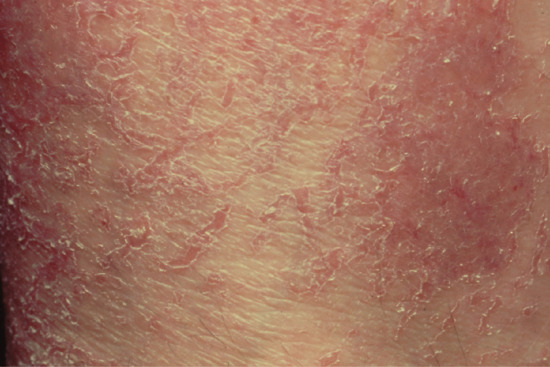
Affected skin looks dull, dry and covered with fine scale which sheds readily. The surface may become crazed with criss-cross superficial cracks in the stratum corneum giving the skin a crazy-paving look (Figure 87.25).
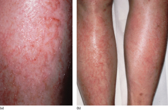
Asteatotic eczema (eczéma craquelé). Xerosis cutis may progress to an eczematous inflammation in which the fissures in the skin surface become red, inflamed and itchy (Figure 87.26). Eczéma craquelé may sometimes be seen after episodes of acute distension of the skin, usually due to acute oedema (see Figure 87.28).
Dry scaly skin is a component of many inflammatory skin diseases. These include atopic eczema (see Chapter 41), hereditary (see Chapter 65) and acquired ichthyosis (see earlier). Apart from the latter, these generally manifest at an early age.
Xerosis cutis is often seasonal, being worse or occurring only in the winter; it tends to worsen and become more persistent with age.
Removal of precipitating factors: patients should be instructed to avoid applying potential irritants to the skin. Simple measures to increase the humidity of centrally heated rooms (e.g. placing a bowl of water close to radiators) may help.
Emollients and moisturisers are the treatment of choice and usually result in rapid improvement.
If asteatotic eczema develops, anti-inflammatory treatment with a mild corticosteroid ointment may be necessary but long-term topical corticosteroids should be avoided, as these may further damage the skin barrier. Topical tacrolimus and pimecrolimus have also been advocated for the treatment of asteatotic eczema [11, 12].
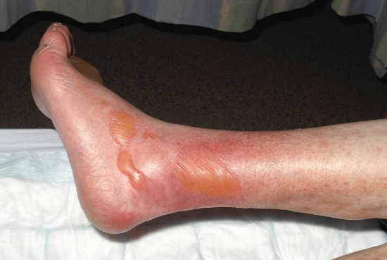
There are certain situations where the epidermis is unable to withstand forces which would normally not affect its integrity. Acute distension of uninflamed skin in the absence of disordered epidermal integrity may at times result in epidermal separation or disruption. Most commonly, this is due to acute dependent oedema, as from acute congestive heart failure, and results in large bullae which may be mistaken for bullous pemphigoid. Alternatively, the distension may result in disruption of epidermal cohesion manifesting as eczéma craquelé.
Subepidermal blistering is a well-recognized component of many skin disorders in which there are hereditary or acquired abnormalities affecting the normal adhesion of the epidermis to the dermis. It is also a normal reaction to excessive thermal or frictional trauma.
Bullae may also occur in otherwise normal skin as a consequence of the rapid onset of oedema. The speed of development of such acute oedema appears to be a more important risk factor than the degree of oedema. The blisters respond rapidly to reduction of oedema [1]. It has been described after attacks of angio-oedema as well as in the elderly following the sudden onset or exacerbation of oedema, e.g. after acute congestive heart failure [1, 2, 3, 4].
Reported only in case series, but no structured studies available.
May appear in any person who develops oedema acutely, though the elderly appear to be more susceptible [2].
Little is known about the pathophysiology of this disease. Ultrasound studies of acute oedema suggests that fluids accumulate more superficially in the dermis than following lymphoedema or chronic stasis [5, 6] which may sometimes be a contributory factor.
Biopsies have shown marked epidermal spongiosis, and a slight lymphohistiocytic inflammatory infiltrate. Direct immunofluorescence was negative [2].
Acute onset of oedema without itching.
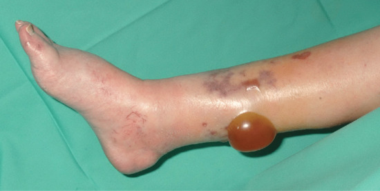
Tense unilocular non-pruritic bullae appearing in areas of acute oedema (Figure 87.27).
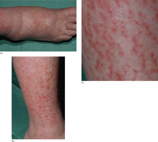
Acute cutaneous distension may alternatively result in disruption of the epidermis as seen in asteatotic eczema with crazing and fissuring of the epidermis and leakage of oedema fluid to the surface (Figure 87.28) [1, 7, 8].
In the elderly population, autoimmune bullous diseases, particularly bullous pemphigoid, are an important differential diagnosis. The close temporal association with acute oedema, the absence of itching and the lack of spread to other areas of the body form important clues to the diagnosis.
Diabetic bullae (see Chapter 64) may resemble acute oedema blisters but are generally not so tense and they are not associated with acute oedema (Figure 87.29).
The lesions resolve rapidly as the oedema resolves, leaving no scars.
This is a clinically defined disease without known pathognomonic tests. In some cases, the attending dermatologist may wish to rule out autoimmune bullous disorders through direct immunofluorescence or identification of circulating antibodies.
Reduction of oedema through appropriate management of the underlying disease. Aseptic puncture of individual lesions to reduce risk of ulceration.