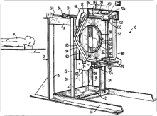
The CT scan goes into use in the United States.
Originally known as a CAT scan—for computed (or computerized) axial tomography, or computer-aided (or assisted) tomography—it uses a series of X-rays to create sequential images of slices of body tissue. Those can be integrated into a 3-D X-ray, so doctors know the precise position of diseased or otherwise abnormal tissue.

Tufts University physics professor Allan Cormack theorized in the 1960s that you could take X-rays from varying angles; account for differences in the density of bone, muscle, and organs; and program a computer to assemble 3-D images.
English electrical engineer Godfrey Hounsfield was working on similar research at EMI, funded by massive profits from the Beatles’ 1960s hits on EMI. Hounsfield began testing a CT brain scanner in 1971, sometimes carrying bulls’ brains across London on public transit. His prototype took five minutes for the scan, and two and a half hours for the computer-processed image. The first EMI production model took four minutes to scan, and its Data General Nova minicomputer needed seven minutes per picture.
Meanwhile, in 1973, dentist-physicist Robert Ledley built a whole-body scanner at Georgetown University. It saved its first life while still in development: a pediatric neurosurgeon used it while Ledley was away for the weekend. Minnesota’s Mayo Clinic and Boston’s Mass. General also began using CT in 1973.
Hounsfield and Cormack shared the 1979 Nobel Prize in Physiology or Medicine. Ledley was inducted into the National Inventors Hall of Fame. CT scanners today are faster—four to ten images a second. Instead of taking discrete, individual slices as images, they use spiral, or helical, tomography, like a virtual HoneyBaked ham.
That’s a lot of progress in 39 years… which is 273 in cat years.—RA