12
Adaptation to extrauterine life
The transition from intrauterine to extrauterine life involves a complex sequence of physiologic changes that begin before birth. Remarkably, although infants experience some degree of intermittent hypoxemia during labor, most undergo this transition smoothly and uneventfully. If not, cardiorespiratory depression requires prompt and appropriate resuscitation.
Physiologic changes in fetal–neonatal transition
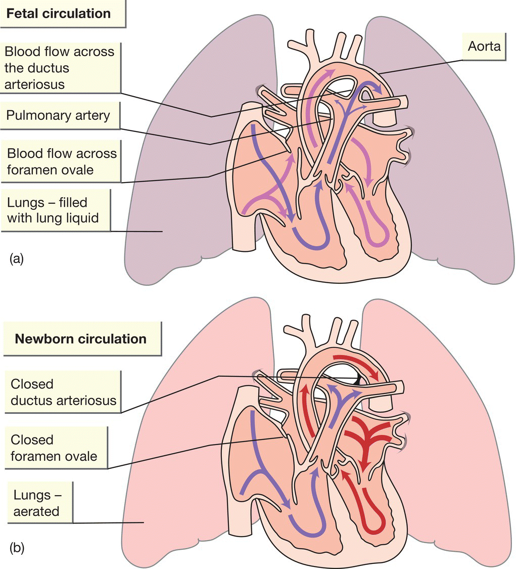
- Before birth, the lungs are filled with fluid. Oxygen is supplied by the placenta. On reaching the right atrium, some of the oxygenated blood from the placenta flows directly to the left atrium via the patent foramen ovale, bypassing the lungs. This ensures that the most oxygenated blood goes to the heart and brain. In addition, the blood vessels that supply and drain the lungs are constricted (providing high pulmonary vascular resistance), so most blood from the right side of the heart bypasses the lungs and flows through the ductus arteriosus into the lower aorta (Fig. 12.1a).
- Shortly before and during labor, lung liquid production is reduced.
- During descent through the birth canal, the infant’s chest is squeezed and some lung liquid exudes from the trachea.
- Multiple stimuli (thermal, chemical, tactile) initiate breathing. Serum cortisol, ADH (antidiuretic hormone), TSH (thyroid-stimulating hormone) and catecholamines dramatically increase.
- The first gasp is usually within a few seconds of birth. A negative intrathoracic pressure is generated to achieve this. Most lung liquid is absorbed into the bloodstream or lymphatics within the first few minutes of birth.
- Aeration of the lungs is accompanied by increased arterial oxygen tension; the pulmonary artery blood flow increases and the pulmonary vascular resistance falls.
- Contraction of the umbilical arteries restricts access to the low resistance placental circulation. This results in increased peripheral vascular resistance and an increase in systemic blood pressure.
- The fall in pulmonary vascular resistance and the rise in systemic vascular resistance result in near equalization of pressures across the duct and virtual cessation of ductal flow and also closure of the foramen ovale (Fig. 12.1b).

Fig. 12.1 Changes in the circulation at birth. (a) Fetal circulation. (b) Newborn circulation.
Abnormal transition from fetal to extrauterine life
The transition may be altered by a variety of antepartum or intrapartum events, resulting in cardiorespiratory depression, asphyxia or both (Table 12.1).
Table 12.1 Conditions assciated with abnormal neonatal adaptation to extrauterine life.
| Fetal | Maternal | Placental |
| Preterm/post-dates | General anesthetic | Chorioamnionitis |
| Multiple birth | Maternal drug therapy, e.g. narcotics, magnesium sulfate | Placenta previa |
| Forceps or vacuum-assisted delivery | Pregnancy-induced hypertension | Placental abruption |
| Breech or abnormal presentation | Chronic hypertension | Cord prolapse |
| Shoulder dystocia | Maternal infection | |
| Emergency cesarean section | Maternal diabetes mellitus | |
| Intrauterine growth restriction (IUGR) | Polyhydramnios | |
| Meconium-stained amniotic fluid | Oligohydramnios | |
| Abnormal fetal heart rate trace | ||
| Congenital malformations | ||
| Anemia, infection |
The Apgar score
The Apgar score, named after Virginia Apgar, an anesthesiologist, is used to describe an infant’s condition during the first few minutes of life (Table 12.2). It is assigned at 1 and 5 minutes of life. If the score is still below 7 or the infant is requiring resuscitation, it is continued every 5 minutes until normal or 20 minutes of age. Although often assigned, few babies truly attain a score of 10, because it is uncommon for the baby to be pink all over. The Apgar score is useful as a shorthand record of the newborn infant’s condition after birth.
Table 12.2 Apgar score.
| Apgar score | |||
| 0 | 1 | 2 | |
| Heart rate | Absent | <100 beats/min | >100 beats/min |
| Respiration | Absent | Slow, irregular | Good, crying |
| Muscle tone | Limp | Some flexion of extremities | Active motion |
| Reflex irritability (response to stimulation) | No response | Grimace | Cough, sneeze, cry |
| Color | Blue or pale | Body pink, blue extremities | Pink |
