18
Neurologic examination
The newborn infant’s neurologic development progresses markedly with gestational age. This needs to be taken into account when performing a neurologic examination, and accounts for many of the components of the neurologic examination used in the clinical assessment of gestational age (Ballard or Dubowitz score; see Chapter 83).
A detailed neurologic examination is performed if there are any concerns about neurologic abnormality. A normal neurologic exam is helpful prognostically, e.g. following hypoxic–ischemic encephalopathy, a normal neurologic examination and normal feeding by 2 weeks of age are associated with a good prognosis. Very low birthweight infants with a normal neurologic examination and intracranial ultrasound at 40 weeks are highly unlikely to develop significant motor disability and the predictive value of combined assessment is better than ultrasound alone.
The neurologic development described here is adapted from that described by Amiel-Tison, who has also devised a standardized examination with 10 components (see video: Neurological examination).
States of alertness
An infant’s state of alertness can be classified (Prechtl scale):
- state 1: eyes closed, regular respiration, no movements
- state 2: eyes closed, irregular respiration, no gross movements
- state 3: eyes open, no gross movements
- state 4: eyes open, gross movements, no crying
- state 5: eyes open or closed, crying.
For satisfactory neurologic assessment infants need to be in state 3, when they are quiet but alert, i.e. able to fix and follow. However, the clinician may have to bring the baby to this state. Inability to do this may occur because the infant is abnormally lethargic or hyperexcitable (or deeply asleep or hungry!). An abnormal cry may also indicate abnormal neurology.
Visual fixing and following
A normal term infant should fix and follow a face or target of concentric black and white circles or a red ball moving from side to side. This starts at about 32 weeks’ gestation. The infant should make eye-to-eye contact when held about 30 cm from the observer.
Hearing
Infants respond to noise with a facial grimace, turning of the head or startle.
Consolability
This is the response of the crying infant to a voice or soothing movements, such as rocking from side to side. It indicates communication between the infant and caregiver.
Head circumference
This is a surrogate measure of brain volume and subsequently of brain growth.
Face (cranial nerves)
There should be normal facial movements, blinking of the eyes and ability to suck strongly.
Posture and spontaneous motor activity
Posture
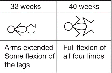
Posture at term is flexed (Fig. 18.1). Movements are smooth, symmetric and varied. The infant can move the fingers and can abduct the thumbs.

Fig. 18.1 Posture.
Passive tone in limbs and trunk
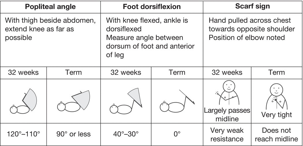
Develops from hypotonia at 24 weeks of gestation to strong flexor tone at 40 weeks, initially in the lower then upper limbs (Fig. 18.2).

Fig. 18.2 Passive tone in limbs and trunk.
Active tone in limbs and trunk
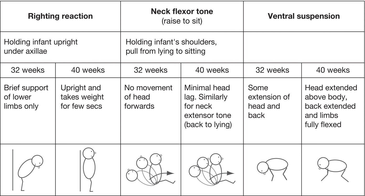
See Fig. 18.3.

Fig. 18.3 Active tone in limbs and trunk.
Primary reflexes
Primary or primitive reflexes reflect brainstem activity (Fig. 18.4). They are a manifestation of central nervous system programming with later suppression by higher cortical function. If they cannot be elicited, suggests central nervous system depression. More important, their persistence suggests damage to upper cortical control (Table 18.1).

Fig. 18.4 Primary reflexes.
Table 18.1 Primary reflexes.
| Reflex | Disappearance (corrected age) |
| Placing | 3 months |
| Palmar grasp | 3 months |
| Plantar grasp | 3 months |
| Moro | 4 months |
| Asymmetric tonic neck reflex (ATNR) | 6 months |
Deep tendon reflexes
May be depressed with lower motor neuron lesions, occasionally increased with upper motor neuron lesions. May reveal asymmetry. Ankle clonus is common and usually of no pathologic significance.
Plantar responses
Elicited by stroking the lateral part of the foot from heel to toe. Unhelpful at this age as normal response may be flexor (toe down) or extensor (toe up).