56
Coagulation disorders
In the newborn, abnormal bleeding may be due to:
- a platelet abnormality (number or function)
- abnormal coagulation system
- vascular endothelial damage/abnormality.
Thrombocytopenia
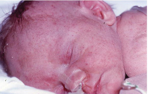
This is the most common platelet disorder. It is defined as a platelet count of less than 150 000/mm3 (150 × 109/L). It is usually identified on the complete blood count (CBC), but, if severe, may cause petechiae (Fig. 56.1) or bleeding.

Fig. 56.1 Petechiae from thrombocytopenia in an infant.
A convenient classification is according to the time of onset (Table 56.1). The most common causes are maternal pre-eclampsia and diabetes mellitus, intrauterine growth restriction and neonatal infection.
Table 56.1 Classification of fetal and neonatal thrombocytopenia (most common causes in bold type).
| Time of presentation | Condition |
| Fetus | Neonatal alloimmune thrombocytopenia (NAITP) |
| Maternal autoimmune thrombocytopenia (ITP, SLE) | |
| Congenital infection (CMV, rubella, herpes, syphilis) | |
| Severe rhesus disease | |
| Chromosome abnormalities (trisomy 21, 18, 13) | |
| Inherited (very rare) | |
| Neonatal (<72 h) | Placental insufficiency (PIH, IUGR, diabetes)Neonatal infectionBirth asphyxiaNeonatal alloimmune thrombocytopenia (NAITP)Maternal autoimmune thrombocytopenia (ITP, SLE)Thrombosis (renal vein, aortic)Congenital infection (CMV, rubella, herpes, syphilis)Inherited (very rare) |
| Neonatal (>72 h) | Late-onset bacterial infection, necrotizing enterocolitisDisseminated intravascular coagulation (DIC)Giant hemangioma (Kasabach–Merritt syndrome) |
ITP, idiopathic thrombocytopenic purpura; SLE, systemic lupus erythematosus; CMV, cytomegalovirus; PIH, pregnancy-induced hypertension; IUGR, intrauterine growth restriction.
Treatment is directed to the underlying cause.
For infants who are sick or septic, where production may be compromised, platelet transfusion is considered if:
- platelets <30 000/mm3 (30 × 109/L) in term infants
- platelets <50 000/mm3 (50 × 109/L) in preterm infants
- if actively bleeding or before surgery, platelets <100 000/mm3 (100 × 109/L).
Abnormal coagulation
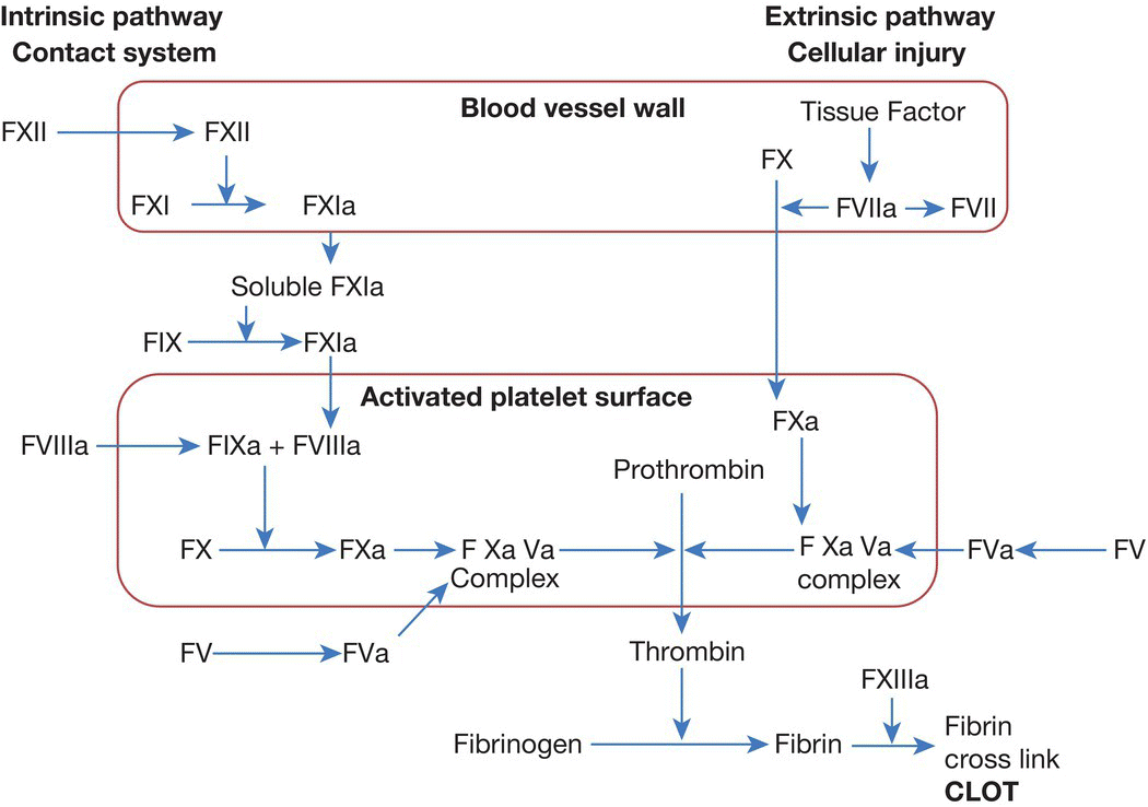
Coagulation factors are a group of proteins that when activated will promote the formation of a fibrin-rich clot or hemostatic plug (Fig. 56.2). These proteins are formed early in gestation in the fetus and do not cross the placenta.

Fig. 56.2 The coagulation pathway.
The most common acquired cause of coagulopathy is a combination of coagulation activation and poor liver reserve in a sick or septic infant.
Deficiency of certain coagulation factors will lead to bleeding disorders (Table 56.2).
Table 56.2 Bleeding disorders.
| Deficiency | Disorder | Comments |
| Vitamin K | Vitamin K deficient bleeding (VKDB) (Hemorrhagic disease of newborn) | Deficiency of vitamin K-dependent coagulation factors (factors II, VII, IX and X) Associated with breast-feeding or severe liver disease in the infant or maternal use of anticonvulsants |
| Factor VIII | Classic hemophilia A | X-linked inheritance – positive family history in 80%. Mild and moderate forms usually asymptomatic during the newborn period, but 20% of cases present in the newborn, usually after circumcision or other surgery. Severe form may result in life-threatening hemorrhage |
| Factor IX | Christmas disease – hemophilia B | X-linked inheritance. Similar presentation to hemophilia A |
| Von Willebrand factor | Von Willebrand disease | Most common inherited bleeding disorder Autosomal dominant inheritance Only rare subtypes present in the newborn period |
Indications for performing clotting studies
These are:
- family history of bleeding disorder
- clinical signs of abnormal bleeding:
- – oozing from venepuncture/surgical sites
- – bleeding umbilical cord stump
- – extensive bruising or large cephalhematoma or subgaleal (subaponeurotic) bleed
- – gastrointestinal bleeding
- septic infant
- necrotizing enterocolitis
- rapidly falling platelet counts in a sick infant
- severe hypoxic–ischemic encephalopathy.
Investigations
Coagulation screen consists of:
- PT (prothrombin time)
- APTT (activated partial thromboplastin time)
- TT (thrombin time).
May include:
- fibrinogen
- D-dimers – a measure of fibrin breakdown, may be useful for diagnosis of disseminated intravascular coagulation (DIC).
Interpretation of abnormal clotting studies (Table 56.3)
Table 56.3 Interpretation of abnormal clotting studies.
| Test | Vitamin K deficiency | DIC | Liver impairment | Hemophilias |
| Platelets | Normal | Reduced | Normal | Normal |
| PT | Prolonged | Prolonged | Prolonged | Normal |
| PTT | Prolonged | Prolonged | Prolonged | Prolonged |
| TT | Normal | Prolonged | Prolonged | Normal |
| Fibrinogen | Normal | Reduced | Reduced | Normal |
The prothrombin time (PT) may be reported by some laboratories in the form of an INR (international normalized ratio). Although this may be useful for adults, the INR is not a reliable measure of coagulation for neonates. PTT, partial thromboplastin time; TT, thrombin time.
The normal values for preterm and term infants are derived locally, as different hospitals use different assays.
The coagulation values in preterm and term neonates differ significantly from older children and adults:
- Prothrombin time tends to be a few seconds longer at birth but will reach the normal adult range within a week.
- Activated partial thromboplastin time may not reach adult normal range for several months because of low levels of the ‘liver’ factors (e.g. IX, XI, XII).
- Thrombin time may be slightly prolonged in early life due to the presence of a fetal form of fibrinogen. This is of no clinical significance.
Management of abnormal clotting
If there is active bleeding a correct diagnosis must be established. Vitamin K should be given while results are awaited, and fresh-frozen plasma (FFP) if there is severe bleeding. Intramuscular injections must not be given to any neonate with a known or suspected major coagulation disorder (e.g. hemophilia), and care must also be taken after venepuncture and/or heelprick testing in such babies – pressure for >5 minutes is recommended.
If there is disseminated intravascular coagulation (DIC), treat the underlying cause. In the interim, platelets, FFP and cryoprecipitate (only if the fibrinogen level is low) may be indicated. Their need is determined by the coagulation tests, which should be repeated regularly as this is an evolving disorder.
FFP contains all coagulation factors and is suitable for emergencies, but does not contain sufficient of any single factor for severe single factor deficiencies. Replacement by a suitable concentrate is optimal, once a firm diagnosis has been established.
Severe congenital coagulation factor deficiencies – consult pediatric hematologist.