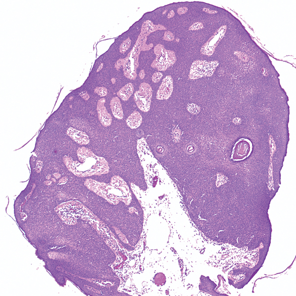
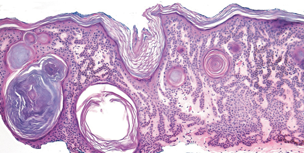
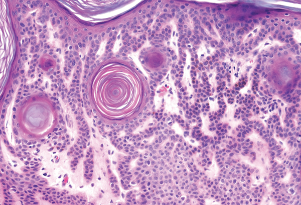
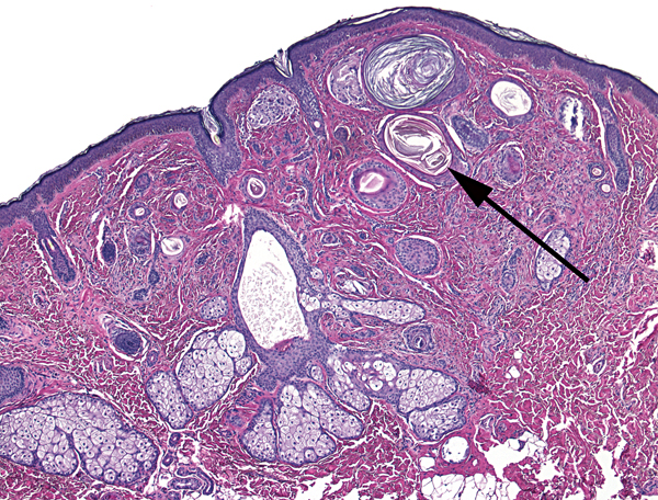
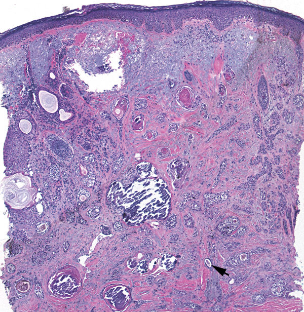
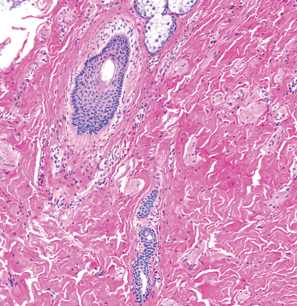
Numerous horn cysts (long arrow) in fibrotic stroma
Tubules of two-layered epithelium (short arrow)
Calcification often present
Confined to dermis
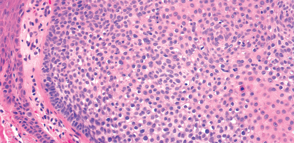
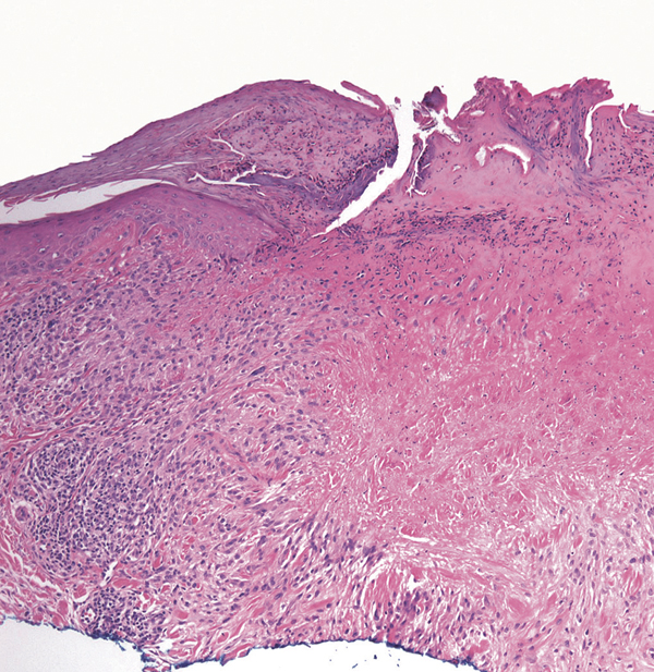
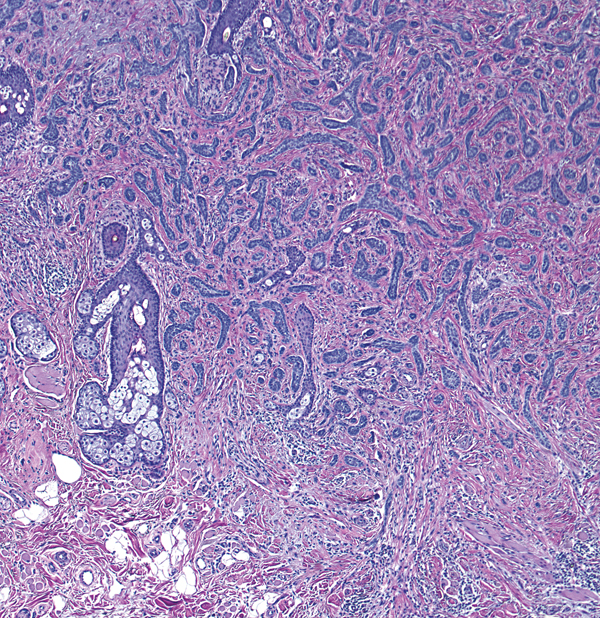
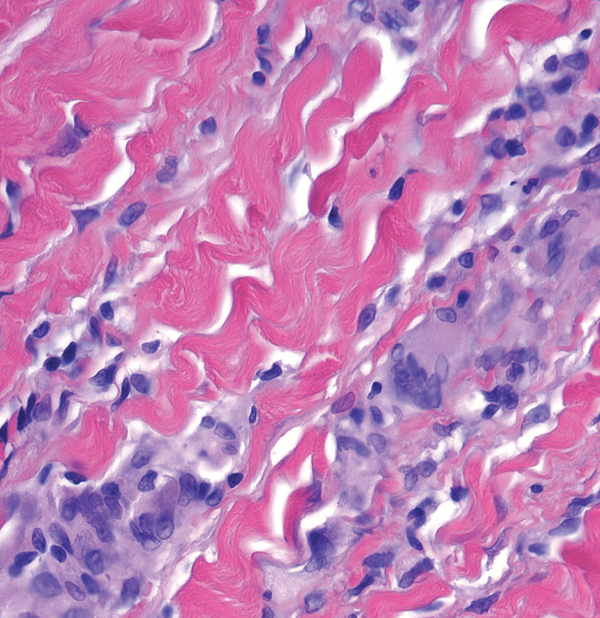
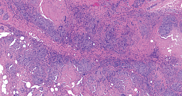
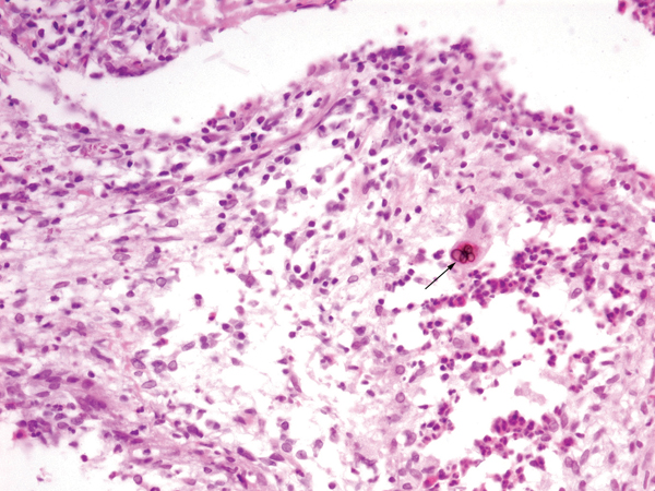
Metastatic breast carcinoma
Cords/tubules and comma shapes in dermis and below
Tubules of single-layered (“Indian filing” – long arrow) and multi-layered epithelium
Some cells forming gland-like structures (short arrow)
Other metastatic carcinomas may look like this – need clinical history; immunohistochemistry may be helpful
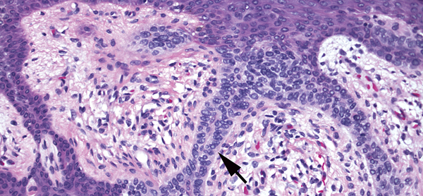
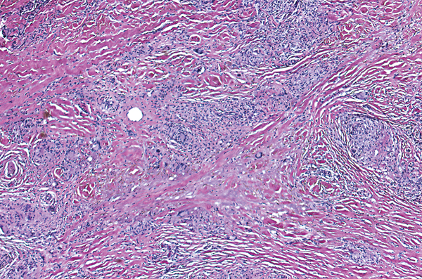
Microcystic adnexal carcinoma
Cords/tubules and comma shapes in dermis
Comma shapes with duct-like spaces
Deeply infiltrative (fills dermis)
Perineural involvement
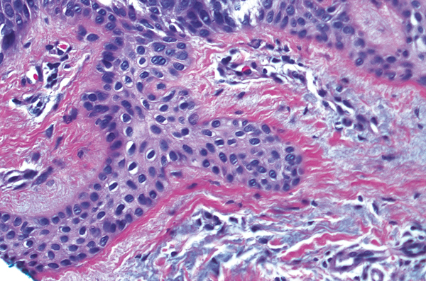
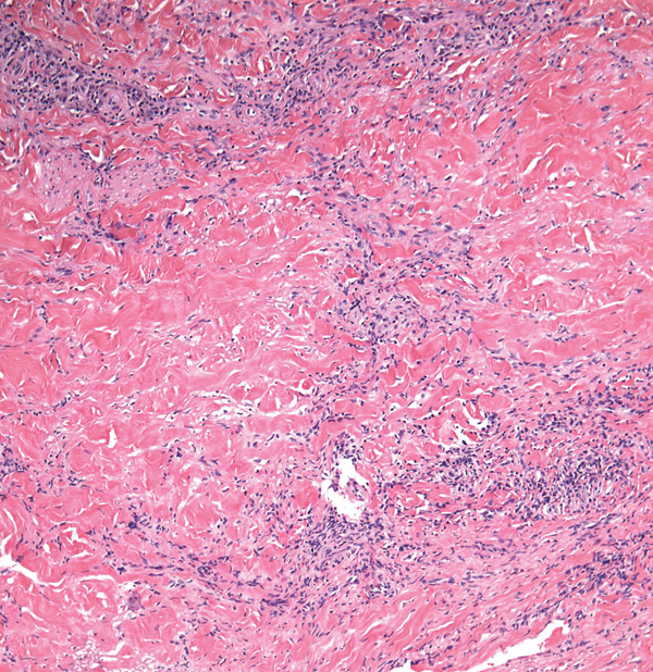
Morpheaform basal cell carcinoma
Cords/tubules and comma shapes in dermis
Tubules of epithelium composed of basaloid cells with hints of peripheral palisading
New collagen forming around islands (arrow)
Deeply infiltrative
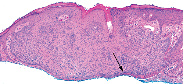
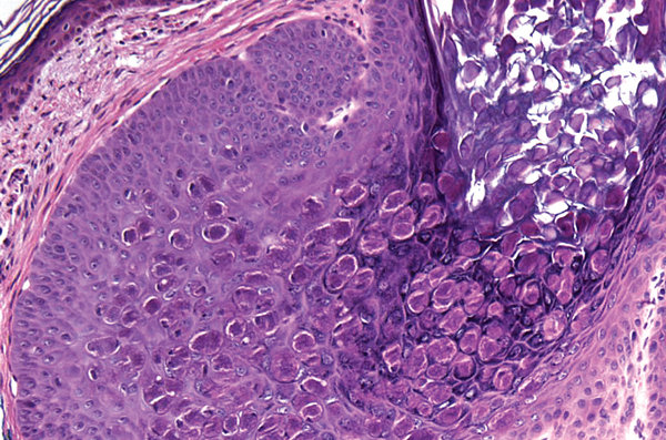
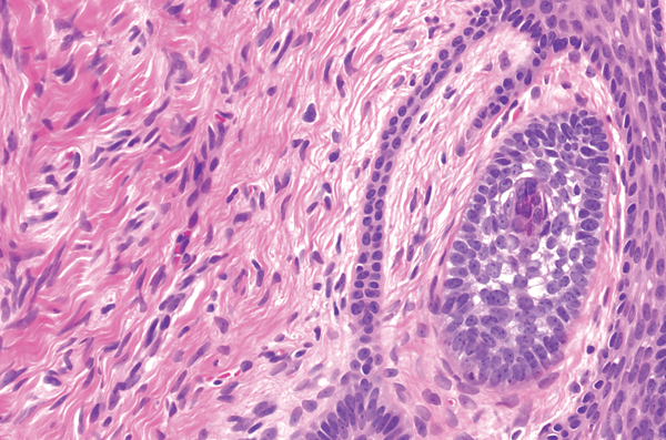
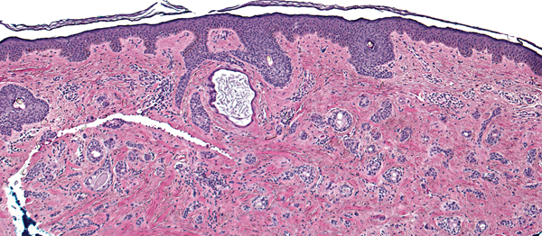
Syringoma
Cords/tubules and comma shapes in dermis
Restricted to upper dermis
“Tadpoles” of epithelium with duct-like structures in heads (arrow)
Darker cells at periphery, clear cells in center
Eosinophilic cuticle lining lumina
No horn cysts
Key differences
(a)
(b)
(c)
(d)
(e)
Desmoplastic trichoepithelioma: horn cysts, no clear cells, circular areas of epithelium surround keratin
Metastatic breast carcinoma: single filing of atypical cells, deeply infiltrative
Microcystic adnexal carcinoma: like syringoma with tadpole-like structures but deeply infiltrative, perineural involvement
Morpheaform basal cell carcinoma: infiltrative cords of basaloid cells with hints of peripheral palisading; may have some duct-like structures (but less than c)
Syringoma: superficial tadpoles with clear cells
Apocrine hidrocystoma
Space with a lining
Lining composed of an inner layer of cells with decapitation secretion (long arrow) and a compressed layer of myoepithelial cells (short arrow)
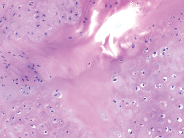
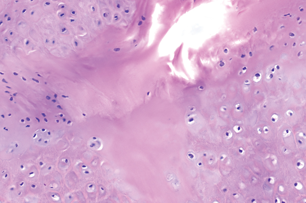
Auricular pseudocyst
Space with a lining
“Lining” is not a true epithelial layer but is cartilage
Centrally, there is degeneration of cartilage
Branchial cleft cyst
Space with a lining
Lining composed of squamous or sometimes cuboidal/columnar epithelium often with squamous metaplasia
Prominent lymphoid follicles in wall
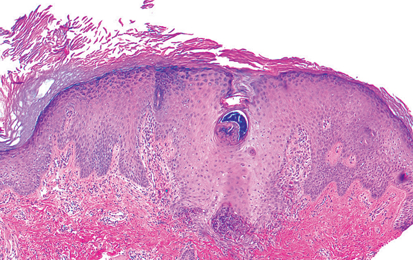
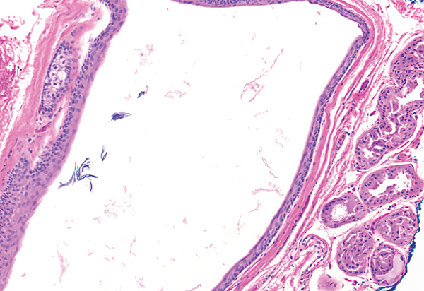
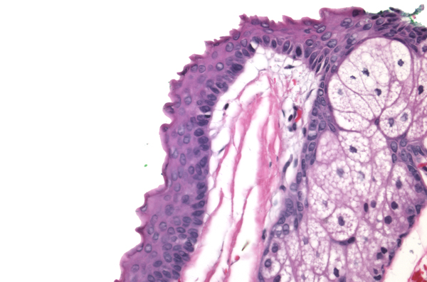
Cutaneous ciliated cyst
Space with a lining
Lining composed of cuboidal/columnar epithelium with cilia (arrows)
Cutaneous endometriosis
Space with a lining
Spaces embedded in a fibrovascular stroma (endometrial stroma)
Lining composed of crowded blue cells
Hemosiderin deposits common in stroma
Dermoid cyst
Space with a lining
Lining composed of squamous epithelium
Walls contain adnexal structures
Epidermoid cyst
Space with a lining
Lining composed of squamous epithelium with a granular layer (arrow)
Cyst contents composed of flakes of keratin
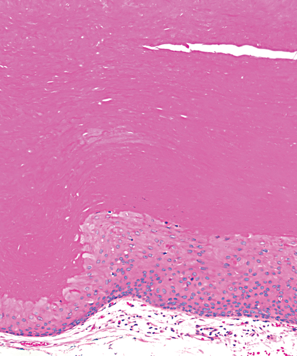
Pilar cyst
Space with a lining
Lining composed of squamous epithelium without a granular layer
Cyst contents composed of dense pink keratin
Steatocystoma
Space with a lining
Lining composed of layered epithelium with a bright pink crenulated keratin (arrow)
Sebaceous glands in wall
Key differences
(a)
(b)
(c)
(d)
(e)
(f)
(g)
(h)
(i)
(j)
Apocrine hidrocystoma: decapitation secretion
Auricular pseudocyst: degeneration surrounded by cartilage
Branchial cleft cyst: prominent lymphoid follicles in wall
Cutaneous ciliated cyst: columnar epithelium with cilia; no structures in wall
Cutaneous endometriosis: fibrovascular stroma with glands
Dermoid cyst: sebaceous glands and other adnexal structures in wall
Epidermal inclusion cyst: epithelium with granular layer, flakes of keratin in center
Glomuvenous malformation (glomangioma): monomorphous, cuboidal blue cells (see also glomus tumor on page 282)
Pilar cyst: epithelium without granular layer, dense keratin in center
Steatocystoma: crenulated keratin lining the cyst; sebaceous glands in wall
Note Bronchogenic cysts are uncommon, and are diagnosed by clinical history and the presence of columnar epithelium +/− cilia, +/− cartilage in wall; venous lakes are common and are composed of flattened endothelial cells with erythrocytes in the space.
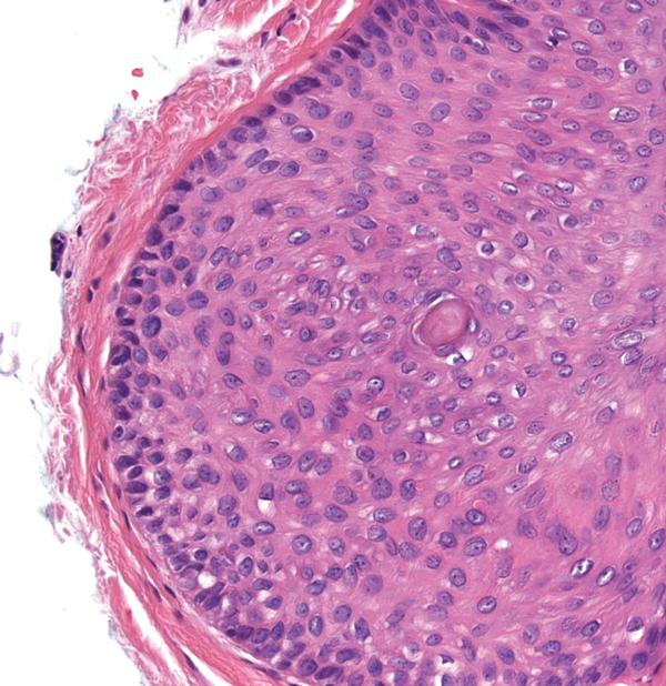
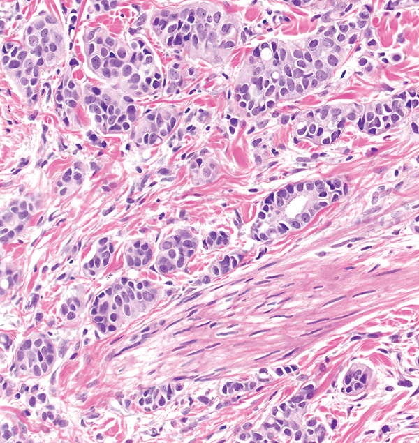
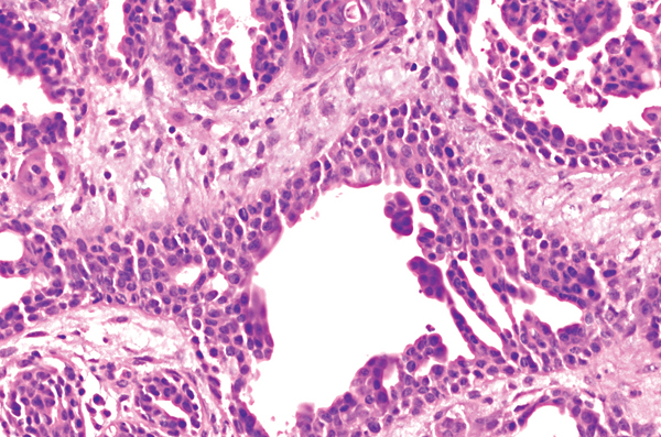
Aggressive digital papillary adenocarcinoma
Papillated dermal tumor
Disordered layers of epithelium in large papillations with some tubules
Variable cytological atypia and mitotic figures
Acral location
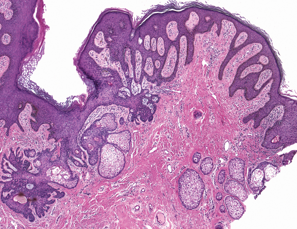
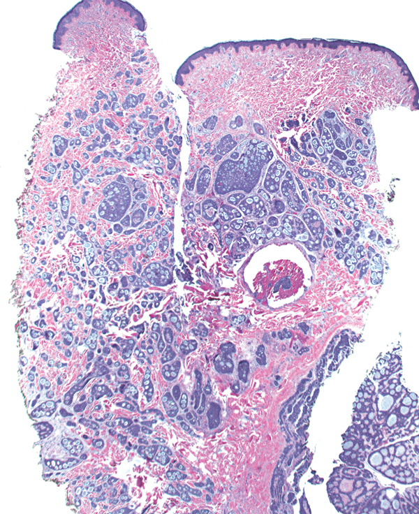
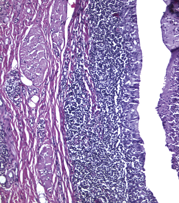
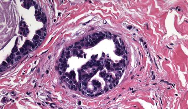
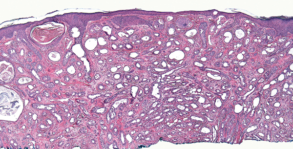
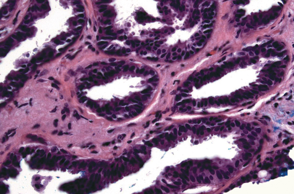
Erosive adenomatosis of the nipple (nipple adenoma)
Papillated dermal tumor
Nipple can sometimes be identified by fascicles of smooth muscle in dermis
Circular islands, some cystic, and tubules
Compressed myoepithelial cells at periphery of islands/tubules
Aggressive digital papillary adenocarcinoma: large tumor, atypical cells and mitoses piled up
Florid papillomatosis (erosive adenomatosis) of the nipple (nipple adenoma): resembles syringocystadenoma papilliferum but fewer plasma cells; nipple may be identified by smooth muscle bundles in dermis
Hidradenoma papilliferum: thin papillations with fibrovascular cores
Papillary eccrine adenoma: islands of epithelium with papillated areas
Syringocystadenoma papilliferum: fat papillations with plasma cells in cores
Tubular apocrine adenoma: decapitation secretion and papillations within islands
Accessory digit
Polypoid shape
Acral skin (thick stratum corneum with stratum lucidum [long arrow])
Dermal nerve bundles (short arrows)
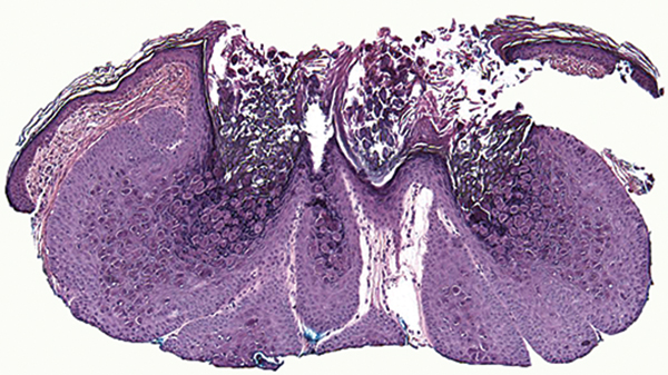
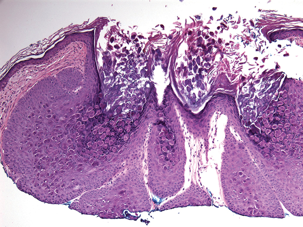
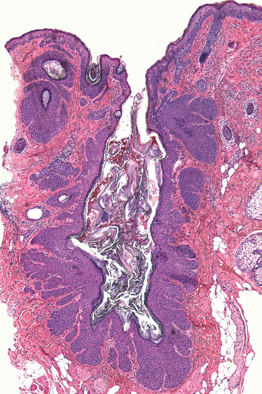
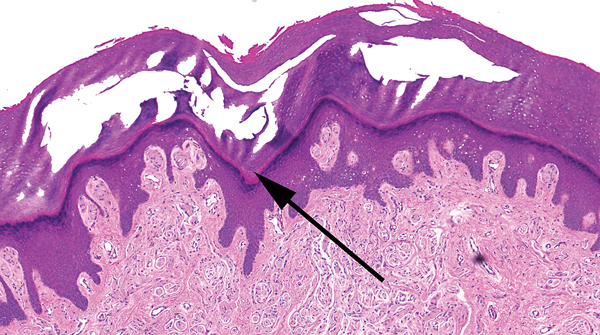
Accessory nipple
Polypoid shape
May see a slight invagination of surface epidermis with underlying sebaceous glands
Surface epidermis often slightly acanthotic and hyperpigmented
May see mammary ducts or apocrine glands deep
Dermis with numerous smooth muscle bundles (arrows)
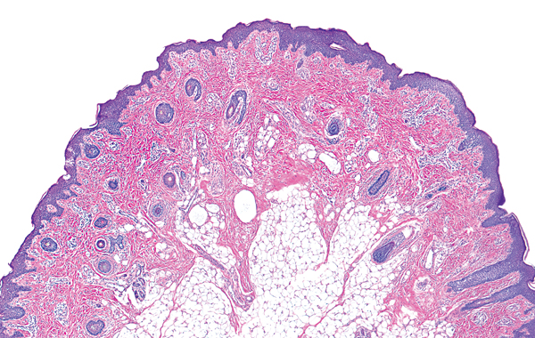
Accessory tragus
Polypoid shape
Thin epidermis
Vellus hairs (arrows)
Cartilage not always present
Differential diagnosis of numerous vellus hairs
– Eyelid/earlobe/sometimes facial skin
– Vellus hair nevus
Digital fibrokeratoma
Polypoid shape
Acral skin
Fibrovascular stroma (thick collagen [arrows])
Key differences
(a)
(b)
(c)
(d)
Accessory digit: nerve bundles in the dermis
Accessory nipple: sebaceous glands, mammary ducts or apocrine glands, smooth muscle bundles in the dermis
Accessory tragus: vellus hairs in the dermis
Digital fibrokeratoma: collagen in the dermis
Note Other entities may also be polypoid; for example, intradermal nevus, neurofibroma, fibrous papule, etc.
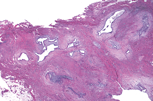
Morphea
Square/rectangular shape
Thick, pink smudgy collagen in dermis
Plasma cells around vessels
Atrophic or absent adnexal structures
Necrobiosis lipoidica
Square/rectangular shape
Altered, reddened collagen (necrobiosis) layered with inflammation
Giant cells and plasma cells are prominent
Normal back skin
Square/rectangular shape
Normal-appearing collagen bundles in dermis
No increased mucin
Scleredema
Square/rectangular shape
Slight widening of space between collagen due to mucin (arrow)
No increase in fibroblasts
Scleromyxedema
Square/rectangular shape
Slight widening of space between collagen due to mucin (long arrow)
Increased fibroblasts (short arrows)
Note Lichen myxedematosus is histologically similar but clinically different.
Note Nephrogenic systemic fibrosis may show similar findings but may have deeper involvement.
Key differences
(a)
(b)
(c)
(d)
(e)
Morphea: thickened bundles of collagen with loss of fenestrations between collagen bundles
Necrobiosis lipoidica: reddened collagen sandwiched between layers of inflammatory cells (giant cells, plasma cells)
Normal back: normal-sized collagen bundles, no increased mucin
Scleredema: mucin between collagen
Scleromyxedema: mucin and increased fibroblasts
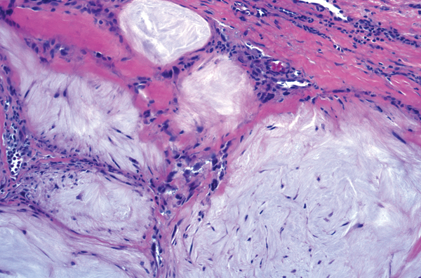
Gout
Palisading of histiocytes around amorphous white-gray substance with a feathery edge
Granuloma annulare
Palisading of histiocytes around altered collagen, basophilic mucin (long arrow)
Lymphocytes around vessels (short arrow)
Necrobiotic xanthogranuloma
Palisading of histiocytes and bizarre, multinucleated giant cells around foci of necrosis
Scattered Touton giant cells
Cholesterol clefts, plasma cells, and/or lymphoid follicles may be present
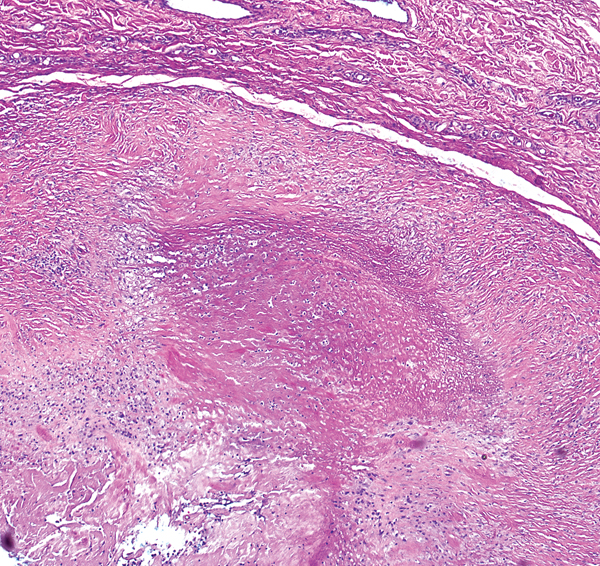
Rheumatoid nodule
Palisading of histiocytes around central pink fibrin
The reaction is often deep
Key differences
(a)
(b)
(c)
(d)
(e)
Gout: central white-gray feathery material
Granuloma annulare: central altered collagen interspersed with blue mucin
Rheumatoid nodule: central pink fibrin
Necrobiosis lipoidica: altered “red” collagen surrounded by giant cells, plasma cells (see page 83)
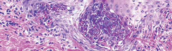
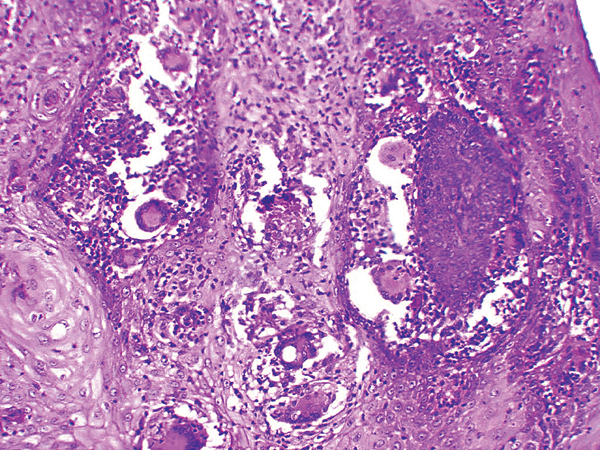
Large (~80-200 micron) spherules containing endospores (arrows)
Key differences
(a)
(b)
(c)
Blastomycosis: ~8–30 micron yeast form (arrow)
Chromomycosis: ~5–12 micron Medlar bodies
Coccidioidomycosis: ~80-200 micron spherules with endospores
Note Paracoccidioidomycosis (~6–60 micron Mariner’s wheel; an uncommon infection in the United States), sporotrichosis (organisms usually not evident in biopsies), and tuberculosis verrucosa cutis may also show this pattern.