Chapter 6
Painting A Gruesome Picture: Bloodstain Analysis
IN THIS CHAPTER
 Diving into the characteristics of blood
Recognizing bloodstain patterns
Checking out a hypothetical case
No doubt you’d prefer to keep your blood inside your body, but it’s somewhat reassuring to know that blood can be extremely useful on the outside, as well. Forensic investigators can figure out a great deal about a crime from blood found at the crime scene. Bloodstains left at an accident, suicide, or crime scene may be the key to determining what happened, helping investigators solve a crime, or, for that matter, determining whether a crime was even committed.
The shape and location of bloodstains provide clues about where the victim and suspect were when the crime took place and where they went afterward. Blood also reveals the presence of disease, drugs, or alcohol, and it can be used to determine the identity of the victim and the suspect through DNA analysis.
Understanding Blood’s Character
In addition to the revealing blood type and DNA evidence it leaves, blood is important to crime scenes because of the way it moves and clots. These characteristics stem from blood’s composition. Knowing how blood operates, inside and outside the body, enables investigators to get to the bottom of how a bloodstain got there.
Thicker than water
Blood is a complex substance, consisting of liquid and solid components. As a liquid, blood shares many of the same physical properties as other liquids, including water. It moves and flows as gravity dictates and tends to pool in low-lying areas. Blood spreads to cover a surface or to conform to the shape of a container. It possesses viscosity (a measure of its thickness) and surface tension (an elastic-like property that results from the attraction of a liquid’s molecules to each other). Surface tension holds a liquid together and pulls a falling drop of that liquid into a spherical shape.
Unlike water, blood is a living, breathing liquid. It really is thicker than water, and, of course, it clots. You can find cellular elements, such as red blood cells, white blood cells, and platelets, as well as various proteins, in blood’s liquid plasma. The clotting process involves many of these elements. When blood clots, it separates into a solid dark-red clot and a clear yellow liquid known as serum.
 Plasma and serum look similar to the naked eye, but they differ in important ways:
- Plasma is the liquid portion of whole, unclotted blood that contains proteins involved in the clotting process. You can separate plasma from whole blood in a centrifuge, a device that rapidly spins a test tube of blood, causing cells to settle in the bottom, leaving the plasma on top.
- Serum is the liquid that remains after the blood’s proteins have done their job and the blood has clotted and retracted into a clump.
 Blood remains a liquid as long as it’s moving inside the body. At death, the heart stops pumping blood through the body, and the blood stagnates and clots. When blood leaves the body, it clots within a few minutes.
Looking into blood clotting
As you’ve no doubt seen after a mishap with a bread knife or a fall from your bike, whenever blood leaves your body, it begins to pool and clot. Normal clotting time for blood is from 3 to 15 minutes, but clotting time is extremely individual and can be affected by certain diseases, such as hemophilia and some types of leukemia, and various medications, including blood thinners.
When blood first begins to clot, it forms a dark, shiny, jelly-like mass. With time, the clot begins to contract and separate from the yellowish serum. Investigators use the state of blood clotting as a rough guide to estimate how much time has passed since the blood was shed. If the blood is still liquid, the bleeding occurred only a few minutes before. If it’s a shiny, gelatinous pool, bleeding occurred from less than half an hour to an hour earlier. And if the blood is separated into clot and serum, an hour or more has probably passed. These are very broad and general timelines, as each situation is different.
Bloodstains that resulted from blood spurting or gushing must have happened before death, when blood was still moving through the body. Impact spatters and splashes can occur after death, but only the assailant (or a clumsy investigator) can cause these kinds of stains, for example by continuing to strike the victim after that victim is dead or stepping into a pool of blood.
Oozing, gushing, and dripping
Blood can leave your body in many ways. Depending on the situation, blood can drip, ooze, flow, gush, or spurt. Each kind of blood movement leaves a recognizable bloodstain pattern or spatter. I tell you more about each of these in the later section “Analyzing Bloodstain Patterns.” Left unchecked, any continuous blood loss can lead to death from exsanguination (bleeding to death).
The mechanisms by which blood leaves the body can be divided into two categories: passive and projected. Passive bleeding depends upon the action of gravity alone. This kind of bleeding includes oozes and drips. Blood is projected when a person or object applies some force other than gravity. Arterial spurts, cast-off blood, and impact spatter are examples of projected blood. The next section tells you more about each of these patterns.
Analyzing Bloodstain Patterns
Investigators can learn a lot from the pattern of bloodstains at a crime scene. Who shed the blood? How did it get where it was? What was the sequence of events that must have occurred for this particular pattern to be present? A careful analysis of the pattern can answer these questions and help solve the crime.
The information bloodstain patterns provide includes
- The origin of the bloodstains
- The type of instrument that caused the bloodstains
- The direction from which an object struck the victim
- The relative positions of the victim, assailant or assailants, and bystanders
- The locations and movements of the victim and assailant during the attack
- The number of blows or gunshots the victim received
- The truthfulness of any suspects and witnesses
Finding clues in passive bloodstains
Passive bloodstains form not because of force but because of the laws of gravity. Blood that oozes, drips, or gushes from the body moves downhill and collects in the lowest areas on or near the injured or deceased person. Stairs, ramps, or even slight inclines can carry blood considerable distances before it clots. Gushing or fast-flowing blood obviously gathers in larger amounts and can travel farther from the body than dripping or oozing blood. A slow ooze clots before blood moves too far from the body.
Blood can drip from an injured person’s wounds, a blood-covered weapon, the assailant’s hands, a tabletop, or any elevated object. Getting shot or stabbed in the shoulder can cause blood to run down your arm and drip from your fingers. Similarly, an escaping assailant can drip blood from a knife, bludgeon, or other weapon.
Typically, a drop forms when a small amount of blood breaks away from a larger blood source. Because of surface tension, drops remain spherical until they strike a surface or until they’re struck by another object. Drops don’t break into smaller drops simply by falling through the air. If a drop hits the edge of a table, or if a swinging arm or weapon strikes it, the drop breaks apart. Otherwise, it falls as a sphere until it reaches the floor or some other surface.
When a falling drop of blood strikes a surface, it splashes in all directions, spattering in a circular pattern around the point of impact. The shape and size of the blood spatter pattern depend upon the size of the drop, how fast it falls, at what angle it hits the surface, and the kind of surface it strikes.
 A blood drop picks up speed as it falls until it reaches terminal velocity, its maximum free-fall speed. The terminal velocity of blood is approximately 25 feet per second, and a drop can reach that speed only after a fall of 20 to 25 feet. But the circular spatter pattern produced by a drop of blood increases in size when it falls from an inch up to about 7 feet. The diameters of spatter patterns from drops falling from higher than 7 feet don’t significantly increase. The size of the diameters of spatter patterns for single drops typically varies from 13 millimeters to 22 millimeters, depending on the distances the drops travel and the sizes of the drops.
When a drop strikes a surface at a right angle (90 degrees), the spatter pattern forms an even circle around the point of impact. If the blood strikes from a smaller angle, the spatter creates an elongated oval pattern with the narrow or pointed end aiming in the drop’s direction of travel (see Figure 6-1).

Illustration by Nan Owen
FIGURE 6-1: The size and shape of stain patterns reveal the angle at which they struck the surface.
The surface that the blood hits can change the size and shape of the spatter (see Figure 6-2). Hard, smooth surfaces like glass, tile, or polished marble create much smaller spatters than rough, irregular surfaces like unfinished wood or concrete.

Illustration by Nan Owen
FIGURE 6-2: Blood behaves differently according to the type of surface it strikes.
Secondary or satellite spatters often create confusion when criminalists analyze bloodstains (see the next section). If a large drop of blood falls onto a hard surface, small secondary droplets may surround the original circular stain. Because these droplets hit the surface at angles that are less than 90 degrees, the secondary stains are elongated, but the elongated tails of these satellite droplets tend to point toward the direction from which they came, not the direction in which they were traveling.
Analyzing projected blood spatters
Projected blood spatters happen when something other than gravity applies force to a blood source. This force may be a naturally occurring internal activity like the heartbeat or the victim’s breathing, or it may be an external force like a gunshot or a blunt-force trauma.
Spatters can be produced by several different mechanisms, including stabbings, beatings, gunshots, arterial bleeding, cast-off blood, splashing, and expirated blood. Expirated blood describes blood the victim expels from his lungs or airways. Each time the victim exhales, blood sprays from his mouth and nose.
Determining where the deed went down
Spatter patterns aid the medical examiner (ME) in determining the source of the blood, the source’s location at the crime scene, and the mechanism that produced the bloodstains. This critical information can show investigators the positions of the assailant and the victim when the attack occurred.
Each droplet in a blood spatter strikes the surface from a unique direction and at a unique angle. The impact angle is the slant at which the blood drops strike the surface, and the directionality is the course the blood drop followed. Investigators figure out the impact angle using a protractor or one of the computer programs developed for that purpose.
Investigators use the directionality of each stain to make the following determinations:
- Point of convergence: A two-dimensional representation of the point where lines tracking the pathways of two or more spatters meet (see Figure 6-3), indicating the general location of the blood source in relation to the spatters. At the crime scene, investigators stretch strings from each stain according to the angle of impact; where those strings meet is the point of convergence.
- Point of origin: A three-dimensional representation of the point where lines tracking not only the pathways but also the angles of impact of two or more spatters meet (see Figure 6-4), indicating the general spatial location of the blood source. By stretching strings or pointing laser light beams along the angle of impact of each stain, investigators can find the point of origin.
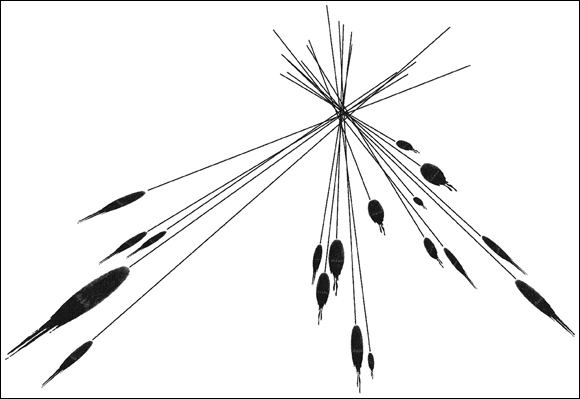
Illustration by Nan Owen
FIGURE 6-3: Lines through the path of each bloodstain meet at the point of convergence.

Illustration by Nan Owen
FIGURE 6-4: Directionality and angle of impact reveal the point of origin of bloodstains.
The crime scene investigator’s report usually provides a range of possible points of origin for the blood source. For example, an analysis of spatter patterns on the floor, wall, and sofa at the scene of a violent attack may indicate a point of origin that was 4 to 6 feet from the wall, 2 to 4 feet from the sofa, and 4 to 6 feet above the floor. These estimates indicate that the victim probably was standing near the wall and the sofa when the trauma that caused the bleeding occurred. Had the analyst found the point of origin to be 4 to 6 feet from the wall and 2 to 4 feet from the sofa, but only 1 to 2 feet from the floor, the victim probably would have been lying on the floor when the blood-shedding trauma occurred.
Interpreting void patterns
A void pattern is an absence of blood spatters in an area where you’d otherwise expect to see them. Often this void indicates where the attacker stood because his body prevented the blood from spattering on the surfaces behind him. For example, if someone is severely beaten, and investigators find blood spatters on the walls or furniture in every direction except to one side of the victim, the attacker probably stood in that position during the attack and intercepted the spattered blood.
Finding blood spatters on a suspect’s body or clothing confirms for investigators that the suspect was at the scene at the time of the attack. Spatterings of blood occur only during the impacts that produce them. As a result, finding spatters on a suspect’s clothes, arms, or face means he must have been in close proximity to the victim at the time of the attack. Stains found on someone who came along after the attack and accidentally got blood on her clothing wouldn’t show a spatter pattern. This information helps investigators confirm or refute statements about whether a suspect was at the scene during the attack.
Classifying projected spatters
Investigators classify projected blood spatters in one of two ways:
- By velocity: This method of classifying blood spatters looks not only at the velocity at which the impacting object strikes the blood source but also the velocity at which the blood leaves the blood source when it’s struck. This system divides spatters into low-, medium-, and high-velocity spatters. These subcategories give an indication of the object and the mechanism that created the spatter.
- By type of spatter: This classification system divides spatters into these three major types:
- Impact spatters typically occur with beatings, stabbings, gunshots, or any other circumstance where a foreign object impacts the victim.
- Projection spatters result from arterial bleeding, cast-off blood, and expirated, or exhaled, blood. The next section provides details about these patterns.
- Combination spatters, which include impact and projection spatters, often are found at crime scenes. A victim who gets stabbed in the chest or neck may leave a combination of impact spatters from the force of the attack and projection spatters from arterial bleeding, expirated blood, and cast-off blood.
Investigators find both methods of spatter classification useful because these methods overlap in many areas.
Low-velocity spatters
Low-velocity spatters occur when an object moving less than 5 feet per second strikes a surface. This impact results in fairly large spatters, typically 4 millimeters or greater in diameter.
Several mechanisms produce low-velocity spatters. Common examples include drops dripping under the influence of only gravity from a wound or a blood-soaked weapon. If the dripping blood source is standing still, the drops fall vertically and create circular stains. But if the source is moving, such as an injured victim taking flight or an escaping assailant carrying a blood-covered weapon, the drops strike the floor at an angle, producing elongated stains with spines or projections of blood extending in the direction of movement.
Suppose that an assailant with blood on his hands stands near the body of his victim. He drips blood onto the floor, leaving a round bloodstain pattern for each drop. If he begins to move around in the house, the drops that fall no longer strike the floor from a 90-degree angle. The direction in which the assailant moves influences the direction in which the drops leave his hands. These drops hit the floor at an angle, resulting in oval stains with elongated tails that point in the direction of travel, like those shown in Figure 6-1. Criminalists follow the tails to determine the assailant’s movements within the crime scene and perhaps his escape route. Along this path investigators are likely to find other useful evidence, such as a discarded murder weapon or bloodied clothes.
Arterial bleeding is also considered low velocity. If an artery is damaged during an assault, suicide attempt, or accident, the blood loss may take the form of gushes or spurts, depending upon the size of the artery, the extent of the damage, and whether clothing or some other object covers the injury. A freely spurting artery results in a linear and cascading spatter pattern (see Figure 6-5). The pattern’s distance from the wound, length, and volume may decline steadily as the victim continues to lose blood, causing his blood volume and blood pressure to decline.

Illustration by Nan Owen
FIGURE 6-5: The shape and pattern of the drops within a blood spatter reveal how the spatter was produced.
Another low-velocity blood source is cast-off blood, or blood that is flung from an object because of centrifugal force. Cast-off patterns usually occur when an attacker uses a weapon to deliver a series of arcing blows. Investigators typically find these patterns on walls and ceilings.
The spatter pattern associated with cast-off blood tends to be a fairly uniform trail of droplets, which reflects the arc that the object traveled in (see Figure 6-5). Determining the point of convergence and the angle of impact of these cast-off stains reveals the assailant’s position at the time he swung the weapon. In some cases, estimating the perpetrator’s height and even whether the assailant is right- or left-handed — or at least which hand struck the blows — is possible.
Also, the number of these cast-off patterns investigators find indicates the minimum number of blows to the victim. Because not every swing of the weapon necessarily produces cast-off stains, the assailant may have delivered more blows, but fewer can’t have been delivered.
Medium-velocity spatters
Medium-velocity spatters come from objects moving between 5 and 100 feet per second. These spatters are typically smaller than spatters from low-velocity droplets and vary from 1 to 4 millimeters in diameter. Medium-velocity spatters come from impacts with blunt or sharp objects and from expirated blood.
Spatters from impacts with a blunt object distribute blood in all directions from the area of impact. As with low-velocity spatters, an analysis of directionality and impact angle of the stains can help locate the point of origin. See the section “Determining where the deed went down” earlier in this chapter for a discussion of directionality and impact angle.
If the wounds are to the face, throat, or lungs, blood mixes with the exhaled air, creating a fine spray and producing a mist spatter pattern. This mist pattern may be found on and around the victim and on the attacker.
High-velocity spatters
High-velocity spatters result when an object strikes the victim at a speed faster than 100 feet per second. The resulting spatters tend to be very small, usually less than 1 millimeter in diameter, and appear as mist-like stains.
A bullet travels at high velocity and thus produces a high-velocity spatter pattern. These patterns show up near entrance and exit wounds, but blood behaves differently according to whether the bullet is coming into the body or heading back out.
A bloodstain associated with an entrance wound is called blowback or back spatter, meaning the droplets travel in a direction opposite to the path of the bullet. Blowback often is found on the shooter or the weapon, even inside the barrel of the gun in very close-range shots. A bloodstain found near the exit wound is called forward spatter. In the case of forward spatter, the blood droplets follow the direction of the bullet.
If you tossed a glass of water through a window screen, the water would keep right on going through the screen. That’s forward spatter. Toss that same glass of water against a wall, and the water splashes back toward you, just like back spatter.
Latching on to transfer patterns
Transfer patterns result when an object soaked with blood comes into contact with an unstained object. Bloody fingerprints and shoeprints fall into this category. The perpetrator may brush against or kneel in a bloodstain, or wipe the weapon or his hands or his shirt, transferring the victim’s blood to his clothing. Matching the blood from such a transfer stain to the victim’s blood may help solve the crime.
Like fingers and shoes, a blood-soaked fabric can leave behind a recognizable pattern. Say a perpetrator knelt on the floor next to his victim and unknowingly transferred blood to the knee of his pants. After his escape, he leaned against his car door and transferred the stain. The weave pattern of his pants may be transferred, also. If investigators find this pattern, they may be able to use the fabric pattern to determine what type of clothing the attacker wore. Regardless, analysis of the transferred blood on a suspect’s clothing can link the suspect to the crime scene and the victim if they can match the DNA of the bloodstain to the suspect. Chapter 15 tells you more about DNA.
Reconstructing the crime scene from bloodstains
Contaminated evidence is no evidence at all, so investigators have to document bloodstain and spatter patterns in a timely and logical fashion. Police, fire, and rescue personnel can alter or contaminate the blood evidence, as can any unnecessary foot traffic at the crime scene. For that reason, investigators need to take control of the scene immediately and consistently. Unless they’re high-traffic public places, indoor scenes usually can be preserved long enough for investigators to obtain needed information. Outdoor scenes, however, are subject to environmental influences, and public places require investigators to gather information more urgently.
Investigators carefully photograph bloodstains. Initial photographs capture an overall view of the scene. Subsequent pictures gradually move in on individual stains. The photographer takes pictures of individual stains close enough to reveal all needed detail, and should include a ruler or other measuring device to provide a scale reference. In homicide cases, investigators check out the body and any associated bloodstains or spatters first. After the body is removed, investigators turn their attention to other spatters.
Some bloodstains are latent (invisible to the naked eye). Investigators often use luminol to expose these hidden stains. Luminol is a chemical that reacts with the hemoglobin in blood to produce a complex substance that luminesces (glows). Luminol is extremely sensitive, detecting blood in concentrations as low as one part per million. Investigators darken the room and spray luminol over areas where they suspect blood to be. When blood is present, stains glow a bluish-white, and the photographer takes pictures of the glowing pattern.
Luminol also can reveal bloody tracks that indicate the perpetrator’s movements or escape route and drag marks that show whether anyone moved the body. Luminol is so sensitive that it can uncover blood in cracks, crevices, and even areas where someone has tried to clean it.
It’s important to note that many substances can interfere with or confuse luminol pattern analysis. Bleach and other cleaning agents, certain paints and varnishes, and even some fruit juices are examples.
After photographers take an adequate number and variety of photographs, crime-scene analysts complete their analyses and create a report that may include implications of the victim’s and assailant’s locations at each stage of the crime, the number and types of injuries inflicted, and the exact sequence of events (see the next section to understand how analysts gather this information).
Putting It All Together: A Hypothetical Case
Joe and Bill have a serious disagreement, and the two men reach a point of true hatred for one another. Bill, carrying a 2-foot piece of metal pipe, kicks in Joe’s door and steps into the entryway of Joe’s home. But Joe is expecting him and shoots Bill in the left arm with a .32 caliber pistol.
Angered more than injured, Bill strikes Joe in the face with the pipe. Joe staggers, drops the gun, and turns to run away. Bill hits him several more times in the head with the pipe as Joe retreats across his living room and down a hallway, both of which have hardwood flooring.
The blows finally bring Joe to his knees at the far end of the hall, where he crawls into his bedroom, which is carpeted. He collapses facedown near his bed and a nightstand. Bill continues his attack until Joe is dead. He then runs back up the hallway, through the kitchen and out the back door. Gruesome, but all too common.
After photographers take pictures of the scene and forensic pathology technicians (see Chapter 22) remove the body, the crime scene investigator examines each and every piece of evidence available to him. Here’s what he finds and where he finds it:
- The entryway: High-velocity blood spattering from the gunshot to Bill’s arm is on the entry wall and the front doorjamb. Later DNA analysis reveals that this blood matches a sample obtained from Bill. The blow to Joe’s face causes bleeding from his nose and mouth, and blood falls to the floor, producing circular stains. Joe also expirates blood in a fine mist over the wall, the floor, and Bill.
- The living room and hallway: Joe continues to expirate a mist of blood as he runs. The spray is found on the carpet, walls, and furniture he passes on his retreat toward his bedroom. He also continues to bleed from his facial wounds. However, the drops that reach the floor no longer leave circular stains because he’s now on the move. The stains become oblong and have spikes or projections pointed along his line of retreat. As Bill continues to strike Joe in the head, medium-velocity impact spatters radiate in all directions. Some reach the floor, and others pepper the wall, the furniture, and Bill. The directionality and impact angle of the drops show that the blood source (Joe’s head) was 5 to 6 feet above the floor and moving toward the bedroom. At the end of the hallway where Joe collapses, the point-of-origin analysis shows that the blood source is less than 3 feet off the floor.
- The bedroom: The carpet shows dripped and expirated mist patterns, but they are less clear because of the texture of the carpet. The same is true for the continued impact spatter from Joe’s blows. The bed, wall, and nightstand show medium-velocity impact and expirated spatter as Bill continues his assault and Joe breathes his last breaths. Investigators find the expirated pattern only in the direction Joe’s head is turned, although the impact spatters occur in all directions. Investigators find a void area where Bill stands over the fallen Joe and delivers his final blows.
- The escape route: Investigators find bloody shoe prints leading back down the hallway, through the kitchen, and out the back door. Low-velocity spatters with projections indicating movement toward the back door show where blood dripped from Bill’s hands and the pipe during his escape.
- Bill: If the police apprehend Bill before he can clean up and change his clothing, he will have blood on his hands and probably his face. Even if he has time to clean himself, his clothing may be recovered, and it will show impact spatter stains over the sleeves and front of his shirt, the front of his pants, and the tops of his shoes. His shoe soles retain blood in the crevices and treads, and the tread pattern matches those shoe prints left at the scene in blood. Then, of course, Bill has his gunshot wound to explain away.
Based on the bloodstain evidence, the criminalist can reconstruct the scene and determine the exact sequence of events. If Bill tells a story that varies from the truth, the spatters will trip him up.