ALMOST ALL CELLS have hairlike appendages called cilia. Human cells have both mobile and immobile cilia. Mobile cilia move fluid, such as mucus in lung cells, with large numbers of cilia in rows engaging in synchronized beating to move the fluid through.
Cilia move eggs along the fallopian tube to the uterus, where they meet sperm. Sperm themselves move with beating cilia. In the early embryo, where cilia beat clockwise, they cause the movement of water to the left, creating a left-right symmetry as the fetus develops. In cerebrospinal fluid, highly organized cilia movements, directed by choroid lining cells, produce currents that wash out waste products, including misfolded proteins and toxins.
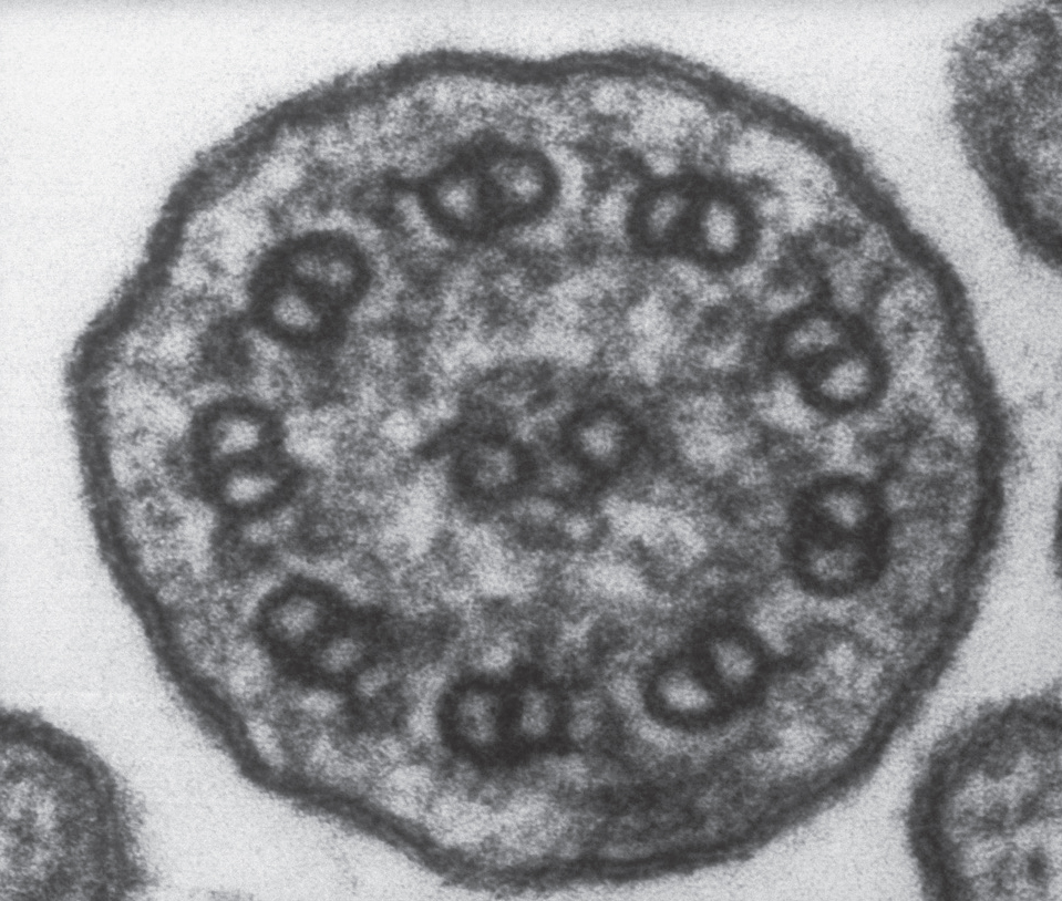
All stationary cilia consist of a cylinder made of nine units of two microtubules each in a circle with a surrounding membrane. Mobile cilia have a somewhat similar structure. As well as the nine doublets of microtubules forming the cylinder’s outer edge, mobile cilia have two additional single microtubules in the middle of the cylinder to generate movement.
Microbes have multiple versions of mobile cilia, including long, thin tails that undulate for swimming. Microbes also have larger and stronger such appendages for swimming, called flagella, which rotate like propellers. Swimming algae synchronize two large flagella in a breaststroke movement. While appearing simple, in fact this movement involves thousands of complex molecular machines working together. Besides being used for movement, microbe cilia also send and receive signals and inject toxins into their host cells and enemies.

Cilium. (LadyofHats/Wikimedia Commons)
PRIMARY CILIA AS A CENTRAL CONTROL CENTER
One type of dynamic immobile cilium, called the primary cilium, sticks out of all human cells. Research now shows that this cilium serves as a central control center antenna for signals both inside and outside a cell. Primary cilia can be quite specialized, such as in sensory neurons that pick up light in the eye and smells in the nose. In the ear, multiple primary cilia in rows of hair cells pick up variations of pressure and frequencies of sound. In kidney cells, cilia gauge liquid flow.
Recently, primary cilia in T cells were found to help regulate other immune cells. When material is presented to the T cell by another immune cell, immune synapses are formed between the two cells using their primary cilia (immune synapses are described in chapter three). With this joining of cilia, T cells receive molecular particles for evaluation before they attack microbes and abnormal cells. Transport systems in the cilia of a T cell carry vesicles filled with molecules that are used to kill microbes and abnormal cells. Receptors in these cilia can also trigger the T cell to morph into an army of killer cells.
The Centriole Spindle and Primary Cilia Link
Although first observed in 1887, primary cilia have been difficult to study because they are ten thousand times smaller than a human cell. As with biological nanotubes, only recent research techniques have enabled closer observation of their functions.
The cylindrical structure of cilia is similar to that of centrioles, which produce and direct microtubules in a cell. In cilia, nine units of two microtubules form the cylinder. In centrioles, nine triplets of microtubules form the cylinder. As well as the central centriole near the nucleus, another centriole sits right next to each primary cilium, producing microtubules to build the cilium structure and to transport material from the cell into the cilium’s long cylinder.
As we know, centrioles are involved in producing the spindle that directs cell division. The spindle is part of a vast structure stabilized by two centrioles. The spindle, sometimes called the most complex machine in nature, consists of microtubule scaffolding with motors that orchestrate cell division by moving chromosomes and organelles. Four thousand microtubules participate in synchronized movements, along with multiple varied motors and pumps, to isolate and separate human chromosomes during cell division.
Recently, a close link was found between the spindle and primary cilia. Protein material was observed being placed in a vesicle at the primary cilium and sent to the spindle during cell division. This vesicle is passed on to one of the two daughter cells during division. It gives to that particular daughter cell’s primary cilium the exact signaling information and material from the mother cell. This connection bolsters the notion that primary cilia are more than antennae for intercellular communications; rather, they serve as a control center, or “brain,” for the entire cell.
Complex Environment and Function
The cylinder of the primary cilium is a long, thin tube that is anchored deep inside a cell and emerges far outward. This provides a unique environment for promoting conversations inside and outside the cell. Because cilia interiors are sheltered from the rest of the cell, they can accumulate a much higher concentration of diverse proteins than other parts of the cell, making signaling more efficient. Transport systems bring molecules into the cilium, then lift them up and down the cylinder like an elevator, with motors in particular locations along with imbedded receptors and signaling apparatus.
Multiple receptors that line the cilium tube have various physiological functions. Primary cilia sense chemicals, concentrations of ions, light, temperature, mechanical forces, and gravity. In the nose, primary cilia become olfactory receptors. In the eye, light-sensing receptors are an outgrowth of the tip of a primary cilium. Cilia sense pressure in cartilage and blood flow in heart cells. Recently, the locations of multiple previously well-known cellular signaling pathways were found to reside in the cilium.
Historically, the first major discovery of a vital function for the primary cilium was in polycystic kidney disease, in which two abnormal cilia proteins were found. In kidney cells, cilia respond to liquid flowing through tubules and send signals related to water pressure and calcium levels vital to kidney regulation.
Various brain functions are tied to primary and moving cilia. A recent study in mice showed that blocking receptors in primary cilia causes memory loss. Both mobile and stationary cilia are critical for brain development in the fetus via conversations among neurons, glia, and choroid lining cells. In developing brains, signals from primary cilia to stem cells are necessary for production of new neurons in the hippo-campus memory centers. Stationary primary cilia are vital for communication in the developing brain during neuronal migrations. Moving cilia help cells such as neurons and glial cells travel in the fetus. Brain disease can result if cilia are unable to send certain molecules through their transport mechanisms.
CILIA TRANSPORT SYSTEMS
Primary cilia use elaborate transport systems. Early observations showed that construction of a cilium starts at the base and then moves up the cylinder, with microtubule scaffolding and other proteins placed in order as it is built. Vesicles were observed transporting material for cilia construction, including pieces of proteins, which are released near the base.
Recently, the complexity of the cilium’s structure and transport system has begun to be unraveled. First, tagged material is brought from other parts of the cell to the base of the cilium with microtubule-based motors. Proteins attach to these molecules and drag them to the membrane near the base of the cilium. Motors then pull the transported material into the cylinder.

Cross section of a cilium showing the nine doublets of microtubule columns. Unlike primary cilia, moving cilia also have one doublet in the middle. Electron micrograph. (Biophoto Associates/Science Source)
Particular motors are built at the base of the cilium to lift molecules up the long, thin cylinder. Motors pull up molecules to produce various receptors and scaffolding along the way. Upon reaching the top, motors are modified to bring other material down. This trip down to the base includes carrying signals that have been received from other cells, which are then transported to the nucleus for response. At the base of the cylinder, the motor again rearranges itself to bring the next cargo up.
Motors for cilia transport are not simple. The train that pulls material to the top consists of at least four motors. Each motor works in sequence, serving not just as a motor but also interacting with the membrane, helping to regulate receptor and signaling functions, such as making decisions about building the fetal brain. Motor molecules are also able to reach through the membrane to the external environment and receive signals or attach to objects. When the motor attaches externally, it can anchor mobile adult cells, as well as help migration during brain development.
CILIA-RELATED DISEASE
Abnormal cilia cause multiple diseases. One type of mobile cilia disease alters synchronized activity in the lungs, where rows of cilia normally beat twenty times per second to sweep out mucus and debris. This disease can affect cilia in the nose and middle ear as well. With smoking, cilia are damaged, causing mucus accumulation.
Multiple diseases have been traced to defects in primary cilia, such as disorders of the retina and kidneys; defects in primary cilia transport motors can even cause blindness. Eye cells have a large bulb at the tip of the cilium with multiple light receptors. Signals from these receptors travel through the narrow cilium cylinder to the body of the cell, where they are sent for responses. With motor problems, signals don’t arrive and receptors aren’t restocked, causing the disease known as retinitis pigmentosa.