
All living things are composed of cells. According to the cell theory, the cell is life’s basic unit of structure and function. This simply means that the cell is the smallest unit of living material that can carry out all the activities necessary for life.
It may seem strange that we are still made of tiny little cells. Why not just be a living thing that is one giant cell?
One reason for this is because compartments allow more specialization. As we will see, compartmentalization is an important part of the organization of unicellular and multicellular organisms.
The second reason we can’t just be giant cells is the surface area-to-volume ratio. There is always lots of exchange going on between the inside of things and the outside. This ratio must be kept large so that there is lots of space to do these exchanges. Some parts of living things are special because they have lots of folds to increase the surface area.
Therefore, even the largest things are still made of cells.
For centuries, scientists have known about cells. However, it wasn’t until the development of the electron microscope that scientists were able to figure out what cells do. We now know that there are two distinct types of cells: prokaryotic cells and eukaryotic cells.
Cells are studied with different types of microscopes. Light microscopes (for example, the compound microscopes commonly found in labs) are used to study stained or living cells. They can magnify the size of an organism up to 1,000 times. Electron microscopes are used to study detailed structures of a cell that cannot be easily seen or observed by light microscopy. They are capable of resolving structures as small as a few nanometers in length, such as individual virus particles or the pores on the surface of the nucleus.
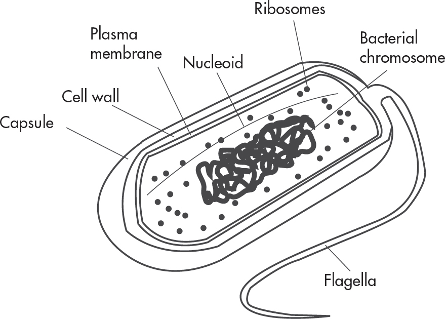
A prokaryotic cell, which is a lot smaller than a eukaryotic cell, is relatively simple. Bacteria and archaea are examples of prokaryotes. The inside of the cell is filled with a substance called cytoplasm. The genetic material in a prokaryote is one continuous, circular DNA molecule that is found free in the cell in an area called the nucleoid (this is not the same as a nucleus!). Most prokaryotes have a cell wall composed of peptidoglycans that surrounds a lipid layer called the plasma membrane. Prokaryotes also have ribosomes (though smaller than those found in eukaryotic cells). Some bacteria may also have one or more flagella, which are long projections used for motility (movement) and they might have a thick capsule outside their cell wall to give them extra protection.
Eukaryotic cells are more complex than prokaryotic cells. Fungi, protists, plants, and animals are eukaryotes. Eukaryotic cells are organized into many smaller structures called organelles. Some of these organelles are the same structures seen in prokaryotic cells, but many are uniquely eukaryotic. A good way to remember the difference is that prokaryotes do not have any membrane-bound organelles. Their only membrane is the plasma membrane.
A eukaryotic cell is like a microscopic factory. It’s filled with organelles, each of which has its own special tasks. Let’s take a tour of a eukaryotic cell and focus on the structure and function of each organelle. Here’s a picture of a typical animal cell and its principal organelles.

The cell has an outer envelope known as the plasma membrane. Although the plasma membrane appears to be a simple thin layer surrounding the cell, it’s actually a complex, double-layered structure made up of mostly phospholipids and proteins. The hydrophobic fatty acid tails face inward and the hydrophilic phosphate heads face outward. It is called a phospholipid bilayer since two lipid layers are forming a hydrophobic sandwich.
The plasma membrane is important because it regulates the movement of substances into and out of the cell. The membrane itself is semipermeable, meaning that only certain substances, namely small hydrophobic molecules (such as O2 and CO2), pass through it unaided. Anything large and/or hydrophilic can pass through the membrane only via special tunnels (discussed more later in this chapter).
Many proteins are associated with the cell membrane. Some of these proteins are loosely associated with the lipid bilayer (peripheral proteins). They are located on the inner or outer surface of the membrane. Others are firmly bound to the plasma membrane (integral proteins). These proteins are amphipathic, which means that their hydrophilic regions extend out of the cell or into the cytoplasm, while their hydrophobic regions interact with the tails of the membrane phospholipids. Some integral proteins extend all the way through the membrane (transmembrane proteins).
This arrangement of phospholipids and proteins is known as the fluid-mosaic model. This means that each layer of phospholipids is flexible, and it is a mosaic because it is peppered with different proteins and carbohydrate chains. Remember, anything hydrophilic should not go through the hydrophobic interior. This means that the phospholipids on one side should never flip-flop to the other side of the membrane (because that would require their polar heads to pass through the hydrophobic area).

Why should the plasma membrane need so many different proteins? It’s because of the number of activities that take place in or on the membrane. Generally, plasma membrane proteins fall into several broad functional groups. Some membrane proteins (adhesion proteins) form junctions between adjacent cells. Others (receptor proteins), such as hormones, serve as docking sites for arrivals at the cell. Some proteins (transport proteins) form pumps that use ATP to actively transport solutes across the membrane. Others (channel proteins) form channels that selectively allow the passage of certain ions or molecules. Finally, some proteins (cell surface markers), such as glycoproteins, are exposed on the extracellular surface and play a role in cell recognition and adhesion.
Attached to the surface of some proteins are carbohydrate side chains. They are found only on the outer surface of the plasma membrane. As mentioned above, cholesterol molecules are also found in the phospholipid bilayer because they help stabilize membrane fluidity in animal cells.
The nucleus is usually the largest organelle in the cell. The nucleus not only directs what goes on in the cell, but is also responsible for the cell’s ability to reproduce. It’s the home of the hereditary information—DNA—which is organized into large structures called chromosomes. The most visible structure within the nucleus is the nucleolus, which is where rRNA is made and ribosomes are assembled.
The ribosomes are sites of protein synthesis. Their job is to manufacture all the proteins required by the cell or secreted by the cell. Ribosomes are round structures composed of two subunits, the large subunit and the small subunit. The structure is composed of rRNA and proteins. Ribosomes can be either free floating in the cell or attached to another structure called the endoplasmic reticulum (ER).
The endoplasmic reticulum (ER) is a continuous channel that extends into many regions of the cytoplasm. The region of the ER that is attached to the nucleus and “studded” with ribosomes is called the rough ER (RER). Proteins generated in the rough ER are trafficked to or across the plasma membrane, or they are used to build Golgi bodies, lysosomes, or the ER. The region of the ER that lacks ribosomes is called the smooth ER (SER). The smooth ER makes lipids, hormones, and steroids and breaks down toxic chemicals.
The Golgi bodies, which look like stacks of flattened sacs, also participate in the processing of proteins. Once the ribosomes on the rough ER have completed synthesizing proteins, the Golgi bodies modify, process, and sort the products. They’re the packaging and distribution centers for materials destined to be sent out of the cell. They package the final products in little sacs called vesicles, which carry products to the plasma membrane. Golgi bodies are also involved in the production of lysosomes.
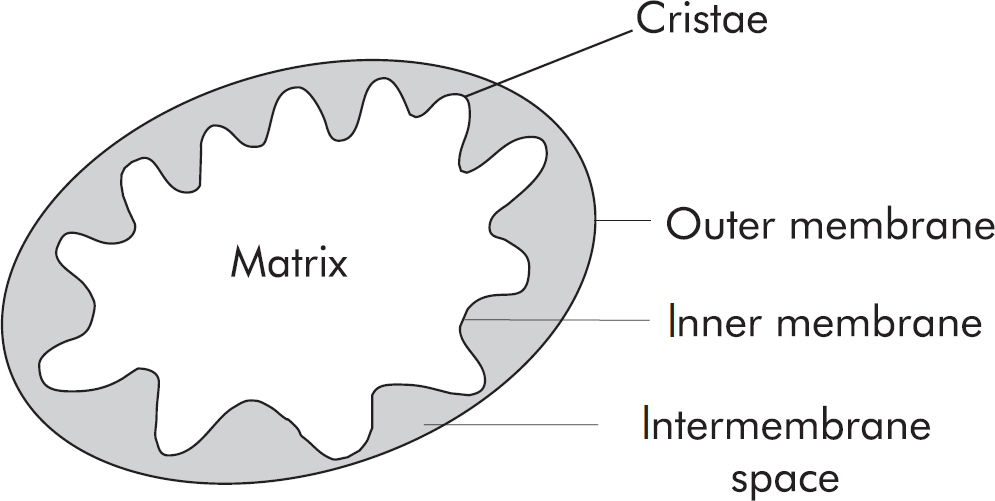
Another important organelle is the mitochondrion. Mitochondria are often referred to as the “powerhouses” of the cell. They’re power stations responsible for converting energy from organic molecules into useful energy for the cell. The most common energy molecule in the cell is adenosine triphosphate (ATP).
The mitochondrion is usually an easy organelle to recognize because it has a unique oblong shape and a characteristic double membrane consisting of an inner portion and an outer portion. The inner mitochondrial membrane forms folds known as cristae and separates the innermost area called the matrix from the intermembrane space. The outer membrane separates the intermembrane space from the cytoplasm. As we’ll see later, most of the production of ATP is done on the cristae. Having folds in the membrane increases the surface area for making ATP.
Throughout the cell are small, membrane-enclosed structures called lysosomes. These tiny sacs carry digestive enzymes, which they use to break down old, worn-out organelles, debris, or large ingested particles. Lysosomes make up the cell’s clean-up crew, helping to keep the cytoplasm clear of unwanted flotsam. Lysosomes contain hydrolytic enzymes that function only at acidic pH, which is enclosed inside the lumen of the lysosome. Lysosomes are made when vesicles containing specific enzymes from the trans Golgi fuse with vesicles made during endocytosis (which you’ll learn about in a bit). Lysosomes are also essential during programmed cell death called apoptosis.
The centrioles are small, paired, cylindrical structures that are often found within microtubule organizing centers (MTOCs). Centrioles are most active during cellular division. When a cell is ready to divide, the centrioles produce microtubules, which pull the replicated chromosomes apart and move them to opposite ends of the cell. Although centrioles are common in animal cells, they are not found in most types of plant cells.
In Latin, the term vacuole means “empty cavity.” But vacuoles are far from empty. They are fluid-filled sacs that store water, food, wastes, salts, or pigments.
Peroxisomes are organelles that detoxify various substances, producing hydrogen peroxide (H2O2) as a byproduct. They also contain enzymes that break down hydrogen peroxide into oxygen and water. In animals, they are common in liver and kidney cells.
Have you ever wondered what actually holds the cell together and enables it to keep its shape? The shape of a cell is determined by a network of protein fibers called the cytoskeleton. The most important fibers you’ll need to know are microtubules and microfilaments.
Microtubules, which are made up of the protein tubulin, participate in cellular division and movement. These small fibers are an integral part of three structures: centrioles, cilia, and flagella. We’ve already mentioned that centrioles help chromosomes separate during cell division.
Microfilaments, like microtubules, are important for movement. These thin, rodlike structures are composed of the protein actin. Actin monomers are joined together and broken apart as needed to allow microfilaments to grow and shrink. Microfilaments assist during cytokinesis, muscle contraction, and formation of pseudopodia extensions during cell movement.
Cilia and flagella are threadlike structures best known for their locomotive properties in single-celled organisms. The beating motion of cilia and flagella structures propels these organisms through their watery environments.
The two classic examples of organisms with these structures are the Euglena, which gets about using its whiplike flagellum, and the Paramecium, which is covered in cilia. The rhythmic beating of the Paramecium’s cilia enables it to motor about in waterways, ponds, and microscope slides in your biology lab. You may have seen these in lab, but here’s what they look like:

Though we usually associate such structures with microscopic or unicellular organisms, they aren’t the only ones with cilia and flagella. As you probably know, these structures are also found in certain human cells. For example, the cells lining your respiratory tract possess cilia that sweep constantly back and forth (beating up to 20 times per second), helping to keep dust and unwanted debris from descending into your lungs. And every sperm cell has a type of flagellum, which enables it to swim through the female reproductive organs to fertilize the waiting ovum.
Plant cells contain most of the same organelles and structures seen in animal cells, with several key exceptions. Plant cells, unlike animal cells, have a protective outer covering called the cell wall (made of cellulose). A cell wall is a rigid layer just outside the plasma membrane that provides support for the cell. It is found in plants, protists, fungi, and bacteria. Cell walls are important for protection against osmotic changes as well. In fungi, the cell wall is usually made of chitin, a modified polysaccharide. Chitin is also a principle component of an arthropod’s exoskeleton. In addition, plant cells possess chloroplasts. Chloroplasts contain chlorophyll, the light-capturing pigment that gives plants their characteristic green color. Because chloroplasts are involved in photosynthesis, we will discuss them in more detail in Chapter 6.
Another difference between plant and animal cells is that most of the cytoplasm within a plant cell is usually taken up by a large vacuole—the central vacuole—that crowds the other organelles. In mature plants, this vacuole contains the cell sap. A full vacuole in a plant is a sign that it is not dehydrated. Dehydrated plants cannot fill their vacuoles and they wilt. Plant cells also differ from animal cells in that plant cells do not contain centrioles.

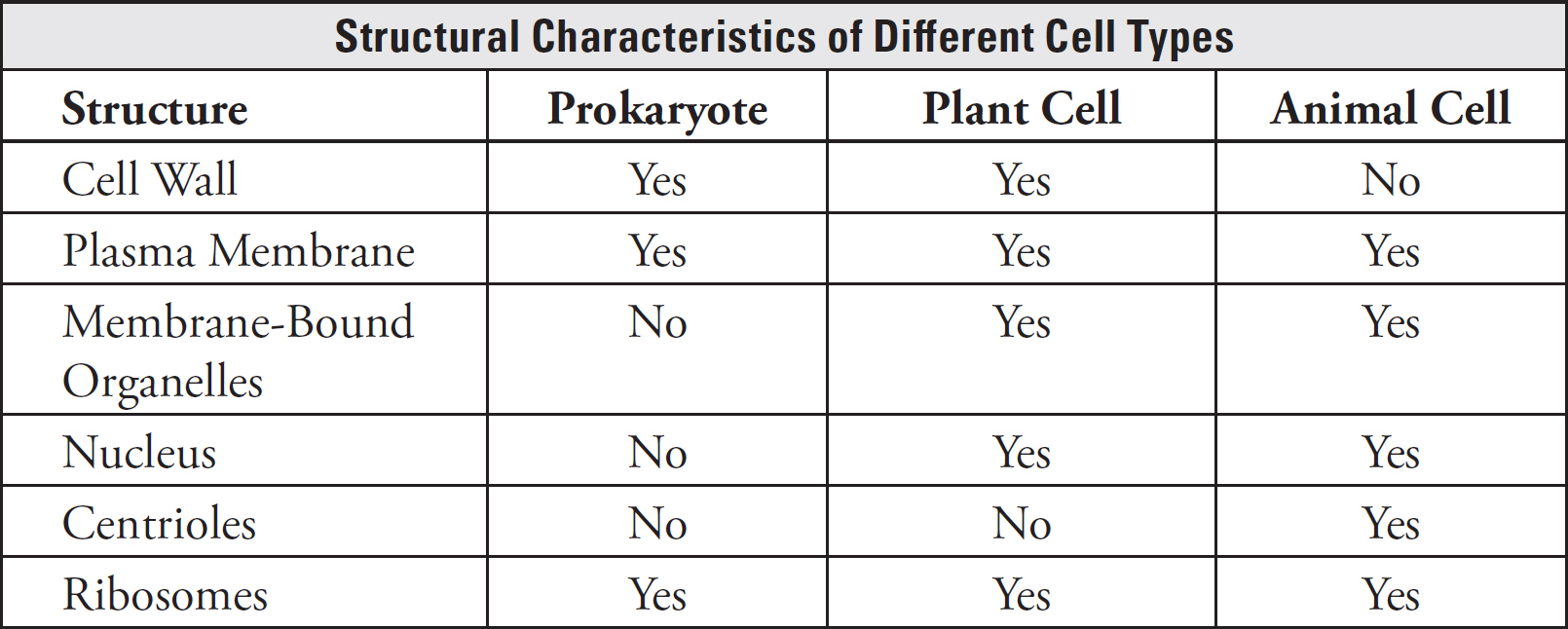
To help you remember the differences among prokaryotes, plant cells, and animal cells, we’ve put together a simple table (on the next page). Make sure you learn it! The testing board is bound to ask you which cells contain which structures.
Why do we need to know about the structure of cells? Because biological structure is often closely related to function. (Watch out for this connection: it’s a favorite theme for the AP Biology Exam.) And, more importantly, because the testing board likes to test you on it!
We’ve talked about the structure of cell membranes; now let’s discuss how molecules and fluids pass through the plasma membrane. What are some of the patterns of membrane transport? The ability of molecules to move across the cell membrane depends on two things: (1) the semipermeability of the plasma membrane and (2) the size and charge of particles that want to get through.
Since the plasma membrane is composed primarily of phospholipids, small, lipid-soluble substances cross the membrane without any resistance. Why? Because “like dissolves like.” The lipid bilayer is a hydrophobic sandwich (hydrophilic outside and hydrophobic interior), and only hydrophobic things can pass that central zone. If a substance is hydrophilic, the bilayer won’t let it pass without assistance, called facilitated transport.
Facilitated transport depends upon a number of proteins that act as tunnels through the membrane. Channels are very specialized types of tunnels that let only certain things through.
The most famous of these are aquaporins, which are water-specific channels. Although water is polar, there are typically sufficient aquaporins for water to traverse the membrane whenever it wishes without forming traffic jams. However, without aquaporins, no water would be able to cross the membrane. Glucose and ions such as Na+ and K+ are also transported across the plasma membrane via membrane proteins.
Now we know how things cross the membrane, but let’s talk about why they cross the membrane. The simple answer is “To get to the other side.” If there is a high concentration of something in one area, it will move to spread out and diffuse into an area with a lower concentration, even if that means entering or exiting a cell. In other words, the substance moves down a concentration gradient. This is called diffusion. When the molecule that is diffusing is hydrophobic, the diffusion is called simple diffusion because the small nonpolar molecule can just drift right through the membrane without trouble. When the diffusion requires the help of a channel-type protein, it is called facilitated diffusion. Anytime that a substance is moving by diffusion, it is called passive transport because there is no outside energy required to power the movement. It’s like riding a bicycle downhill. The molecules are just “going with the flow.”

When the molecule that is diffusing is water, the process is called osmosis. Water always wants to move from an area where it is most concentrated to an area where it is least concentrated. Water is not usually pure water; since it is so great at dissolving things, it usually exists as a solution. A solution is made when a liquid solvent dissolves solute particles. So, if we rethink water moving down its concentration gradient, this is the same as saying that it wants to go from where there is less solute (ie. watery solution) to where there is more solute (ie. concentrated solution). It’s as if the water is moving to dilute the concentrated solute particles.
Often, situations discussing osmosis set it up so that there are two areas and the water can flow freely, but the solute cannot. For example, if a chamber containing water and a chamber containing a sucrose solution are connected by a semipermeable membrane that allows water but not sucrose to cross, diffusion of sucrose between the chambers cannot occur. In this case, osmosis draws water into the sucrose chamber to reduce the sucrose concentration. This will reduce the total volume of the water chamber. Water will flow into the sucrose chamber until the concentration is the same across the membrane.

In both diffusion and osmosis, the final result is that solute concentrations are the same on both sides of the membrane. The only difference is that in diffusion the membrane is usually permeable to solute, and in osmosis it is not.
In plants, the cell wall is important to protect it against osmotic changes. The cell wall stays relatively the same size all the time. On the other hand, the cell membrane can shrink away from the wall (a process called plasmolysis) if it loses water and can expand and squeeze tightly against the cell wall if it takes in water.
The term tonicity is used to describe osmotic gradients. If an environment is isotonic to the cell, the solute concentration is the same inside and outside. A hypertonic solution has more total dissolved solutes than the cell, while a hypotonic solution has less. The tendency of water to move down its concentration gradient (into or out of cells) can be a powerful force; it can cause cells to explode and can overcome gravity. You may also hear the terms isosmotic, hyperosmotic, and hypoosmotic. These words are generally used when comparing two solutions, while tonicity terms are used when comparing a solution to a cell. Other than this, the definitions are similar; isotonic and isosmotic both describe situations in which the concentration is the same on either side of a membrane, for example. The only difference is what type of membrane.
Water potential (Ψ) is the measure of potential energy in water and describes the eagerness of water to flow from an area of high water potential to an area of low water potential. It is affected by two factors: pressure potential (Ψp) and solute potential (Ψs). The equation for solute (osmotic) potential is listed on the AP Biology Equations and Formulas sheet and describes the effect of solute concentration on water flow. Adding a solute lowers the water potential of a solution, causing water to be less likely to leave this solution and more likely to flow into it. This makes sense because the added solute makes the solution more concentrated, and the water is now unlikely to diffuse away. The more solute molecules present, the more negative the solute potential is.
A red blood cell dropped into a hypotonic solution (such as distilled water) will expand because water will move into the cell, an area of lower water potential. Eventually, the red blood cell will pop. If a similar experiment is done with a plant cell, water will still move into the cell, but the cell wall will exert pressure, increasing the water potential and limiting the gain of water. Water potential also explains how water moves from soil into plant roots and how plants transport water from roots to leaves to support photosynthesis.
The concentration of a solution can be calculated by dividing the number of moles of solute by the volume (in liters) of solution. Highly concentrated (or stock) solutions can be diluted to make less concentrated solutions. The equation to do this is on the AP Biology Equations and Formulas sheet.
CiVi = CfVf
This equation tells you how much (Vi) of a concentrated solution (at Ci) is required to make a more dilute solution (Cf) of a certain volume (Vf). The amount of solvent added will be the final volume of the solution (Vf) minus whatever amount of stock solution you need to add. For example, if you have a bottle of 2 M solution of Tris buffer and would like to make 50 mL of 0.1 M Tris:
CiVi = CfVf
(2 M)(Vi) = (0.1 M)(0.050 L)
Vi = 0.0025 L, or 2.5 mL
Thus, you would add 2.5 mL of the concentrated (stock) Tris solution to a new tube or bottle and then add water (the solvent) up to a final volume of 50 mL. In other words, you should add 47.5 mL of water.
Suppose a substance wants to move in the opposite direction—from a region of lower concentration to a region of higher concentration. A transport protein can help usher the substance across the plasma membrane, but it’s going to need energy to accomplish this. This time it’s like riding a bicycle uphill. Compared with riding downhill, riding uphill takes a lot more work. Movement against the natural flow is called active transport.
But where does the protein get this energy? Some proteins in the plasma membrane are powered by ATP. The best example of active transport is a special protein called the sodium-potassium pump. It ushers out three sodium ions (Na+) and brings in two potassium ions (K+) across the cell membrane. This pump depends on ATP to get ions across that would otherwise remain in regions of higher concentration. Primary active transport occurs when ATP is directly utilized to transport something. Secondary active transport occurs when something is actively transported using the energy captured from the movement of another substance flowing down its concentration gradient.
We’ve now seen that small substances can cross the cell membrane by:
simple diffusion
facilitated transport
active transport
When the particles that want to enter a cell are just too large, the cell uses a portion of the cell membrane to engulf the substance. The cell membrane forms a pocket, pinches in, and eventually forms either a vacuole or a vesicle. This process is called endocytosis.

Three types of endocytosis exist: pinocytosis, phagocytosis, and receptor-mediated endocytosis. In pinocytosis, the cell ingests liquids (“cell-drinking”). In phagocytosis, the cell takes in solids (“cell-eating”). A special type of endocytosis, receptor-mediated endocytosis, involves cell surface receptors that work in tandem with endocytic pits that are lined with a protein called clathrin. When a particle, or ligand, binds to one of these receptors, the ligand is brought into the cell by the invagination, or “folding in” of the cell membrane. A vesicle then forms around the incoming ligand and carries it into the cell’s interior.
Other substances move by bulk flow. Bulk flow is the one-way movement of fluids brought about by pressure. For instance, the movement of blood through a blood vessel and the movement of fluids in xylem and phloem of plants are examples of bulk flow.
Dialysis is the diffusion of solutes across a selectively permeable membrane. Special membranes that have holes of a certain size within them can be used to sort substances by using the process of diffusion. Kidney dialysis is a specialized process by which the blood is filtered by using machines and concentration gradients. Things present at high levels will naturally diffuse out of the blood; dialysis just gives them the opportunity.
Sometimes large particles are transported out of the cell. In exocytosis, a cell ejects waste products or specific secretion products, such as hormones, by the fusion of a vesicle with the plasma membrane, which then expels the contents into the extracellular space. Think of exocytosis as reverse endocytosis.
cells
surface area-to-volume ratio
light microscopes
electron microscopes
prokaryotic cells
eukaryotic cells
cytoplasm
nucleoid
cell wall
plasma membrane
flagella
capsule
organelles
phosholipid bilayer
peripheral proteins
integral proteins
transmembrane proteins
fluid-mosaic model
adhesion proteins
receptor proteins
transport proteins
channel proteins
cell surface markers
carbohydrate side chains
nucleus
chromosomes
nucleolus
ribosomes
endoplasmic reticulum (ER)
rough ER
smooth ER
Golgi bodies
vesicles
mitochondria
adenosine triphosphate (ATP)
cristae
lysosomes
centrioles
microtubule organizing centers (MTOCs)
vacuoles
peroxisomes
cytoskeleton
microtubules
microfilaments
tubulin
cilia
Euglena
Paramecium
cell wall
chitin
chloroplasts
central vacuole
cell sap
facilitated transport
aquaporins
simple diffusion
facilitated diffusion
passive transport
osmosis
tonicity
isotonic
hypertonic
hypotonic
water potential
solute (osmotic) potential
solutes
active transport
sodium-potassium pump
endocytosis
pinocytosis
phagocytosis
receptor-mediated endocytosis
bulk flow
dialysis
exocytosis
Living things are composed of cells, the smallest unit of living organisms. Cells may be categorized into prokaryotes, which do not have a nucleus or membrane-bound organelles, and eukaryotes, which have a nucleus and membrane-bound organelles. Features of prokaryotic cells (like bacteria, for example) can include the following:
plasma membrane
flagella and cilia
cell wall
nucleoid region
ribosomes
circular chromosome
cytoplasm
Components of the eukaryotic cells include the following:
nucleus
nucleolus
rough endoplasmic reticulum
smooth endoplasmic reticulum
Golgi body
ribosomes
mitochondria
vacuole
lysosome
peroxisomes
centrioles (animals only)
cytoplasm
plasma membrane
cytoskeleton (microtubules, microfilaments)
central vacuole (plants only)
cell wall (plants, fungi, and some protists)
The plasma membrane is composed of a phospholipid bilayer. The hydrophobic nature of the inside of the membrane is responsible for its selective permeability. Transport through the membrane is dependent on size and polarity of the molecules and concentration gradients. There are a few different types of cellular transport:
simple diffusion
facilitated transport
active transport
bulk flow
dialysis
endocytosis (pinocytosis, phagocytosis, receptor-mediated endocytosis)
exocytosis
Answers and explanations can be found in Chapter 15.
1. Movement of substances into the cell is largely dependent on the size, polarity, and concentration gradient of the substance. Which of the following represents an example of active transport of a substance into a cell?
(A) Diffusion of oxygen into erythrocytes (red blood cells) in the alveolar capillaries of the lungs
(B) Influx of sodium ions through a voltage-gated ion channel in a neuron cell during an action potential
(C) The sodium-potassium pump, which restores resting membrane potentials in neurons through the use of ATP
(D) Osmosis of water into an epithelial cell lining the lumen of the small intestine
2. The development of electron microscopy has provided key insights into many aspects of cellular structure and function, which had previously been too small to be seen. All of the following would require the use of electron microscopy for visualization EXCEPT
(A) the structure of a bacteriophage
(B) the matrix structure of a mitochondrion
(C) the shape and arrangement of bacterial cells
(D) the pores on the nuclear membrane
3. Vibrio cholerae (shown below) are highly pathogenic bacteria that are associated with severe gastrointestinal illness and are the causative agent of cholera. In extreme cases, antibiotics are prescribed that target bacterial structures that are absent in animal cells. Which of the following structures is most likely targeted by antibiotic treatment?
(A) Cytoplasm
(B) Plasma membrane
(C) Ribosomes
(D) Cell wall
Question 4 is based on the information and table below.
A new unicellular organism has recently been identified living in thermal pools in Yellowstone National Park. The thermal pools have average temperatures of 45°C, a pH of approximately 3.2, and high concentrations of sulfur-containing compounds. To identify the organism, a microbiologist performs a series of tests to evaluate its structural organization. The table below summarizes the microscopy data of the newly identified organism.
|
Cellular Structure |
Analysis |
|
Plasma Membrane |
Present |
|
Cell Wall |
Present, very thick |
|
Mitochondria |
Absent |
|
Ribosomes |
Present, highly abundant |
|
Flagella |
Present, peritrichous organization |
4. This organism is most likely a new species of which of the following?
(A) Algae
(B) Protozoa
(C) Bacteria
(D) Fungi
5. In a eukaryotic cell, which of the following organelles directly work together?
(A) Nuclear envelope, nucleolus, vacuoles, centrioles
(B) Ribosomes, rough endoplasmic reticulum, Golgi bodies, plasma membrane
(C) Mitochondria, ribosomes, lysosomes, chloroplasts
(D) Centrioles, nucleolus, smooth endoplasmic reticulum, lysosomes
6. A lipid-soluble hormone, estrogen, is secreted from the ovaries. This molecule travels through the body via the bloodstream. A researcher was interested in reducing estrogen’s effect in order to determine the response of decreased estrogen on the organism. Which of the following is a valid strategy for reducing effects of estrogen on the whole research organism?
(A) Treat with a competitive inhibitor drug that blocks all receptors at the plasma membrane
(B) Treat with lipid-soluble testosterone
(C) Treat with a lipid-soluble noncompetitive inhibitor that specifically reduces estrogen binding to the intracellular receptor
(D) Remove ovaries of the organism
7. Which type of cell would be the most useful for studying the rough endoplasmic reticulum?
(A) Neurons firing action potentials
(B) Insulin-making cells in the pancreas
(C) Bacterial cells in a colony
(D) White blood cells
8. Paramecium is a single-celled protist that lives in freshwater habitats. In these conditions, Paramecium has evolved strategies to handle the potential consequences of inhabiting this hypotonic environment. One of these strategies could be
(A) contractile vacuoles, which expel water forcefully
(B) increased aquaporins in its cellular membrane
(C) many cilia covering its surface
(D) salt receptors on its surface to seek out less concentrated areas
9. A cotransporter is something that moves two substances across a membrane, one passively and the other actively. The Na+K+ ATPase transports sodium and potassium ions across the plasma membrane against their concentration gradients. This pump is not considered a cotransporter because
(A) ATP is produced through this transporter
(B) both ions are moved via active transport
(C) both ions are moved via passive transport
(D) ATP hydrolysis does not occur during transport
Respond to the following questions:
Which topics from this chapter do you feel you have mastered?
Which content topics from this chapter do you feel you need to study more before you can answer multiple-choice questions correctly?
Which content topics from this chapter do you feel you need to study more before you can effectively compose a free response?
Was there any content that you need to ask your teacher or another person about?