This section includes detailed scientific and clinical toxicological information. For non-health professionals this scientific and medical information should be utilized for informational purposes only; certain medical terms utilized in this section are not defined and may require a medical background to undertand. For trained medical personnel clinical care should not simply focus on the potential plant exposure, although this information is obviously helpful in many situations. Rather, clinical care should take into account the patient’s current and prior medical history, physical examination, appropriate diagnostic testing, response to therapy, and all other factors normally utilized in the provision of clinical care. That is, the patient should be managed based on his or her clinical condition rather than just on the knowledge of an exposure or suspicion of a toxin.
Appropriate clinical judgment should be exercised in the management of all patients. The information in this section should be supplemented by consultation with a Poison Control Center, medical toxicologist, other appropriate expert, or the use of a medical textbook or other authoritative reference.
General initial medical management strategies that are required for all plant-exposed patients include, but are not necessarily limited to, vital sign assessment, consideration of the need for immediate interventions (e.g., ventilation and oxygenation, blood glucose), determination of the need for laboratory or other diagnostic testing, and the consideration of the need for gastrointestinal decontamination (see Section 4). Intervention at any point that is deemed appropriate to correct or prevent progression of a clinical abnormality is critical. Specific considerations and interventions follow. Additional information and references are found in the individual plant descriptions in Section 5.
Examples of plant genera associated with this syndrome:
Atropa | Brugmansia | Datura |
Hyoscyamus | Solandra | Solanum |
Competitive antagonism of acetylcholine at the muscarinic subtype of the acetylcholine receptor, which is primarily located in the parasympathetic nervous system and the brain. Because of the specificity of the toxin for the muscarinic receptor, this is often referred to as the antimuscarinic syndrome.
The classically described anticholinergic syndrome includes dry, warm, and flushed skin, parched mucous membranes, garbled speech, sinus tachycardia, adynamic ileus (absent bowel motility), urinary retention, seizure, and delirium often with hallucinations. The hallucinations may be quite troubling to the patient, and patients may develop severe dysphoria or agitated delirium along with their sequelae. The patient’s temperature may be slightly elevated, and is rarely above 102 °F unless he or she is severely agitated or convulsing. Seizures generally only occur in patients who have other clinical findings consistent with anticholinergic poisoning. Complete clinical recovery even in the absence of complications may take many hours to days.
Given the common clinical presentation of altered mental status in association with elevated body temperature, patients should be evaluated for other medical problems, including sepsis, meningitis, and serotonin syndrome unless the diagnosis is certain. Patients who are seriously poisoned by an antimuscarinic agent, particularly those with an appropriate confirmatory history, should receive either sedation with a benzodiazepine or reversal of their clinical syndrome with physostigmine. This antidote, a cholinesterase inhibitor, raises intrasynaptic levels of acetylcholine by preventing the neurotransmitter’s metabolism by the enzyme cholinesterase and allows acetylcholine to successfully compete with the toxin for the muscarinic receptor. The initial dose of physostigmine is 1–2 mg in adults (0.02 mg/kg in children) administered intravenously over no less than 5 minutes. Lack of clinical improvement suggests that either the diagnosis is incorrect or the dose of physostigmine is insufficient. Failure to develop cholinergic findings (e.g., salivation, diaphoresis, rales, bradycardia) following physostigmine raises the likelihood of the diagnosis of anticholinergic poisoning, and cautious administration of increasing doses of the drug (up to 5 mg total dose in adults over 30 minutes) may be appropriate. The duration of action of some of the antimuscarinic alkaloids may be longer than that of physostigmine, and repeated administration of the latter may be required; alternatively, once the diagnosis is confirmed by an appropriate response to antidote, the patient may be sedated with a benzodiazepine, as needed, and observed.
Burns MJ, Linden CH, Graudins A, Brown RM, Fletcher KE. A comparison of physostigmine and benzodiazepines for the treatment of anticholinergic poisoning. Ann Emerg Med. 2000;35:374–81.
Glatstein M, Alabdulrazzaq F, Scolnik D. Belladonna alkaloid intoxication: the 10-year experience of a Large Tertiary Care Pediatric Hospital. Am J Ther. 2016;23(1):e74–7. https://doi.org/10.1097/01.mjt.0000433940.91996.16.
Howland MA. Physostigmine salicylate. In: Nelson LS, Howland MA, Lewin NA, Smith SW, Goldfrank LR, Hoffman RS, editors. Goldfrank’s Toxicologic Emergencies. 11th ed. New York: McGraw Hill; 2018. pp. 755–8.
Examples of plant genera associated with this syndrome:
Alocasia | Arisaema | Brassaia |
Caladium | Caryota | Colocasia |
Dieffenbachia | Epipremnum | Monstera |
Philodendron | Spathiphyllum |
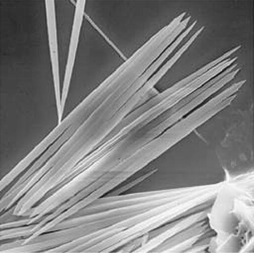
Calcium oxalate crystals at high magnification (Franceschi and Nakata 2005)
Upon mechanical stimulation, as occurs with chewing, insoluble crystalline calcium oxalate needles, bundled in needle-like raphides, release from their intracellular packaging (idioblasts) in a projectile fashion. These needles penetrate the mucous membranes and are associated with the release of histamine and other inflammatory mediators.
After biting or chewing, there is rapid onset of local oropharyngeal pain, which typically limits continued exposure, as well as local swelling and garbled speech. If swallowed, inflammation of the posterior oropharynx or larynx may rarely produce oropharyngeal edema and airway compromise. Endoscopic evaluation of the patient’s airway, esophagus, or stomach may be necessary. Ocular exposure produces extreme pain, keratoconjunctival injection, and chemosis, with the potential for severe ocular damage and vision loss. Extensive dermal contact may produce pain and signs of irritation. In contrast to that occurring with soluble oxalate ingestion, after which profound hypocalcemia may occur, there is generally no associated systemic toxicity from this insoluble form.
Airway assessment and management is of the highest priority following oral exposure. Oropharyngeal or dermal pain may be managed with appropriate demulcents, viscous lidocaine, analgesics, or with copious irrigation. Further evaluation of the patient’s pharyngeal, respiratory, and gastrointestinal tract may be necessary. Eye exposure generally requires extensive irrigation and analgesia. Ophthalmologic consultation should be considered as needed.
Altin G, Sanli A, Erdogan BA, Paksoy M, Aydin S, Altintoprak N. Severe destruction of the upper respiratory structures after brief exposure to a dieffenbachia plant. J Craniofac Surg. 2013;24(3):e245–7.
Franceschi VR, Nakata PA. Calcium oxalate in plants: formation and function. Annu Rev Plant Biol. 2005;56:41–71.
Nelson LS, Goldfrank LR. Plants. In: Nelson LS, Howland MA, Lewin NA, Smith SW, Goldfrank LR, Hoffman RS, editors. Goldfrank’s Toxicologic Emergencies. 11th ed. New York: McGraw Hill; 2018. pp. 1597–616.
Examples of plant genera associated with this syndrome:
Acokanthera | Adenium | Adonis |
Calotropis | Cryptostegia | Digitalis |
Helleborus | Ornithogalum | Convallaria |
Nerium | Pentalinon | Thevetia |
Urginea | Strophanthus | Scilla |
Cardioactive steroids, termed cardiac glycosides when sugar moieties are attached, inhibit the cellular Na+/K+-ATPase. The effect is to indirectly increase intracellular Ca2+ concentrations in certain cells, particularly myocardial cells. Therapeutically, this both enhances cardiac inotropy (contractility) and slows the heart rate. However, excessive elevation of the intracellular Ca2+ also increases myocardial excitability, predisposing to the development of ventricular dysrhythmias. Enhanced vagal tone, mediated by the neurotransmitter acetylcholine, is common with poisoning by these agents, and produces bradycardia and heart block.
Ingestion of plants containing cardioactive steroids may cause abdominal pain and vomiting, the latter of which serves both as an early sign of toxicity and a mechanism to limit poisoning. Cardiovascular and electrocardiographic effects include sinus and junctional bradycardia as well as ventricular tachydysrhythmias, most concerningly ventricular tachycardia and ventricular fibrillation. Hyperkalemia may develop and is associated with poor patient outcome. Serum digoxin concentrations may be obtained but should not be relied upon to exclude toxicity as other cardioactive steroids will have unpredictable assay cross-reactivity. Consequently, treatment, if clinically indicated, should not await laboratory confirmation.
Most of the available clinical experience with cardioactive steroid poisoning is related to digoxin toxicity. In these patients, standard supportive medical management is often inadequate. Therefore, any patient with consequential digoxin poisoning should receive digoxin-specific Fab. This product contains the antigen-binding regions (Fab) of antidigoxin antibodies. Although specifically designed for the management of digoxin poisoning, digoxin-specific Fab appears to have sufficient cross-recognition with other cardioactive steroids to warrant its administration in other nondigoxin cardioactive steroid poisoned patients. The empiric dose is 10 vials (400 mg) administered intravenously in both adults and children, with additional dosing based on clinical response or additional information. Indications for its use include significant bradycardia, tachydysrhythmias, or hyperkalemia, with or without a markedly elevated serum digoxin concentration, in any patient seriously believed to be poisoned by a cardioactive steroid-containing plant.
Bandara V, Weinstein SA, White J, Eddleston M. A review of the natural history, toxinology, diagnosis and clinical management of Nerium oleander (common oleander) and Thevetia peruviana (yellow oleander) poisoning. Toxicon. 2010;56(3):273–81. https://doi.org/10.1016/j.toxicon.2010.03.026.
Eddleston M, Persson H. Acute plant poisoning and antitoxin antibodies. J Toxicol Clin Toxicol. 2003;41:309–15.
Hack JB. Cardioactive steroids. In: Nelson LS, Howland MA, Lewin NA, Smith SW, Goldfrank LR, Hoffman RS, editors. Goldfrank’s Toxicologic Emergencies. 11th ed. New York: McGraw Hill; 2018. pp. 969–76.
Roberts DM, Gallapatthy G, Dunuwille A, Chan BS. Pharmacological treatment of cardiac glycoside poisoning. Br J Clin Pharmacol. 2016;81(3):488–95. https://doi.org/10.1111/bcp.12814.
Smith SW, Howland MA. Digoxin-specific antibody fragments. In: Nelson LS, Howland MA, Lewin NA, Smith SW, Goldfrank LR, Hoffman RS, editors. Goldfrank’s Toxicologic Emergencies. 11th ed. New York: McGraw Hill; 2018. pp. 977–84.
Examples of plant genera associated with this syndrome:
Aethusa | Anemone | Blighia |
Caltha | Caulophyllum | Cicuta |
Clematis | Conium | Coriaria |
Gymnocladus | Hippobroma | Laburnum |
Lobelia | Menispermum | Myoporum |
Nicotiana | Pulsatilla | Ranunculus |
Sophora | Spigelia | Strychnos |
A convulsion is the rhythmic, forceful contraction of the muscles, one cause of which are seizures. Seizures are disorganized discharges of the central nervous system that generally, but not always, result in a convulsion. There are various toxicological mechanisms that result in seizures including antagonism of gamma-aminobutyric acid (GABA) at its receptor on the neuronal chloride channel (e.g., cicutoxin, picrotoxin), imbalance of acetylcholine homeostasis, excitatory amino acid mimicry, sodium channel alteration, or hypoglycemia. Strychnine and its analogues (e.g., brucine) antagonize the postsynaptic inhibitory activity of glycine at the spinal cord motor neuron. Strychnine results in hyperexcitability of the motor neurons, which manifests as a convulsion but does not cause seizure.
Unless an underlying central nervous system lesion exists, patients with plant-induced seizures generally present with generalized, as opposed to focal, seizures. Most patients develop generalized tonic-clonic convulsions, in which periods of shaking movement (convulsion or clonus) are interspersed with periods of hypertonicity. Occasionally, patients may not have overt motor activity (i.e., nonconvulsive seizure), or may present in the postictal period (partially or fully recovered from their seizure). The diagnosis in this situation may be difficult to determine. Patients who are having a generalized seizure should have loss of consciousness as a result of central nervous system dysfunction, and often have urinary or fecal incontinence, tongue biting, or other signs of trauma.
Conscious patients who are manifesting what appear to be generalized convulsions may have myoclonus or strychnine poisoning. Strychnine-poisoned patients manifest symmetrical convulsive activity, but because the activity is the result of spinal cord dysfunction, there is no loss of consciousness (i.e., no seizure) until metabolic or other complications intercede.
Once hypoglycemia and hypoxia have been excluded (or treated), a rapidly acting anticonvulsant benzodiazepine (e.g., diazepam, 5–10 mg intravenously (IV) in adults (0.1–0.3 mg/kg in children) or lorazepam, 2 mg IV in adults, or 0.1 mg/kg in children), should be administered parenterally for persistent seizures. Although diazepam and lorazepam are nearly equivalent in time to onset, lorazepam has a substantially longer duration of anticonvulsant effect. Lorazepam can be administered intramuscularly, though this route is not ideal because of a slow absorptive phase. Dosing may be repeated several times as needed. Inability to expeditiously control seizures with benzodiazepines may necessitate the administration of barbiturates, propofol, or another anticonvulsant medication. There is generally no acute role for phenytoin, levetiracetam, or other maintenance anticonvulsants in patients with toxin-induced seizures.
Chen H-Y, Horng H, Rowley F, Smollin C. Rapid respiratory arrest after ingestion of poison hemlock mistaken for wild celery. Clin Toxicol. 2016;55(2):155–6.
Chan YC. Strychnine. In: Nelson LS, Howland MA, Lewin NA, Smith SW, Goldfrank LR, Hoffman RS, editors. Goldfrank’s Toxicologic Emergencies. 11th ed. New York: McGraw Hill; 2018. pp. 1536–9.
Hotti H, Rischer H. The killer of Socrates: coniine and related alkaloids in the plant kingdom. Molecules. 2017;22(11). https://doi.org/10.3390/molecules22111962.
Singhapricha T, Pomerleau AC. A case of strychnine poisoning from a Southeast Asian Herbal Remedy. J Emerg Med. 2017;52(4):493–5.
Examples of plant genera associated with this syndrome:
Eriobotrya | Hydrangea | Malus |
Prunus | Sambucus |
Cyanogenic compounds, most commonly glycosides, must be metabolized to release cyanide. Cyanide inhibits the final step of the mitochondrial electron transport chain, resulting rapidly in cellular energy failure.
Because the cyanogenic glycosides must be hydrolyzed in the gastrointestinal tract before cyanide ion is released, the onset of toxicity is commonly delayed. Abdominal pain, vomiting, lethargy, and sweating develop initially, followed shortly by altered mental status, seizures, cardiovascular collapse, and with initial survival, multisystem organ failure. Laboratory testing should reveal an elevated blood lactic acid; cyanide levels are not generally available rapidly. Thiocyanate, a metabolite of cyanide, may be measured in the patient’s blood, and although often confirmatory in retrospect, immediate results are not readily available.
Initial management includes aggressive supportive care, intravenous fluid therapy, and correction of life-threatening metabolic acidosis using intravenous sodium bicarbonate as appropriate. Antidotal therapy, available in two distinct formulations, should be administered to any patient believed to be suffering from cyanide poisoning. The preferred antidote is intravenous hydroxocobalamin due to its safety and simplicity. This compound is a precursor of vitamin B12 (cyanocobalamin), which forms once the antidote scavenges the cyanide from the mitochondria. The alternative antidote utilizes the combination of a nitrite and sodium thiosulfate to remove cyanide from the mitochondria (nitrite-induced methemoglobin) and detoxify it (thiosulfate). Before the establishment of an intravenous line, an amyl nitrite pearl may be broken and held under the patient’s nose for 30 seconds each minute. In patients with intravenous access, 10 ml of 3% sodium nitrite in an adult, or in an appropriate pediatric dose (guidelines supplied with the kit), should be administered intravenously; this should be followed rapidly by 50 ml of 25% sodium thiosulfate intravenously in an adult, or 1.65 ml/kg in children. The nitrite portion of the antidote is associated with hypotension and methemoglobin formation which is therapeutic as noted above. However, in certain circumstances, for example, when the diagnosis is uncertain, administration of only the sodium thiosulfate component of the antidote kit may be appropriate.
Abraham K, Buhrke T, Lampen A. Bioavailability of cyanide after consumption of a single meal of foods containing high levels of cyanogenic glycosides: a crossover study in humans. Arch Toxicol. 2016;90(3):559–74. https://doi.org/10.1007/s00204-015-1479-8.
Bebarta VS, Tanen DA, Lairet J, Dixon PS, Valtier S, Bush A. Hydroxocobalamin and sodium thiosulfate versus sodium nitrite and sodium thiosulfate in the treatment of acute cyanide toxicity in a Swine (Sus scrofa) model. Ann Emerg Med. 2010;55(4):345–51.
Holstege CP, Isom G, Kirk MA. Cyanide and hydrogen sulfide. In: Nelson LS, Howland MA, Lewin NA, Smith SW, Goldfrank LR, Hoffman RS, editors. Goldfrank’s Toxicologic Emergencies. 11th ed. New York: McGraw Hill; 2018. pp. 1684–93.
Vetter J. Plant cyanogenic glycosides. Toxicon. 2000;38:11–36.
Many and various plant genera are associated with this syndrome.
Several different mechanisms are utilized by plant toxin to produce gastrointestinal effects, generally described as either mechanical irritation or a pharmacologic effect. Irritant toxins indirectly stimulate contraction of the gastrointestinal smooth muscle. The pharmacologically active agents most commonly work by stimulation of cholinergic receptors in the gastrointestinal tract to induce smooth muscle contraction (e.g., cholinergic (including nicotine-like)) alkaloids. Some plant toxins (e.g., mitotic inhibitors, toxalbumins) alter the normal development and turnover of gastrointestinal lining cells and induce sloughing of this cellular layer. Hepatotoxins may directly injure the liver cells, commonly through the production of oxidant metabolites. Indirect hepatotoxicity may occur, as with the pyrrolizidine alkaloids (see “Poisoning by Plants with Pyrrolizidine Alkaloids”, p 31).
Nausea, vomiting, abdominal cramping, and diarrhea are the hallmarks. Vomiting may be bloody or may contain acid-degraded blood (“coffee grounds”) leaked secondary to gastric irritation. Extensive diarrhea and vomiting may produce acid–base, electrolyte, and fluid abnormalities, leading to hypokalemia and profound volume depletion. Small children in particular may become rapidly volume depleted and it may be more difficult to diagnose than in adults. Certain plant toxins that produce prominent gastrointestinal findings may subsequently produce systemic toxicity following absorption. For agents in this group (e.g., mitotic inhibitors, toxalbumins), the gastrointestinal manifestations serve as a warning for potential systemic toxicity.
Vomiting may be mitigated by antiemetic agents such as metoclopramide; occasionally, resistant emesis may require a serotonin antagonist such as ondansetron. Specific treatment for a patient’s diarrhea (e.g., loperamide) is generally unnecessary. Assessment for and correction of volume depletion and metabolic changes are critical. For most patients, intravenous rehydration should be initiated using normal saline or lactated Ringer’s solution and adjusted based on clinical or laboratory criteria. Oral rehydration therapy may be attempted in patients with minor clinical abnormalities. Electrolyte and acid–base derangements usually resolve with supportive care but may occasionally require specific therapy. Pharmacotherapies for the prevention of treatment of hepatotoxicity are varied, but empiric therapy with N-acetylcysteine is often suggested.
Zuckerman MD, Church RJ. Gastrointestinal principles. In: Nelson LS, Howland MA, Lewin NA, Smith SW, Goldfrank LR, Hoffman RS, editors. Goldfrank’s Toxicologic Emergencies. 11th ed. New York: McGraw Hill; 2018. pp. 287–96.
Examples of plant genera associated with this syndrome:
Bulbocodium | Catharanthus | Colchicum |
Gloriosa | Podophyllum |
These agents interfere with the polymerization of microtubules, which must polymerize for mitosis to occur, leading to metaphase arrest. Rapidly dividing cells (e.g., gastrointestinal or bone marrow cells) typically are affected earlier and to a greater extent than those cells that divide slowly. In addition, microtubules are important in the maintenance of proper neuronal function.
Patients typically have early gastrointestinal abnormalities, including vomiting and diarrhea. Oral ulcers and frank gastrointestinal necrosis can occur. Multisystem organ failure may follow. Bone marrow toxicity typically manifests as an initial leukocytosis, due to release of stored white blood cells, followed by leukopenia. Death may occur from direct cellular toxic effects or from sepsis. Nervous system toxicity, including ataxia, headache, seizures, and encephalopathy, may develop initially, and peripheral neuropathy may develop in patients who survive.
Initial management includes aggressive supportive and symptomatic care. In patients with profound bone marrow toxicity, colony-stimulating factors may be beneficial. Consultation with appropriate specialists, such as a hematologist, should be strongly considered.
Eddleston M, Fabresse N, Thompson A, et al. Anti-colchicine Fab fragments prevent lethal colchicine toxicity in a porcine model: a pharmacokinetic and clinical study. Clin Toxicol (Phila). 2018;17:1–9.
Galland-Decker C, Charmoy A, Jolliet P, Spertini O, Hugli O, Pantet O. Progressive organ failure after ingestion of wild garlic juice. J Emerg Med. 2016;50(1):55–60.
Reijnen G, Bethlehem C, van Remmen JMBL, Smit HJM, van Luin M, Reijnders UJL. Post-mortem findings in 22 fatal Taxus baccata intoxications and a possible solution to its detection. J Forensic Leg Med. 2017;52:56–61.
Santos CD, Schier J. Colchicine, Podophyllin, and the Vinca Alkaloids. In: Nelson LS, Howland MA, Lewin NA, Smith SW, Goldfrank LR, Hoffman RS, editors. Goldfrank’s Toxicologic Emergencies. 11th ed. New York: McGraw Hill; 2018. pp. 501–10.
Examples of plant genera associated with this syndrome:
Baptisia | Caulophyllum | Conium |
Gymnocladus | Hippobroma | Laburnum |
Lobelia | Nicotiana | Sophora |
These agents are direct-acting agonists at the nicotinic subtype of the acetylcholine receptor in the ganglia of both the parasympathetic and sympathetic limbs of the autonomic nervous system (NN receptors), the neuromuscular junction (NM receptors), and the brain.
Sympathetic stimulation, including hypertension, tachycardia, and diaphoresis, and parasympathetic stimulation, including salivation and vomiting, are common (NN). Hyperstimulation at the NM results in fasciculations, muscular weakness, and, rarely, depolarizing neuromuscular blockade. Seizures may occur as a result of effects at cerebral nicotinic receptors.
Control of the patient’s autonomic hyperactivity is generally not needed unless secondary complications, such as myocardial ischemia, develop or are anticipated. In this case, the vital sign abnormalities may be corrected through the judicious use of antihypertensive drugs, including nitroprusside or diltiazem, as appropriate. Neuromuscular effects cannot be effectively antagonized because effective agents (e.g., curare-like drugs) would also produce neuromuscular blockade. Patients with inadequate ventilatory effort should be managed supportively. Seizures should respond to intravenous benzodiazepine, such as lorazepam or diazepam.
Hotti H, Rischer H. The killer of Socrates: coniine and related alkaloids in the plant kingdom. Molecules. 2017;22(11). https://doi.org/10.3390/molecules22111962.
Rogers AJ, Denk LD, Wax PM. Catastrophic brain injury after nicotine insecticide ingestion. J Emerg Med. 2004;26:169–72.
Fernandez D, Soghoian S. Nicotine. In: Nelson LS, Howland MA, Lewin NA, Smith SW, Goldfrank LR, Hoffman RS, editors. Goldfrank’s Toxicologic Emergencies. 11th ed. New York: McGraw Hill; 2018. pp. 2013–1211.
Vetter J. Poison hemlock (Conium maculatum L.). Food Chem Toxicol. 2004;42:1373–82.
Examples of plant genera associated with this syndrome:
Crotalaria | Echium | Heliotropium |
Senecio | Sesbania |
Pyrrolizidine alkaloids are metabolized to pyrroles, which are alkylating agents that injure the endothelium of the hepatic sinusoids or pulmonary vasculature. Endothelial repair and hypertrophy result in veno-occlusive disease. Centrilobular necrosis may occur following acute, high-dose exposures, presumably caused by the overwhelming production of the pyrrole. Chronic use is also associated with hepatic carcinoma.
Acute hepatotoxicity caused by massive pyrrolizidine alkaloid exposure produces gastrointestinal symptoms, right upper quadrant abdominal pain, hepatosplenomegaly, and jaundice as well as biochemical abnormalities consistent with hepatic necrosis (e.g., aspartate ammotransferase (AST), bilirubin, increased international normalized ratio (INR)). Prolonged, lower-level exposure produces more indolent disease, and patients may present with cirrhosis or ascites caused by hepatic venous occlusion. This syndrome is clinically and pathologically similar to the Budd–Chiari syndrome.
Certain pyrrolizidine alkaloids (e.g., that from Crotalaria spectabilis) produce pulmonary vasculature occlusion and the syndrome of pulmonary hypertension in animals, but it is not known whether there is an analogous human response.
Standard supportive care may allow for some spontaneous repair. There are no known specific therapies. Liver transplantation may be an option for patients with severe hepatotoxicity or cirrhosis.
Nelson LS, Goldfrank LR. Plants. In: Nelson LS, Howland MA, Lewin NA, Smith SW, Goldfrank LR, Hoffman RS, editors. Goldfrank’s Toxicologic Emergencies. 11th ed. New York: McGraw Hill; 2018. pp. 1597–616.
Neuman MG, Cohen L, Opris M, Nanau RM, Hyunjin J. Hepatotoxicity of pyrrolizidine alkaloids. J Pharm Pharm Sci. 2015;18(4):825–43.
Stewart MJ, Steenkamp V. Pyrrolizidine poisoning: a neglected area in human toxicology. Ther Drug Monit. 2001;23:698–708.
Examples of plant genera associated with this syndrome:
Aconitum | Kalmia | Leucothoe |
Lyonia | Pernettya | Pieris |
Rhododendron | Schoenocaulon | Veratrum |
Zigadenus |
These agents stabilize the open form of the voltage-dependent sodium channel in excitable membranes, such as neurons and the cardiac conduction system. This causes persistent sodium influx (i.e., persistent depolarization) and prevents adequate repolarization leading to seizures and dysrhythmias, respectively. In the heart, the excess sodium influx activates calcium exchange, and the intracellular hypercalcemia increases both inotropy and the potential for dysrhythmias.
Vomiting, dizziness, weakness, and blurry vision are very common and occur through a central nervous system-mediated mechanism. In some parts of the world (e.g., Turkey), this syndrome is associated with the consumption of “mad honey”, which is derived from Rhododendron flowers. Sodium channel effects on sensory neurons may produce paresthesias in a perioral and distal extremity distribution. Persistent depolarization of motor neurons produces fasciculations, motor weakness, and ultimately paralysis. In the heart, the effects of sodium channel opening have been compared to that of the cardioactive steroids: atropine-sensitive sinus bradycardia, atrioventricular blocks, repolarization abnormalities, and, occasionally, ventricular dysrhythmias. However, although the clinical findings are similar, the underlying mechanisms and treatments may differ.
Normal saline should be rapidly infused into patients with hypotension, and atropine is often therapeutic for sinus bradycardia and conduction blocks. Hypotension may require pressor agents such as norepinephrine. Mechanism-based therapy suggests the use of sodium channel blocking drugs such as lidocaine or amiodarone. None has been proven superior, and the agent used should probably be based on the comfort level of the provider. Although the clinical presentation is similar to poisoning by cardioactive steroids, there is no defined role for digoxin-specific Fab.
Amirshahi MA, Nelson LS. Antidysrhythmics. In: Nelson LS, Howland MA, Lewin NA, Smith SW, Goldfrank LR, Hoffman RS, editors. Goldfrank’s Toxicologic Emergencies. 11th ed. New York: McGraw Hill; 2018. pp. 865–75.
Coulson JM, Caparrotta TM, Thompson JP. The management of ventricular dysrhythmia in aconite poisoning. Clin Toxicol (Phila). 2017;55(5):313–21. https://doi.org/10.1080/15563650.2017.1291944.
Lin CC, Chan TY, Deng JF. Clinical features and management of herb-induced aconitine poisoning. Ann Emerg Med. 2004;43:574–9.
Silici S, Atayoglu AT. Mad honey intoxication: a systematic review on the 1199 cases. Food Chem Toxicol. 2015;86:282–90.
Examples of plant genera associated with this syndrome:
Abrus | Hura | Jatropha |
Momordica | Phoradendron | Ricinus |
Robinia |
The protein toxins derived from these plants work specifically by inhibiting the function of ribosomes, the subcellular organelle responsible for protein synthesis. The toxins typically have two linked polypeptide chains. One of the chains binds to cell-surface glycoproteins to allow endocytosis into the cell. The other chain upon cell entry binds the 60S ribosomal subunit and impairs its ability to synthesize protein.
Clinical manifestations depend largely on the route of exposure. Following ingestion, local gastroenteritis produces diarrhea and abdominal pain. Because the seed coat of many toxalbumin containing seeds is tough, chewing is generally a prerequisite for toxicity. Absorption of toxin into the systemic circulation allows widespread distribution and multisystem organ failure. Parenteral administration via injection similarly produces diffuse organ dysfunction. Following inhalation of aerosolized toxin, localized pulmonary effects are of greatest concern, although systemic toxicity is possible. Depending on the dose and route administered, the development of findings may be delayed.
Death from multisystem organ failure is best prevented through aggressive support of vital organ function and prevention of infection. Work is progressing on the use of antiricin antibodies and vaccine, but they are not in current clinical use.
Audi J, Belson M, Patel M, Schier J, Osterloh J. Ricin poisoning: a comprehensive review. JAMA. 2005;294:2342–51.
Bradberry SM, Dickers KJ, Rice P, Griffiths GD, Vale JA. Ricin poisoning. Toxicol Rev. 2003;22:65–70.
Lopez Nunez OF, Pizon AF, Tamama K. Ricin poisoning after oral ingestion of castor beans: a case report and review of the literature and laboratory testing. J Emerg Med. 2017;53(5):e67–71.
Smith SW, Graber NM, Johnson RC, Barr JR, Hoffman RS, Nelson LS. Multisystem organ failure after large volume injection of castor oil. Ann Plast Surg. 2009;62(1):1214. https://doi.org/10.1097/SAP.0b013e31817763bd.