3
Cell Division
CELLULAR REPRODUCTION
To create more cells and more living organisms, cells reproduce. However, simply dividing in half is not sufficient. Since it is imperative that each cell passes along its genetic information to succeeding generations of cells when a cell reproduces, each daughter cell must receive more than just a portion of the vital information; a complete copy of all the essential genetic material is necessary.
Some mechanism is needed to pass on all a cell's chromosomal information to its daughter cells. Therefore, before dividing, the parent cell must produce a copy of all the required information. This duplication is also called replication; both words are used interchangeably when discussing cell division.
Cells, though similar, aren't all identical, and neither are the mechanisms they use to divide. Prokaryotes are thought to represent the most primitive living forms of cells. They divide in a manner that is less complex than the more advanced eukaryotes.
Prokaryotic Cell Division: Binary Fission
Prokaryotic cells have one circular chromosome. When the chromosome duplicates, each of the resulting copies moves to a separate end of the cell, and a membrane forms in the middle of the parent cell, dividing it into two parts. Those two parts then separate, creating two daughter cells; each daughter cell contains an entire set of genetic material. This process of prokaryotic cell division is called binary fission (see Figure 3.1).
Eukaryotic Cell Division
Eukaryotic cell division involves a series of steps that are distinct from the process that occurs in prokaryotes. These steps include the division of the nucleus, known as karyokinesis, as well as the division of the rest of the cell, which is called cytokinesis.
Before the cell begins to divide, the genetic information located in the nucleus in the form of chromosomes must be duplicated. The chromosomes are the structures in the cells that contain the greens. All the instructions concerning the life processes of a cell emanate from the chromosomes. At the ends of the chromosomes are regions of repetitive DNA called telomeres that protect the chromosomes from fusing with neighboring chromosomes, and the telomeres protect the chromosomes from deteriorating. Many people who live over 100 years have longer telomeres than people who don't live as long.

Figure 3.1 (a) Generalized cyanobacterium (or blue-green bacterium or blue-green algae) undergoing cell division; and (b) electron photomicrograph of an actual cyanobacterium dividing in two.
It is only when the two sets of chromosomes are segregated in separate parts of the cell that the cytoplasm and other requisite materials may divide and the parent cell can complete its division.
Interphase
Compared to the rest of the cell's life, cell division is a brief and distinct stage in the cell's life history. Interphase, although the longest and most physiologically dynamic part of the cell's life history, is not considered part of cell division. Rather, this is the stage during which the cell is growing, metabolizing, and maintaining itself.
During interphase, the nucleus exists as a distinct organelle, bound by the nuclear membrane. Inside the nucleus are long, thin, unwound strands of chromosomes. While unwound throughout interphase, the chromosomes influence the activities of the cell. It is during interphase that the cell's single set of chromosomes replicates.
Cell division may occur by either mitosis or meiosis, depending on what type of cell is involved (see Figure 3.2).
MITOSIS
Prophase
Mitosis has begun when the unwound chromosomes begin to coalesce. During their first stage of mitosis, called prophase, two small cylindrical bodies become very important when present (see Figure 3.3). They help organize much of what is about to happen. Located just outside the nucleus in animal cells (though not present in most higher plants), the centrioles begin to move to opposite ends of the cell. As this happens, the DNA, which is the primary constituent of the chromosomes, recoils. During this process, the chromosomes become more distinct, while the nucleoli become less distinct.
It is in the nucleoli that ribosome production occurs; during mitosis, when the chromosomes condense, ribosome production ceases.
When unraveled, the chromosomes interact with their surrounding medium. However, for cell division to occur, the already replicated chromosomes must be pulled apart. Yet this cannot happen until the chromosomes have condensed, making it possible for cell division to proceed.
Once recoiled, the chromosomes become “X” shaped (see Figure 3.4). The Xs are composed of two identical chromatids, each held to the other by a single centromere (see Figure 3.4). One of these chromatids was copied from the other during interphase, when replication occurred.
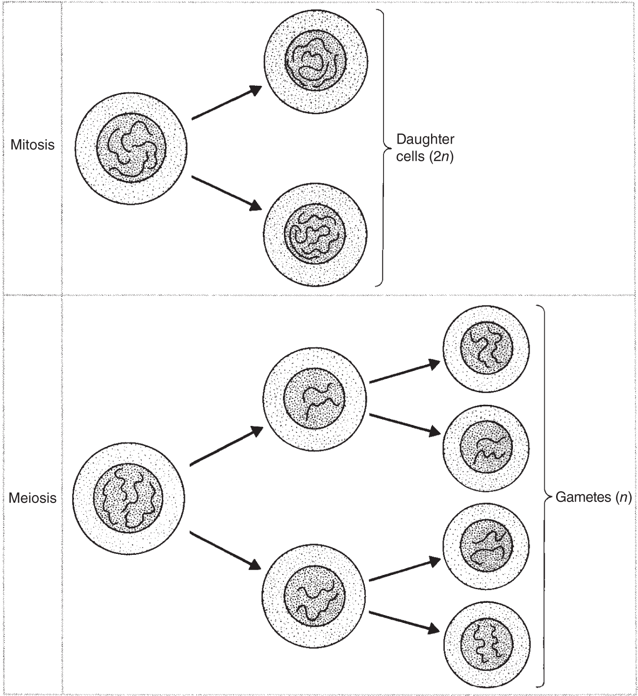
Figure 3.2 Generalized and somewhat simplified representations of two types of cell division: mitosis and meiosis. In mitosis the two daughter cells have the same number of chromosomes as the parent cell. In meiosis the gametes have half as many chromosomes as the original parent cell.
While the chromosomes are recoiling, the nucleoli and nuclear envelope are disappearing. And at the same time, a series of microtubules are forming the spindle. The spindle is the characteristic grouping of microtubules that occurs during nuclear division. It helps to align and move the chromosomes. During the early stages of spindle composition, microtubules radiate around each centriole, creating formations collectively known as the asters. Although most higher plants do not have centrioles, they still develop spindle fibers at prophase (but asters do not form). The spindle fibers are the microtubules that, together, constitute the mitotic apparatus, called the spindle.
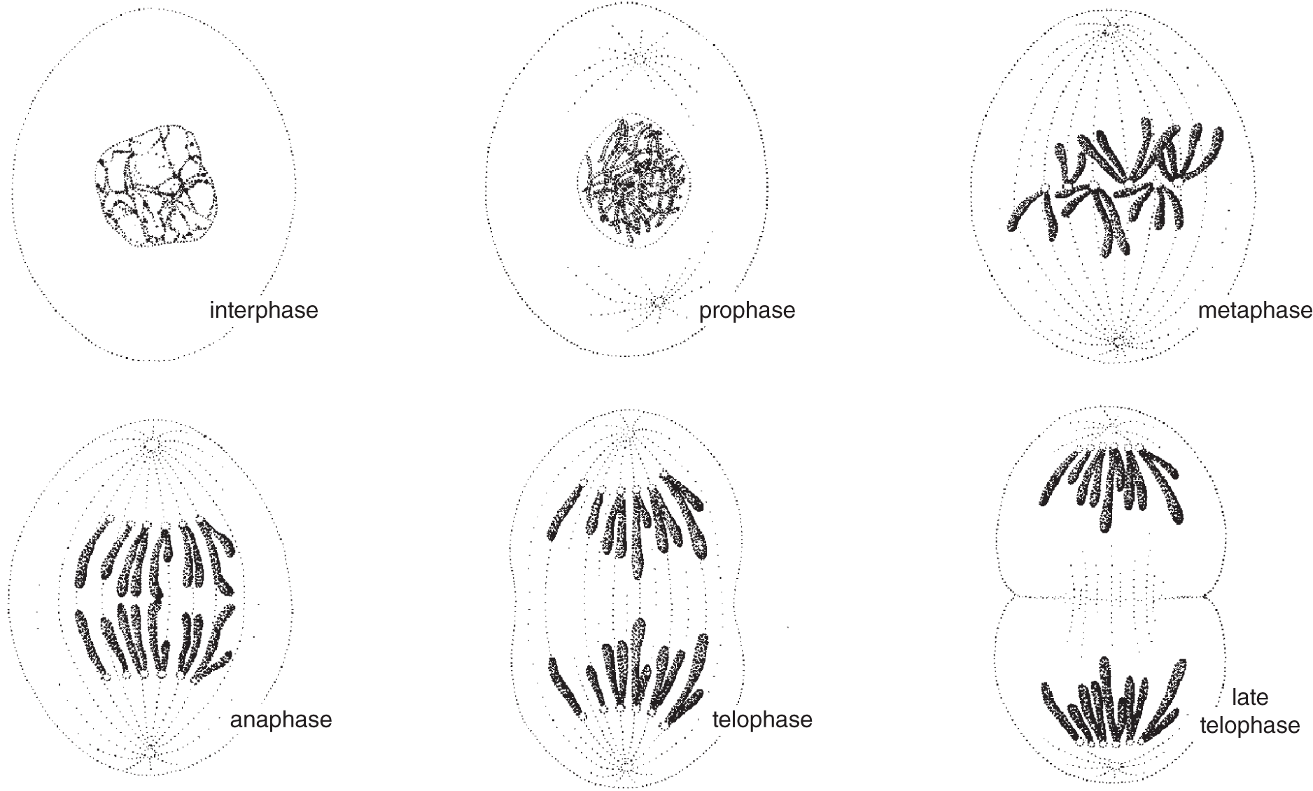
Figure 3.3 Schematic drawings of an animal cell with six chromosomes undergoing the different stages of mitosis.
During prophase, the chromosomes move toward the middle or the equator of the cell. By the end of prophase, the nuclear membrane is no longer visible; it has broken down.
Metaphase
Metaphase lasts only as long as all the chromosomes remain lined up along the equator. The centromeres have divided in two. Each is attached to one of the two corresponding chromosomes from the pair. The individual chromosomes are called homologs, and together, both chromosomes in each pair are called homologous chromosomes.
Anaphase
Anaphase begins when the two complete sets of chromosomes start moving toward opposite ends of the spindle. Each chromosome appears to be dragged along by its centromere, which is attached to a spindle fiber. The division of the cytoplasm, or cytokinesis, begins at the end of anaphase.

Figure 3.4 Drawings depicting an animal cell undergoing meiosis. For simplicity and clarity, this cell has been shown with one pair of chromosomes. The actual number of chromosomal pairs depends on the species involved. Humans have 23 pairs of chromosomes. Each chromosome is composed of two identical chromatids.
Telophase
Telophase is the last stage of mitosis. This is when the cytoplasm separates in two parts of the cell, while the cell's plasma membrane pinches in from both sides, creating two distinct cells. While this is occurring, each set of chromosomes reaches its respective pole, where the nuclear membrane forms, enclosing the chromosomes. The chromosomes then uncoil, while the nucleolus reappears. Upon completion of cytokinesis, a new centriole is made.
Cytokinesis
Cytokinesis, the division of the cytoplasm, usually begins in late anaphase and is complete in telophase. In animal cells, cytokinesis begins with the formation of an indentation, or cleavage furrow, which forms all the way around the equatorial region, becoming deeper and deeper until it cuts right through, leaving two distinct daughter cells.
Plant cells also have a plasma membrane that divides in the same manner. In addition, plant cells have a rigid cell wall that cannot form a cleavage furrow. Instead, a cell plate forms at the equatorial region, and rather than moving from the outside in, the cell plate begins forming inside and grows toward the periphery.
Syncytia and Coenocytes
As already discussed, the nuclear membrane reforms during telophase and cytokinesis occurs; however, this does not happen in all types of cells. Some tissues have cells that undergo mitotic divisions that are not followed by cytokinesis, so the cell ends up containing many nuclei. When nuclear division is not followed by cytokinesis, the result in animals is called a syncytium, and in plants it is a coenocyte.
SEX AND SEXUALITY
The exchange of genetic material between two cells is a sexual union. With most single-celled organisms, such a sexual union occurs in water. While most prokaryotes reproduce by simple cell division (binary fission), some forms reproduce by budding, in which broken-off cell fragments grow into mature bacterial cells. Binary fission and budding produce groups of genetically identical cells, known as clones.
In some cases, cells exchange or mix their genetic material together, producing populations of unique yet related cells. The transfer is accomplished by a sexual union of two cells that then separate after the genetic exchange occurs. This process of genetic recombination between two cells is known as conjugation. Another way that some bacteria exchange genetic material is accomplished simply through the absorption of bits of DNA that were released in the surrounding medium by dead bacteria. This process is known as transformation. Genetic material may also be carried from one bacterial cell to another by a virus, a process called transduction. Conjugation and transduction occur not only among many bacteria but also among certain algae and protozoa.
Sex can be either a verb or a noun. The noun usually refers to an individual that is either a female or male, depending on the sex cells the individual produces. A female produces eggs, which are sex cells, which are also called gametes. The sex cells (or gametes) are eggs and sperm. The females produce eggs, the males produce sperm. The biological definition of the verb, sex, is the act that involves coupling, mating, amplexus, the goal of which may be for the good feelings, or for joining together of male and female sex cells. When the sex cells join, they go from having been haploid to being diploid.
Gametes
Larger organisms that are composed of many cells, called multicellular organisms, can still accomplish sexual union with single cells. In contrast to cells that contain two of each chromosome, sex cells possess only one of each corresponding pair of chromosomes. Such a composition is known as haploid or monoploid, and it is possible for the two sex cells to unite by forming a single cell, the zygote. The merging of genetic material from both cells is called fertilization. The zygote has twice the number of chromosomes in each sex cell and is called diploid.
Most multicellular organisms have two different types of sex cells, known as gametes, such as eggs and sperm. The sex that produces eggs is female, and the sex producing sperm is male. Sometimes the same individual has both male and female organs. Such an organism is called monoecious or hermaphroditic. Eggs are usually larger than sperm, since they contain nutrients that help nourish the developing embryo. Unlike eggs, sperm are small and motile (can move on their own).
MEIOSIS
The development of gametes, or gametogenesis, occurs through a series of cell divisions known as meiosis. Unlike mitosis, which produces diploid (2n) daughter cells, meiosis produces haploid (1n) cells, which mature into gametes (sex cells such as sperm and eggs). In contrast to cells that contain two of each chromosome, each sex cell, or gamete, possesses only one of each corresponding pair of chromosomes. This haploid composition makes it possible for the genetic material from both sex cells to unite in fertilization, forming a single cell, the zygote, which is diploid, because once again it has twice the number of chromosomes.
In some respects, meiosis is like two back-to-back modified mitotic divisions. For clarity, meiosis has been divided into meiosis I and meiosis II. The steps are described in the rest of this chapter and are illustrated in Figure 3.4.
MEIOSIS I
Prophase I
During the first phase of meiosis, prophase I, the individual chromosomes coil up and condense, while the two homologous chromosomes move next to one another. The process of the pairing of the homologous chromosomes is termed synapsis. Each chromosome that synapses possesses two chromatids, so that together a series of tetrads is formed. Each tetrad consists of four chromatids.
At the time of synapsis, there is an opportunity for genetic material to recombine in new arrangements. This process is called genetic recombination. During meiosis, especially during prophase I, when homologous chromosomes line up in pairs, fragments of DNA may crossover. This is called genetic recombination. During synapsis, the chromatids may exchange segments of genetic material. If this occurs, it is termed crossing over. The recombination of genetic material is a fairly common event.
There is another way that genes (sequences of DNA) move from one location to another. Such movements are made by transposons, or jumping genes. This cut-and-paste process is called transposition. Barbara McClintock, an American scientist, won the Nobel Prize in 1983 for this discovery.
Metaphase I
After prophase I, when the homologous chromosomes have paired up and moved toward the equatorial plane of the spindle, the centromeres line up along the middle, and the centromeres attach to the spindle fibers, each connected to a synaptic pair of chromosomes.
Anaphase I
In anaphase I, the centromeres do not divide. Instead, one homolog from each of the homologous pairs moves toward a separate pole.
Telophase I
During telophase I, the parent cell splits into two, and the double-stranded chromosomes in the new haploid nuclei fade from view.
Interkinesis
Between mitotic divisions, the genetic material replicates. This does not happen during the brief intervening period after meiosis I and before meiosis II because the chromosomes are already double-stranded. This period is called interkinesis.
MEIOSIS II
Prophase II
In meiosis I, the diploid cell produced two haploid cells. During interkinesis, there was no replication of genetic material. In prophase II, each chromosome is double-stranded. The chromosomes condense and move toward the equatorial plane, where their centromeres will attach to the spindle fibers.
Metaphase II
Here, the chromosomes line up along the equatorial plane.
Anaphase II
The centromeres then split, and the sister chromatids move toward opposite poles.
Telophase II
In this phase, the chromosomes unwind, the nuclear membranes re-form, and the cells divide. Both cells from the beginning of meiosis II were products of a single cell that began at the start of meiosis I. Since both of these cells divided again, the end result of meiosis is that from one cell we get four. Each of the four cells is haploid.
KEY TERMS
| anaphase | eggs | prophase |
| anaphase I | fertilization | prophase I |
| anaphase II | gametes | prophase II |
| asters | gametogenesis | replication |
| binary fission | genetic recombination | sex |
| budding | haploid | sex cells |
| cell plate | hermaphroditic | sexual union |
| centrioles | homologous chromosomes | sperm |
| centromere | homologs | spindle |
| chromatids | interkinesis | spindle fibers |
| cleavage furrow | interphase | synapsis |
| clones | karyokinesis | syncytium |
| coenocyte | meiosis | telomere |
| conjugation | metaphase | telophase |
| crossing over | metaphase I | telophase I |
| cytokinesis | metaphase II | telophase II |
| daughter cell | mitosis | tetrads |
| diploid | monoecious | transduction |
| duplication | monoploid | transformation |
| zygote |
SELF-TEST
Multiple-Choice Questions
Cellular Reproduction, Mitosis, and Meiosis
- Mitosis and meiosis are both types of __________.
- prokaryotic cell division
- eukaryotic cell division
- blue-green algae
- cytokinesis
- binary fission
- Nuclear division is characterized by chromosome duplication and the formation of two practically identical daughter nuclei known as __________.
- mitosis
- meiosis
- cytokinesis
- chromatin
- binary fission
- Cytokinesis is the division of the __________.
- nucleus
- centromere
- chromatin
- cytoplasm
- nucleolus
- Before a diploid eukaryotic cell begins to divide, the __________ must divide.
- nucleus
- nuclear membrane
- cell wall
- chromosomes
- buds
- A eukaryotic cell is dividing only during a brief portion of its life. During most of a cell's life it is consuming things, excreting things, growing, and metabolizing. The time when the cell is not dividing is termed __________.
- interphase
- prophase
- metaphase
- anaphase
- telophase
- Mitosis has begun when two small cylindrical bodies, the __________that lie just outside the nucleus, begin to move apart. They are present in animal cells, but they are not present in cells of most higher plants.
- chromosomes
- chromatin
- centrioles
- chlorophylls
- carotenes
- During prophase, all the __________ composing the __________coils and condenses into tighter bundles.
- DNA, centromeres
- DNA, spindle fibers
- DNA, chromosomes
- DNA, asters
- RNA, centromeres
- During mitosis in a diploid cell when the DNA is all wound up, the chromosomes can be seen as two long, distinct __________.
- centromeres
- chromatins
- asters
- chromosomes
- chromatids
- The two identical chromatids are held together by the same __________ during mitosis in a diploid cell.
- chromosome
- chromatid
- centromere
- centriole
- cell membrane
- As the centrioles move, each to its opposite pole, a system of thin strands of __________ form around the centrioles in all directions.
- fibers
- mucus
- syncytia
- coenocytes
- endosperm
- Some centrioles link up with the fibers from the opposite centriole, and these are called the __________. The others radiating around each centriole are collectively called the __________.
- chromatids, centromere
- spindle fibers, chromatids
- asters, spindle fibers
- spindle fibers, asters
- syncytia, coenocytes
- During __________, the chromosomes line up in the middle of the cell.
- interphase
- prophase
- metaphase
- anaphase
- telophase
- Metaphase is very brief, lasting only as long as all the chromosomes are attached to their centromeres while lined up along the __________.
- nuclear membrane
- cell membrane
- opposite poles
- equator
- coenocytes
- The moment each centromere divides and they all begin to move to opposite poles, each carrying one of the chromatids, metaphase is over and the next phase, which is __________, has begun.
- interphase
- prophase
- metaphase
- anaphase
- telophase
- During anaphase, when the centromeres have split, there is now twice the number of independent __________ in the cell.
- centromeres
- centrioles
- zygotes
- spindle fibers
- chromosomes
- During anaphase, each chromosome appears to be dragged along by its __________, which is attached to a spindle fiber.
- centromere
- centriole
- cytokinesis
- syncytia
- coenocytes
- At the end of anaphase, the division of the cytoplasm, or __________, begins.
- syncytia
- coenocytes
- synapsis
- cytokinesis
- interphase
- During telophase, the following happens:
- Cytokinesis is complete, and the nucleolus reappears.
- The nuclear membrane forms, and the chromosomes uncoil.
- Each set of single-stranded chromosomes is at its respective pole.
- Cytokinesis is completed, and the chromosomes uncoil.
- All of the above.
- In animal cells, cytokinesis begins with the formation of an indentation or __________ that forms all the way around the cell.
- cell plate
- equatorial plane
- endosperm
- cleavage furrow
- coenocyte
- When cells come together, exchange genetic material, and then separate, this is known as __________.
- mitosis
- meiosis
- binary fission
- gametogenesis
- conjugation
- Cells specialized for sexual union are known as __________.
- sex cells
- centrioles
- bacteria
- centromeres
- conjugals
- Most multicellular organisms have two different types of sex cells known as __________.
- gonads
- testes
- ovaries
- gametes
- eggs
- Sex cells are produced by a specific series of cell divisions known as __________.
- mitosis
- binary fission
- meiosis
- conjugation
- gonadogenesis
- When a nucleus has two of each type of chromosome, the cell is said to be __________.
- a gamete
- a chromosome
- diploid
- haploid
- polyploid
- Two haploid cells, known as __________, unite in fertilization, forming a __________.
- zygotes, gamete
- zygotes, chromosome
- coenocytes, gamete
- syncytia, zygote
- gametes, zygote
- Cells that undergo meiosis are __________.
- somatic cells
- germ cells
- cheek cells
- hair follicles
- intestine cells
- Examples of cells that undergo mitosis are __________.
- somatic cells
- germ cells
- cheek cells
- a and b
- a and c
- The pairing of homologous chromosomes during prophase I is called __________.
- synapsis
- metaphase
- fission
- parthenogenesis
- telophase
- Four __________ are lined up forming a __________ during synapsis, providing an opportunity for genetic material to recombine in new ways.
- germ cells, genetic recombination
- chromatids, tetrad
- chromatids, synapsis
- tetrad, synapsis
- homologous pairs, parthenogenesis
- During synapsis, it is possible that the chromatids may exchange segments of genetic material, a process called __________.
- synapsis
- parthenogenesis
- homology
- crossing over
- interphase
- During __________, homologous chromosomes pair up and move toward the equatorial plane of the spindle.
- mitosis: telophase
- mitosis: anaphase
- meiosis: prophase I
- meiosis: metaphase II
- meiosis: anaphase II
- In mitosis, __________ begins when each centromere carrying its double-stranded chromosome divides and each single-stranded chromosome starts moving toward the opposite poles of the spindle.
- interphase
- prophase
- metaphase
- anaphase
- telophase
- Instead of an interphase, the brief intervening period after meiosis I and before the commencement of meiosis II is called __________.
- interphase I
- interphase II
- prophase I
- prophase II
- interkinesis
- At the ends of the chromosomes are regions of repetitive DNA called __________ that protect the chromosomes from fusing with neighboring chromosomes, and these regions also protect the chromosomes from deteriorating.
- karotin
- telomeres
- centrioles
- asters
- spindle fibers
ANSWERS
- b
- a
- d
- d
- a
- c
- e
- e
- c
- a
- d
- c
- d
- d
- e
- a
- d
- e
- d
- e
- a
- d
- c
- c
- e
- b
- e
- a
- b
- d
- c
- d
- e
- b
Questions to Think About
- Compare and contrast prokaryotic and eukaryotic cell division. Which is less complex and why?
- In both eukaryotic and prokaryotic cell division, the genetic information must be duplicated before the cell begins to divide. Why?
- Cell division is a brief and distinct stage in a cell's life history, compared to the rest of a cell's life. Describe what happens during interphase.
- Define karyokinesis and cytokinesis.
- Describe, with the use of labeled diagrams, each of the stages in eukaryotic cell division.
- Define syncytium and coenocyte in terms relating them to cell division.
- Compare and contrast binary fission, budding, cloning, conjugation, transformation, and transduction.
- Define haploid and diploid, using the terms fertilization and zygote.
- Define the following terms with labeled illustrations, comparing and contrasting the roles these parts play in different stages of a cell's life history: chromosome, chromatid, and centromere.
- With carefully labeled illustrations, describe all the steps in meiosis.