12
Circulatory System
Like most plants and invertebrates, vertebrates also have a system that delivers nutrients, minerals, and dissolved gases to and from each of their cells and then carries off the metabolites such as carbon dioxide and nitrogenous wastes. Methods of internal exchange used by plants and some invertebrates were discussed in the preceding chapter. This chapter is primarily concerned with the circulatory systems of insects and vertebrates.
Higher animals usually have a closed circulatory system through which their blood circulates, meaning it is repeatedly cycled throughout the body. Blood is the medium that flows through the tubes (arteries, veins, and capillaries), carrying specialized cells, proteins, nutrients, dissolved gases, and metabolites throughout the body. The force maintaining the flow is usually provided by the pumping action of a hollow muscular organ; sometimes there is more than one of these organs, known as hearts, which receive blood in one end and pump it out the other.
The one-way pumping action is often accompanied by a series of one-way valves that help regulate the movement of blood through a designated route. An open circulatory system is characterized by large open sinuses through which the blood flows. Two major groups of animals with open circulatory systems are the mollusks (snails, clams, squid, and octopuses) and arthropods (spiders, ticks, mites, centipedes, millipedes, insects, crabs, and lobsters). In addition to a large open sinus, open circulatory systems also have hearts and vessels.
Animals as varied as annelids (which include earthworms) and vertebrates (fish, amphibians, reptiles, birds, and mammals) have vessels, without an open sinus; such circulatory systems are termed closed. Before discussing closed circulatory systems, open circulatory systems will be described, using insects as the major example.
INSECTS: OPEN CIRCULATORY SYSTEMS
By definition, virtually any physiological system common to insects is evolutionarily successful as insects account for over 80% of all living species. Since insects are in many respects the most important group of invertebrates, and since their circulatory system is so different from ours, it is important to evaluate what makes their system so effective.

Figure 12.1 On the left is the countercurrent exchange mechanism as it works in fish gills. When water and oxygen flow into the gills in opposite directions (top), oxygen will move into the blood. The exchange would be much less efficient if both were to flow in the same direction (bottom). On the right is the insect respiratory system. Insects don't breathe through their mouths; air enters the spiracles and moves to the tissues through the tracheae.
Although one of the main functions of the closed circulatory system is to deliver oxygen to all the tissues, that is not always the primary role of the open circulatory system. Unlike other organisms, insects have a tracheal system, a network of hollow tubes that delivers air through pores in the body wall, or exoskeleton, to the cells throughout the insect's interior. For this reason, the insect's circulatory system does not deal with oxygen exchange (see Figure 12.1).
Without lungs, and with tracheae, the insect's circulatory system has evolved from the ancestral system, which was probably closed, to something new that utilizes an open internal body cavity, the haemocoel.
INSECT BLOOD
Insect blood, haemolymph, is composed of water, plasma, minerals, nutrients, waste products, and blood cells, or haemocytes. Since oxygen is brought to the tissues through small pores in the exoskeleton, it is clear that the insect's circulatory system differs considerably from that of animals with lungs. Because these systems are so different, it is necessary to explain how the insect's circulatory system works and why its design differs so much from the vertebrate's closed circulatory system.
The blood enters a dorsal longitudinal vessel, which is sometimes called the heart, through openings called the ostia, when the vessel is relaxed. When the heart contracts, the blood exits through the anterior opening into the head region. Beyond the dorsal longitudinal vessel, insects have no arteries, veins, or capillaries. The blood simply circulates throughout the haemocoel (see Figure 12.2).
The blood, composed of haemocytes, is bathed in the plasma that carries the nutritive materials secreted from the digestive system to tissues where they are metabolized. Haemocytes appear to be involved in the formation of connective tissue and are also concerned with the activation of certain glands. They have also been shown to assist in coagulation and epidermal regeneration.
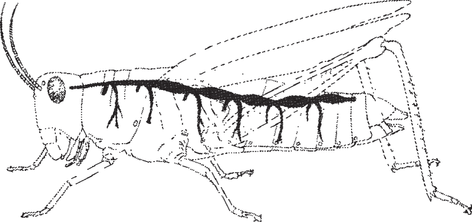
Figure 12.2 Open circulatory system of a grasshopper illustrating blood flow from the dorsal longitudinal vessel.
Metabolic wastes and dead cells have to go somewhere. Since they cannot be passed out through the tracheae, they are passed into the haemocoel, where some are consumed by haemocytes through phagocytosis. The larger particles, such as metazoan, or multicellular, parasites, cannot be consumed by the haemocytes; however, through a process called encapsulation, they are enclosed in a sheath or capsule, and may be stored or eventually passed out through the alimentary canal.
Those waste products that cannot be consumed, recycled, or encapsulated and stored somewhere enter the alimentary canal through the Malpighian tubules. These are blind sacs that absorb water, salt, and nitrogenous wastes at one end, where they are bathed in blood. At the other end of the sacs the contents are passed into the hindgut, from which they continue into the rectum. Here water is resorbed and the drier waste, or fecal matter, is excreted.
The blood of many arthropods and mollusks contains an oxygen-carrying pigment called hemocyanin, which is like the hemoglobin found in vertebrate blood. Unlike hemoglobin, hemocyanin never occurs in cells but is dissolved in the plasma of those animals that have it. Another difference between these two similar oxygen-carrying substances is that hemocyanin contains copper while hemoglobin contains iron.
CIRCULATION IN VERTEBRATES
Insect hearts and other invertebrate hearts are simpler versions of the more complex hearts that evolved in the vertebrates. In vertebrates, the blood is enclosed within a system composed of a heart and vessels. The heart pumps the blood into large arteries, which transmit oxygenated blood away from the heart, toward the cells and tissues throughout the body. The arteries branch into smaller arteries (arterioles), which branch into the smallest blood vessels, the capillaries (see Figure 12.3). It is through the thin walls of the capillaries that nutrients, gases, hormones, waste products, and other molecules are exchanged between the blood and the interstitial fluids that surround and bathe the body's cells. After passing through the capillaries, the blood passes into small veins, or venules, which merge into larger veins. Eventually, these return all the blood to the heart. Therefore, this system, which is known as the cardiovascular system, circulates blood throughout the body, bringing it to and from the capillaries, where the blood is able to accomplish its primary functions.
In addition to the similarities, there are differences that exist among the major groups of vertebrates (fish, amphibians, reptiles, birds, and mammals). From group to group, there is increasing separation between the left and right sides of the heart. This separation decreases the amount of mixing between the oxygenated, or oxygen-rich, blood returning from the gills or the lungs, and the deoxygenated, or oxygen-poor, blood returning to the heart from the tissues.
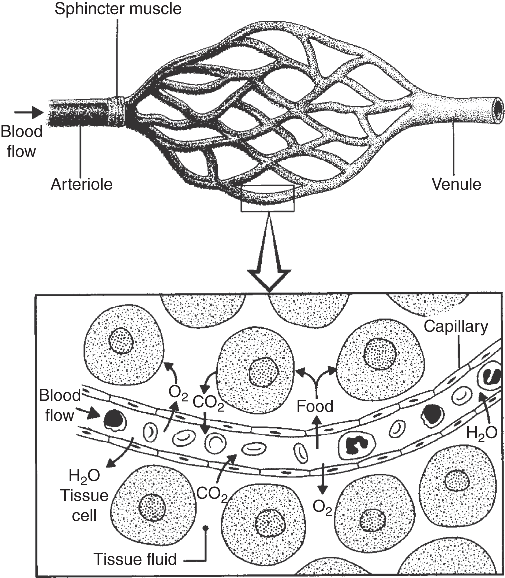
Figure 12.3 Rate of blood flow to capillary bed (top) is regulated by the constriction or relaxation of the arteriole's sphincter muscle. The exchange of water, nutrients, gases, wastes, and other chemicals occurs between the capillary's blood and the surrounding tissue cells (bottom).
Fish hearts have four internal chambers arranged linearly, with one-way valves between each chamber. The blood comes in one end and is pumped out the other. There is no mixing of oxygenated and deoxygenated blood in the fish heart because the aerated blood flows straight from the gills to the rest of the body without first returning to the heart (see Figure 12.1).
Amphibians have a heart composed of three different compartments or heart chambers, known as a three-chambered heart. Two of the chambers are called atria and one is called a ventricle. The deoxygenated blood flows from the body to the heart, first entering a chamber known as the right atrium, which acts as a storage chamber. The thin-walled atrium expands when the blood enters. Then the valves open to allow the blood to flow into the ventricle, a thicker chamber whose walls are made of muscle. When the muscle fibers contract, the blood is forced out through the pulmonary artery to the lungs. From there the oxygenated blood flows back to the heart, entering the storage chamber on the other side, called the left atrium. The blood flows back to the muscular ventricle, which then contracts and pumps the oxygenated blood to the rest of the body.
The only problem with this heart design is that there is some mixing of the oxygenated and deoxygenated blood in the ventricle, despite certain compensating devices, such as the grooves in a frog's heart that channel blood flow and minimize mixing.
Like the amphibians, reptiles have two atria. But rather than just one ventricle, there are two. These ventricles are not completely divided in most reptiles, although the ventricular division is complete in all bird and mammal hearts. This is why bird and mammal hearts are said to be four-chambered (two atria, two ventricles). It is the four-chambered heart that has enabled only birds and mammals to possess completely separate circulation paths for arterial and venous blood.
The human heart, with a right atrium and right ventricle, as well as a left atrium and left ventricle, essentially has two separate hearts inside one (see Figure 12.4). Both beat simultaneously. Blood circulates through a human in the following way. After flowing through all parts of the body except the lungs, the deoxygenated blood enters the heart via the right atrium. From the right atrium the blood is forced through the tricuspid valve into the right ventricle. The right ventricle pumps the deoxygenated blood through the pulmonary semilunar valve into the pulmonary artery, which divides into two main branches, each entering one of the lungs. In the lungs, the pulmonary arteries branch into arterioles, which connect with the capillary beds in the walls of the alveoli, the small air sacs throughout the lungs. This is where gas exchange occurs. Upon inhalation, the oxygen passes from the lungs into the thin layer of fluid lining the alveoli, and then it is picked up by the hemoglobin inside the red blood cells. Also at the alveoli, carbon dioxide is released from the blood to the air in the lungs, which is then exhaled.
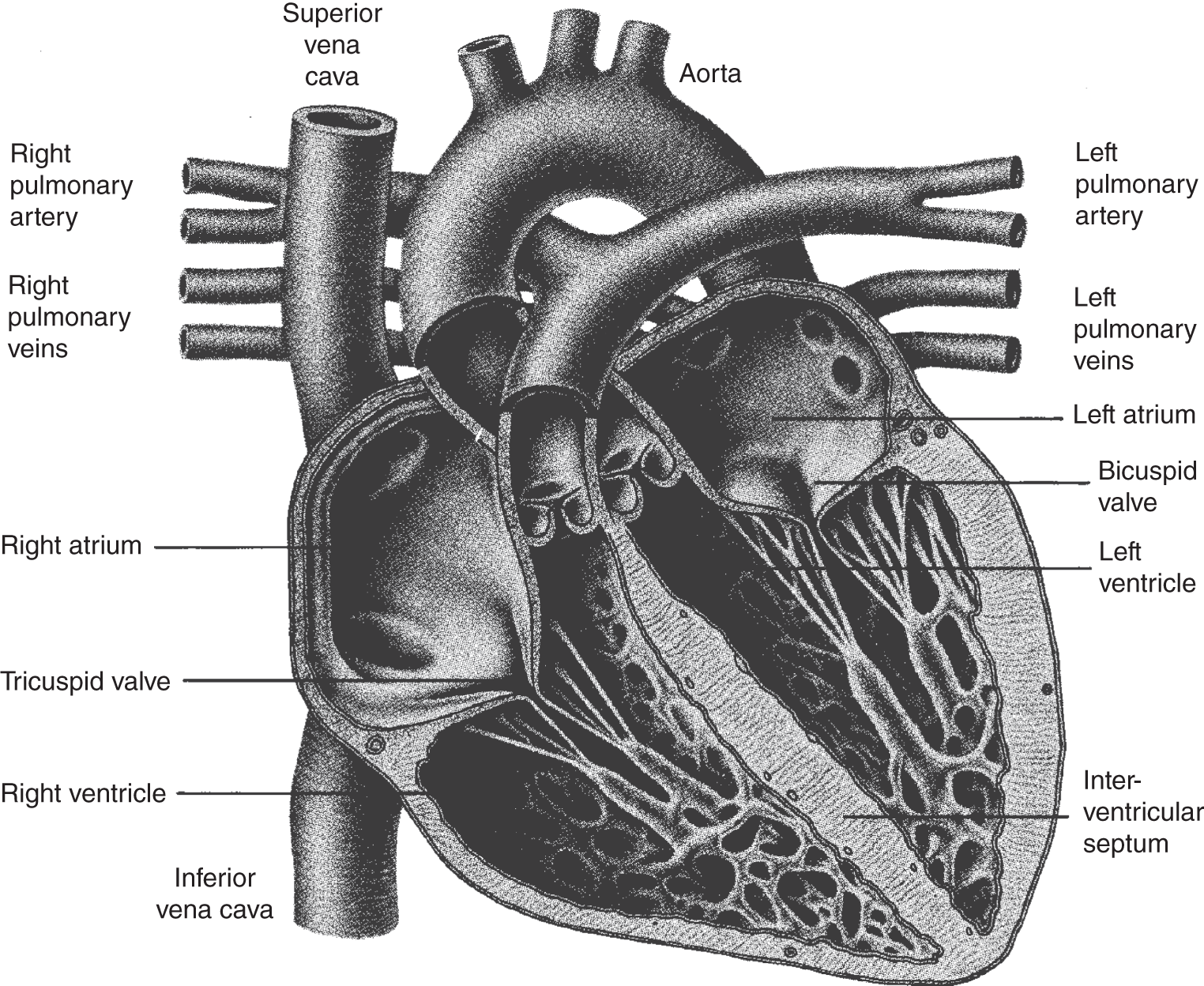
Figure 12.4 Cross section of a human heart showing the four chambers (two atria and two ventricles). The left ventricle has the thickest, most muscular walls, and this is the chamber responsible for pumping blood through the aorta.
From the capillaries in the lungs, oxygenated blood passes into small veins and then into the larger pulmonary veins, two of which lead from each lung back to the heart, where they empty into the left atrium. The blood then passes through the bicuspid valve (also called the mitral valve) and enters the left ventricle. The left ventricle contracts and pumps the blood through the aortic semilunar valve into the aorta, the artery that carries the oxygenated blood out of the heart and to all parts of the body except the lungs (see Figure 12.4).
Oxygenated blood passes through arteries that lead to smaller arteries, then to arterioles, then to capillaries, where oxygen, nutrients, hormones, and other substances move from the blood to the cells. It is through the capillary walls that waste products such as carbon dioxide and nitrogenous wastes are picked up. Other substances enter the blood through the capillary walls, such as hormones that are secreted by specific tissues and nutrients that are released from the intestine and liver. During one's life, due to the pumping action of the heart, blood continually circulates throughout the body, maintaining the constant exchange of materials.
After passing through the capillary beds in the tissues, the blood moves into venules and on to larger veins. These eventually empty into the anterior vena cava, which collects all the blood from the head, neck, and arms, and the posterior vena cava, which drains the blood from the rest of the body. The anterior and posterior vena cavas empty into the right atrium, completing one cycle.
The portion of the circulatory system that travels from the heart to the lungs and back is known as the pulmonary circulatory system. The other portion, which carries blood from the heart to the rest of the body and back, is called the systemic circulatory system (see Figure 12.5).
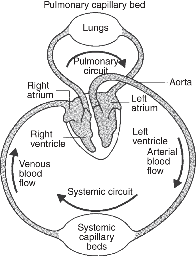
Figure 12.5 A highly simplified representation of the human circulatory system.
CAPILLARIES
Unlike the walls of the larger vessels, capillaries are only one cell thick. Made of endothelial cells, which together compose the endothelium, capillaries form a tube that encloses a passageway, or lumen, that is about 6 micrometers in diameter, wide enough to allow the passage of one red blood cell at a time. While the blood moves through the capillaries, gases, hormones, and other materials diffuse both in and out of the blood and the surrounding tissues through the cytoplasm of the endothelial cells, as well as through junctions between individual endothelial cells. In addition to diffusion, active transport and pinocytosis (cells engulfing small amounts of liquid or very small particles) probably help ferry dissolved materials across the endothelium.
LYMPHATIC SYSTEM
At the arteriolar end of the capillaries, the hydrostatic pressure exceeds the osmotic pressure, forcing some of the water and its dissolved gases, minerals, and protein molecules through the capillary wall, or endothelium. As the blood passes along inside the capillaries, the hydrostatic pressure gradually decreases to the point where it is less than the surrounding osmotic pressure. This allows much of the surrounding fluid that was previously forced out to reenter the blood capillaries. The relatively small amount of fluid that doesn't reenter is brought back to the bloodstream by the lymphatic system.
The lymphatic system drains protein-containing fluids that weren't reabsorbed from the blood capillaries. These escaped fluids become the lymph upon being absorbed by the lymph capillaries. Lymphatic capillaries are located in the interstitial spaces between cells where the fluids accumulate. These capillaries are slightly larger and more permeable than the blood capillaries. The lymph capillaries converge into larger lymph vessels, the lymphatics, which resemble veins but have thinner walls and more valves. The lymphatics just underneath the skin usually run parallel to veins, while those in the viscera generally follow arteries (see Figure 12.6).

Figure 12.6 Ninety percent of the blood plasma that leaks from the tissues passes back into the veins. The remaining 10% passes into the lymphatic system, where it circulates somewhat haphazardly, before being returned to the venous system.
In addition to returning fluids back to the bloodstream, the lymphatic system is also an important factor in the absorption of fat from the intestines. On the other hand, blood seems to be much better at absorbing sugars and amino acids from the intestines.
Located along the major lymph vessels is a series of nodes known as lymph nodes, or lymph glands. These are composed of a matrix of connective tissue harboring phagocytotic cells that filter out and cleanse the lymph by ingesting bacteria, cell fragments, and entire cells. The lymph glands then return these wastes to the blood, where they are carried to the lungs, kidneys, and sweat glands that eliminate them from the body. In addition, these wastes are detoxified when they pass through the liver. When the body is fighting infection, the lymph nodes become swollen and more sensitive.
In addition to filtering and processing lymph, the lymph nodes, along with other lymphoid tissues such as the spleen, thymus, and tonsils, are the sites where certain white blood cells, known as lymphocytes, are formed. The lymph is moved by a process similar to that which moves blood through the veins. A series of one-way valves work in conjunction with the pressure applied by the contraction of nearby skeletal muscles that constantly squeeze the lymph forward in a one-way direction. Some animals, though no mammals, have specialized pumping devices, or lymphatic hearts, located along the major lymphatic vessels to move the lymph along. Ultimately the lymph is emptied into the left and right subclavian veins, both located in the base of the neck.
KEY TERMS
| alveoli | chambers |
| anterior vena cava | closed circulatory system |
| aorta | deoxygenated blood |
| aortic semilunar valve | dorsal longitudinal vessel |
| arteries | encapsulation |
| arterioles | endothelial cells |
| bicuspid valve | endothelium |
| capillaries | haemocoel |
| capillary beds | haemocytes |
| cardiovascular system | haemolymph |
| heart chambers | pinocytosis |
| hearts | posterior vena cava |
| hemocyanin | pulmonary artery |
| interstitial fluids | pulmonary circulatory system |
| left atrium | pulmonary semilunar valve |
| left ventricle | pulmonary veins |
| lumen | right atrium |
| lungs | right ventricle |
| lymph | spleen |
| lymph nodes | systematic circulatory system |
| lymphatic system | thymus |
| lymphatics | tonsils |
| lymphocytes | tracheal system |
| Malpighian tubules | tricuspid valve |
| mitral valve | valves |
| open circulatory | veins |
| ostia | ventricle |
| oxygenated blood | venules |
| phagocytosis | white blood cells |
SELF-TEST
Multiple-Choice Questions
Open Circulatory Systems and Insect Blood
- Animals with open circulatory systems include the following:
- spiders and insects
- crabs and lobsters
- snails and clams
- a and b
- a, b, and c
- Animals with closed circulatory systems include the following:
- annelids
- fish, amphibians, and reptiles
- birds and mammals
- b and c
- a, b, and c
- Unlike most organisms, insects have a network of hollow tubes to carry air through pores perforating the body wall to the cells throughout their interior; these tubes are called ___________.
- exoskeleton
- tracheids
- tracheae
- ostia
- osteids
- Insects have blood that circulates through an open internal body cavity known as the ___________.
- haemolymph
- pericardium
- endocardium
- endometrium
- haemocoel
- Insect blood, the ___________, is composed of water, plasma, minerals, nutrients, waste products, and blood cells, or ___________.
- lymph, nematocysts
- lymphocytes, erythrocytes
- haemolymph, haemocytes
- thromboplasma, thrombocytes
- lymph, erythrocytes
- In insects, blood enters a ___________, which is often called the heart, through openings, or ___________.
- ventral tube, pores
- ventral longitudinal vessel, lenticels
- dorsal longitudinal vessel, ostia
- atrium, ducts
- ventricle, Malpighian tubules
- Waste products enter an insect's alimentary canal through blind sacs known as ___________.
- ostia
- Malpighian tubules
- pores
- lenticels
- dorsal longitudinal vessels
- The blood of many arthropods contains an oxygen-carrying pigment, ___________, which is like that found in vertebrate blood.
- hemoglobin
- hemocyanin
- gammaglobulin
- albumin
- anthocyanin
Circulation in Vertebrates
- Fish hearts have internal chambers arranged linearly.
- two
- three
- four
- five
- six
- Amphibians have a ___________-chambered heart, with ___________ atria and ___________ ventricles.
- two, one, one
- three, one, two
- three, two, one
- four, two, two
- four, one, three
- Reptiles have ___________ atrium(a) and ___________ ventricle(s).
- one, one
- two, one
- one, two
- two, two
- two, three
- The ventricular division is complete in the following hearts:
- birds
- mammals
- reptiles
- amphibians
- a and b
- In the human heart, when the blood is forced from the right atrium to the right ventricle, it moves through the ___________.
- tricuspid valve
- pulmonary semilunar valve
- bicuspid valve
- mitral valve
- aortic semilunar valve
- When the blood moves from the right ventricle into the pulmonary artery, it passes through the ___________.
- tricuspid valve
- pulmonary semilunar valve
- bicuspid valve
- mitral valve
- aortic semilunar valve
- The left ventricle contracts and pumps blood through another valve, called the ___________, into the artery called the ___________.
- mitral valve, right vena cava
- bicuspid valve, inferior vena cava
- mitral valve, superior vena cava
- aortic semilunar valve, aorta
- aortic semilunar valve, inferior vena cava
- After passing through the capillary beds in the tissues, the blood moves into venules and on to larger veins, which eventually empty into the ___________.
- anterior vena cava
- superior vena cava
- aorta
- a or b
- a, b, or c
- The portion of the circulatory system traveling from the heart to the lungs and back is known as the ___________, and the other portion of the circulatory system carrying blood from the heart to the rest of the body and back is called the ___________.
- systemic circulation, pulmonary circulation
- pulmonary circulation, systemic circulation
- cardiac circulation, somatic circulation
- cardio-pulmonary circulation, somatic circulation
- cardio-pulmonary circulation, cardio-systemic circulation
Capillaries and Lymphatics
- In humans and other vertebrates, the blood is enclosed within a system composed of a heart and vessels, where the heart pumps the blood into large ___________ that branch into smaller ___________ that branch into the smallest blood vessels, the ___________.
- arteries, arterioles, capillaries
- veins, venules, capillaries
- capillaries, arterioles, arteries
- capillaries, venules, veins
- a and c
- Capillary walls are the following number of cells thick:
- one
- two
- three
- four
- five
- The walls of the capillaries are made of ___________.
- endothelial cells
- endometrium cells
- endomysium cells
- endoneurium cells
- endocrine cells
- Escaped fluids from the blood capillaries become the ___________ upon being absorbed by the ___________.
- lymph, lymph capillaries
- blood, blood capillaries
- plasma, plasma capillaries
- plasma, lymphatics
- plasma, lymph nodes
- Located along the major lymph vessels is a series of nodes known as ___________.
- plasma nodes
- lymph nodes
- lymph glands
- a and b
- b and c
- Lymph glands do the following:
- filter out and cleanse the lymph
- harbor phagocytotic cells
- contain cells that ingest bacteria
- contain cells that ingest cell fragments and dead cells
- all of the above
ANSWERS
- e
- e
- c
- e
- c
- c
- b
- b
- d
- c
- d
- e
- a
- b
- d
- d
- b
- a
- a
- a
- a
- e
- e
Questions to Think About
- Describe the similarities and differences between closed and open circulatory systems.
- How does the insect tracheal system affect its circulatory system?
- What is the role of the Malpighian tubules, and how do they interact with the circulatory system of what kinds of animals?
- Compare and contrast several different types of hearts.
- Follow the entire route of blood through a vertebrate circulatory system.
- What is the function of a lymphatic system?