13
Blood
Like muscle, bone, cartilage, and nerves, blood is a type of tissue, a collection of similar types of cells and the associated intercellular substances that surround them. Tissues are categorized into four main types: (i) epithelium, (ii) muscle, (iii) nervous, and (iv) connective. Connective tissue includes blood, lymph, bone, and cartilage. In the case of blood and lymph, the base substance, or matrix, is a liquid. What differentiates these two from other tissue types is that they are not stationary. Blood is a fluid flowing through blood vessels throughout the body (lymph runs through the lymph vessels).
Blood accounts for about 8% of a human's total body weight, amounting to an average of 4 to 6 liters per adult (over a gallon), depending on individual size. Blood is thicker (more viscous) and slightly heavier than water. And, depending on the organism, blood is usually slightly warmer than the animal's body temperature. While the core body temperature of most humans is 37 °C (98.6 °F), their blood is about 38 °C (100.4 °F). Blood pH is slightly alkaline, ranging from about 7.35–7.45. Its salt (NaCl) concentration normally varies from about 85 to 90 ppt (parts per thousand), or two to three times the concentration of sea water, which is usually about 34 ppt.
Plasma is the fluid portion of both blood and lymph. Fifty to sixty percent of the blood volume consists of plasma. When the proteins involved in clotting are removed from blood plasma, the remaining liquid is called serum. The plasma, which is over 90% water, carries a variety of ions and molecules. In addition to salts and proteins, there are many nutrients such as amino acids, fats, and glucose. There are also dissolved gases such as carbon dioxide, as well as antibodies, hormones, enzymes, and certain waste products such as urea and uric acid. The relative amount of plasma in the blood depends upon the species, the sex, the organism's health when being examined, and a host of other variables. The remaining 40–50% of the blood volume is composed of cells and cell fragments that can be divided into three main categories: red blood cells, or erythrocytes; white blood cells, or leukocytes; and platelets, or thrombocytes, which are fragments of cells.
ERYTHROCYTES (RED BLOOD CELLS)
The cells in human blood are often referred to as corpuscles. Of these, 99% are red blood cells, which are involved in the transportation of oxygen throughout the body. Quite distinct in appearance, they are biconcave, flattened disks (see Figure 13.1). Birds and reptiles have red blood cells that retain their nucleus throughout the life span of each red blood cell, but when mammalian erythrocytes mature, their nuclei disintegrate.
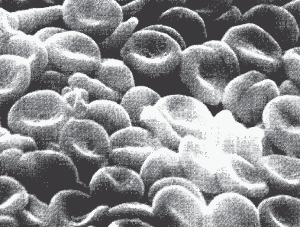
Figure 13.1 A mass of human red blood cells. Their flattened, indented shape enhances their flexibility and surface area/volume ratio, improving the efficiency with which they carry and exchange oxygen and carbon dioxide.
It has been surmised that the loss of the nuclei adds to the cells' oxygen exchange capacity by increasing the area/volume ratio. Area/volume means area to volume ratio. In other words, without a nucleus taking up space in the center of the cell, the cell has a biconcave design (both sides of the cell are indented). This indented surface on the outside, and less volume on the inside, increases the ability for gases to move in and out of the cell, because the volume of the cell is reduced, which has increased the surface-to-area ratio of each red blood cell. Without the nucleus taking up precious space inside the cell, the hemoglobin inside the red blood cells, which is the iron-containing protein that functions in oxygen transport, can fill almost the entire volume of all mammalian red blood cells, enabling them to interact more effectively in a deoxygenated medium. Without a nucleus, red blood cells only have an average life span of about three to four months, and then the red blood cells are consumed by the liver. New red blood cells are constantly produced by the red bone marrow. Birds have nuclei in their red blood cells, so they don't have to produce new red blood cells as often. As a result, they can have hollow bones, which makes them lighter, which has proven important in their ability to fly.
The concentration of cells and particulate matter in the blood plasma, known as the hematocrit value, is usually about 40–45%. If the concentration falls below this level, the individual is in danger of not having enough red blood cells or hemoglobin to supply the proper amount of oxygen to maintain cellular respiration. When the blood's concentration of red blood cells, or the red blood cell's concentration of hemoglobin, drops below normal, a person becomes anemic; the condition is known as anemia. Symptoms often include a general pallor (pale appearance) and lack of energy.
BLOOD TYPES
Everyone has highly individualized blood that is directly attributable to proteins and other genetically determined factors located both on the surfaces of red blood cells and in the plasma bathing the red blood cells. Of all the blood groups (there are over 300), the most widely known is the ABO group. When doctors first began giving blood transfusions (transferring blood from one person to another), it became apparent that while many patients' lives were saved, many other patients died almost immediately. It took years until it was learned that there were different blood types, some of which are incompatible.
When incompatible blood types are mixed, the blood of the patient receiving the transfusion clumps and clots, blocking capillaries, clogging organs, and causing strokes. We now understand that some erythrocyte surfaces contain genetically determined types of proteins known as the agglutinogenic proteins, which function as antigens, substances that stimulate the body to produce antibodies against it. Antibodies are proteins that inactivate or destroy antigens. Antigens, in the form of proteins on the red blood cell surface, and plasma antibodies both account for different blood types.
The ABO group depends on the combination of two alleles at a chromosomal locus, which can be AA, AB, BB, AO, BO, or OO. These are the result of two different antigens, A and B, and their absence, which is called O. Two AA or one A without another antigen (AO) produce type A blood. BB and BO produce type B blood. When A and B are found together, the blood is called type AB, and when neither occurs, the blood is type O.
Blood type A contains antibody B (anti-B); blood type B contains anti-A; blood type O contains anti-A and anti-B; and blood type AB contains neither anti-A nor anti-B. When antibodies are present in the plasma of one blood type, they will react with the antigens on the erythrocytes of another blood type, resulting in clumping. Therefore it is always best, when getting a blood transfusion, to obtain the same blood type of blood as your own. Because type O can be given to people with any blood type, it is often called the universal donor. Unless a transfusion is going to be massive, usually types A or B can be given to a person with type AB. Likewise, people with types A or B can receive blood from a person with AB. Table 13.1 shows the antigens and antibodies found in each blood type, and which types of blood can act as a donor during a blood transfusion.
Table 13.1 Compatibility of blood types.
| Antigens and Antibodies | ||
| Blood type | Antigens on the erythrocytes | Antibodies in the plasma |
| A | A | anti-B |
| B | B | anti-A |
| AB | A and B | None |
| O | None | anti-A and anti-B |
| Donor/Receiver Relationships | ||
| Blood type | Can receive blood from | Can act as donor to |
| A | A, O | A, AB |
| B | B, O | B, AB |
| AB | A, B, AB, O | AB |
| O | O | A, B, AB, O |
Rh
The Rh system derives its name from rhesus monkeys among which it was first studied. Like the ABO system, the Rh system is based on agglutinogens on the erythrocyte surface. People with erythrocytes having the Rh agglutinogens are called Rh+; those without it are Rh−. When blood is transfused, both the ABO blood types and the Rh type must be checked, because Rh incompatibility can produce a severe reaction that is sometimes is fatal.
Rh types are also important in regard to having children. When the father is Rh+, the mother is Rh−, and the fetus is Rh+, fetal Rh+ antigens may enter the mother's blood during delivery. The fetal Rh+ antigens will stimulate the mother's blood to make anti-Rh antibodies. Then, should the woman become pregnant again, her anti-Rh antibodies will cross the placenta, resulting in hemolysis, or the breakage of fetal erythrocytes, which will release the hemoglobin. This condition, known as erythroblastosis fetalis, used to kill the baby. Later, when a baby was born with such a condition, the doctors slowly removed its blood, replacing it with antibody-free blood. Today, erythroblastosis is routinely prevented by injecting an anti-Rh preparation into Rh− mothers right after delivery or an abortion. This injection contains antibodies that rid the mother's blood of the fetal agglutinogens. This prevents the mother's blood from producing its own antibodies and, thus, protects a fetus during the next pregnancy.
WHITE BLOOD CELLS (LEUKOCYTES)
In each organism the mature red blood cells are all basically identical to one another. This is not true of an animal's mature white blood cells (also called leukocytes). These are separated into several distinct categories. There are basophils, eosinophils, neutrophils, lymphocytes, and monocytes (see Figure 13.2). All have nuclei and none contains hemoglobin. Of the five types listed above, the first three develop in the red bone marrow. The other two develop from lymphoid and myeloid tissues.
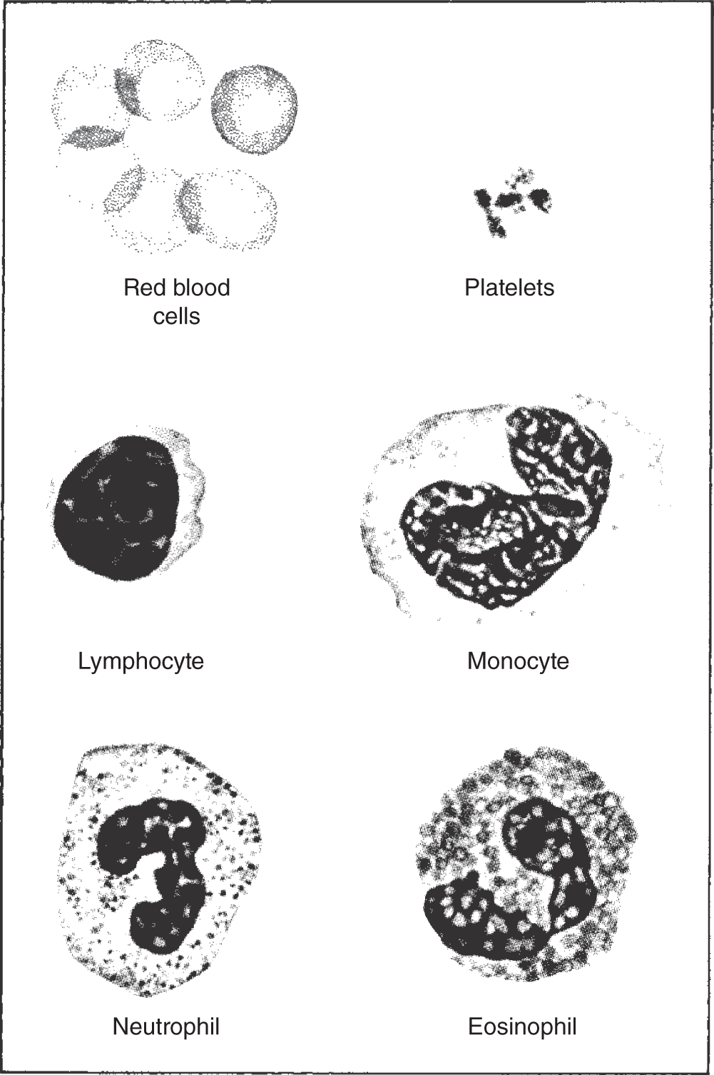
Figure 13.2 Blood cell types are classified according to the shape and size of the cell; absence, presence, shape, and size of the nucleus; and whether the cytoplasm is granular. These blood cell differences reflect their division of labor.
Generally, leukocytes combat inflammation and infection, either by ingesting bacteria and dead matter through phagocytosis or by producing chemicals that inactivate foreign bodies. Important factors in the immune system, lymphocytes are involved in producing antibodies to inactivate antigens. Most antigens are not produced by the body but are released by bacteria or other foreign microorganisms. However, when blood is transfused from one person to another, specific proteins in an erythrocyte's surface can be an antigen.
There are two basic types of lymphocytes. These are differentiated according to their function. Although they originate in the bone marrow, some lymphocytes, while developing, pass through the thymus gland. Therefore, they are called T cells. They specialize in combating foreign cells or tissues introduced into the body such as bacteria, fungi, and cells in transplanted organs. They are also involved in fighting cancer cells and in protecting cells from viruses. T cells are involved both in forming cell-killing chemicals and in attracting other white blood cells that engulf the foreign bodies encountered. They do not release antibodies.
The other lymphocytes, those that don't pass through the thymus gland during their developmental stage, are called B cells. When stimulated, they divide and form plasma cells that manufacture antibodies. When the antibodies encounter antigens, they attempt to destroy them. This is known as the antigen-antibody reaction. In addition to functioning inside the bloodstream, most leucocytes possess the ability to pass through minute spaces in the capillary walls and nearby tissues.
BLOOD TEST
A blood test often involves a differential count, which amounts to counting the number of each kind of white blood cell. A normal differential count contains 60–70% neutrophils, with considerably fewer of the other types of white blood cells. White blood cell counts that vary significantly from the norm often indicate the presence of an injury or infection. Such information is valuable to a doctor attempting to diagnose the cause or nature of a disorder from the symptoms.
Because the leukocytes are constantly involved in fighting unhealthy cells, such as those which may be cancerous, as well as bacteria and other foreign substances that enter the body through such points as the mouth, nose, and skin, leukocytes usually have very short life spans. Their normal life span is considerably shorter than that of the normal red blood cell, generally lasting only a few days. During a period of infection, they may survive only a few hours.
PLATELETS (THROMBOCYTES)
Platelets are small, disc-shaped, anucleate (without a nucleus) cells, or cell fragments, that are crucial to the clotting process. They help minimize the amount of bleeding when a blood vessel is cut or damaged by initiating the steps that result in coagulation. The damaged tissues and ruptured platelets both release substances known as thromboplastins. These are thought to initiate blood clotting in the presence of calcium ions.
Once released, the thromboplastins convert the plasma protein prothrombin into thrombin, which then converts fibrinogen, another plasma protein, into fibrin. Fibrin is fibrous and forms a meshwork that begins to shrink, squeezing out fluids to form a blood clot. These steps are summarized here:


Recently, the blood clotting process has been explained more fully. It has been shown that when blood comes in contact with cells outside the bloodstream, a protein molecule called tissue factor (TF), which is found on the surface of most cells outside the bloodstream, initiates the sequence of events leading to coagulation.
This tissue factor protein combines with a protein that circulates in the blood (factor 7). Instantly this becomes a new tissue factor, 7a (TF/7a), which initiates the clotting process by binding with tissue factor 10 and converting it into factor 10a. This binds to protein factor 5a. Then, these bind to the circulation protein prothrombin, creating thrombin molecules, which, in turn, create fibrin molecules, as explained above.
To aid the clotting process, damaged tissues release histamine, a chemical that expands the diameter of the capillaries and arterioles, thereby increasing the blood supply to the area and permitting more clotting proteins to leak out of the bloodstream into the damaged tissue. In addition to stopping the bleeding, clotting blocks bacteria and other foreign agents from entering a wound. The dead white blood cells that accumulate around the inflamed wound constitute the thick, creamy yellow fluid known as pus. If the wound is near the surface, the damaged area may eventually open, allowing the pus to drain. Otherwise the body slowly resorbs the dead cells.
IONS, SALTS, AND PROTEINS IN THE BLOOD
The inorganic ions and salts comprise approximately 0.9% of a human's blood plasma by weight. More than two-thirds of this is sodium chloride (NaCl, ordinary table salt). The most abundant positively charged inorganic ions (cations) found in the plasma are sodium (Na+), calcium (Ca2+), potassium (K+), and magnesium (Mg2+). The primary negatively charged inorganic ions are chloride (Cl−), bicarbonate (HCO−), phosphate ( and H2
 ), and sulfate (
 ).
).
The plasma proteins contribute another 8–9% of the plasma by weight. Most of these, such as the albumins, globulins, and fibrinogen, are synthesized by the liver. They are important in maintaining the blood's osmotic pressure, which is particularly important in the capillary beds. They are also crucial for maintaining the viscosity of the plasma, which is vital to the heart for maintaining a normal blood pressure.
HEARTBEAT
The heartbeat is initiated in its unique tissue, the nodal tissue, which has characteristics similar to both muscles and nerves, being able to contract like muscles and transmit impulses like nerves. There are two nodes, or areas composed of nodal tissue, in the heart. The heartbeat begins in the sinoatrial node (or S-A node), also called the pacemaker, which is located in the wall of the right atrium near the anterior vena cava. At regular intervals, a wave of contraction spreads from the S-A node across the walls of the atria to the atrioventricular node(A-V node), located in the lower part of the partition between both atria. As the wave of contraction reaches the A-V node, this node transmits impulses to the ventricles via a bundle of branching fibers known as the bundle of His. The impulse is slowed down in the bundle of His. During atrial relaxation, ventricular contraction occurs (see Figure 13.3).
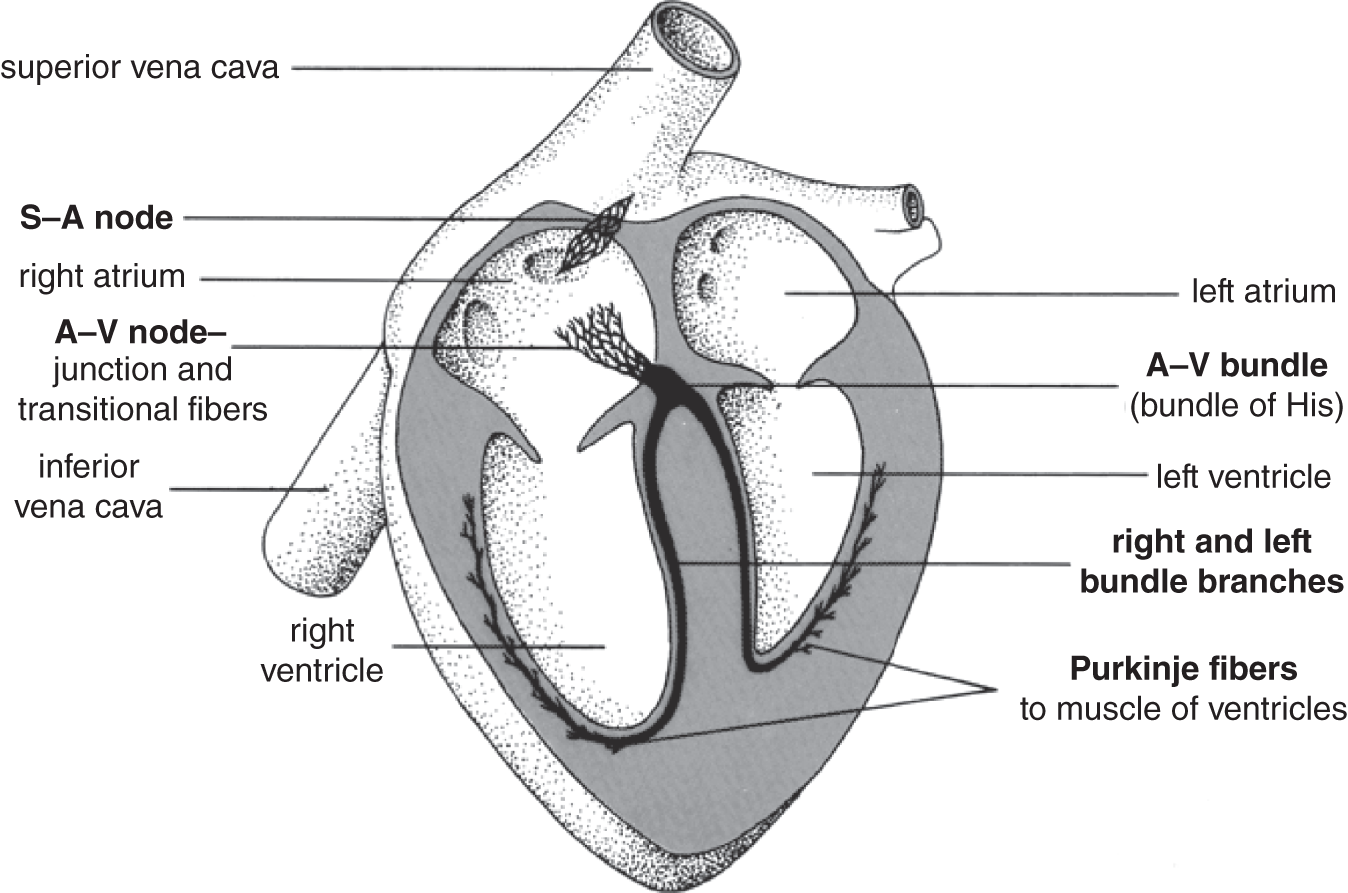
Figure 13.3 Pacemaker tissues in the human heart. Heartbeat is initiated by the S-A node, which stimulates the atria to contract. The A-V node coordinates these contractions with those of the ventricles.
These contractions occur at regular intervals, which are modified according to the physiology of the organism at the time. The speed at which these contractions occur is measured by counting the number of cycles per minute; this is known as the pulse rate, which in adult humans averages 70 beats per minute, though it varies considerably among individuals.
BLOOD PRESSURE
Blood pressure is defined as the pressure exerted by blood on the wall of any blood vessel. Two pressures are measured, that during the systole, when the heart's ventricles are contracting, and that during the diastole, when the heart's ventricles are relaxed (see Figure 13.4). Blood pressure is measured with a mechanical device called a sphygmomanometer. The average systolic blood pressure of a young adult male is about 120 mm of mercury; the average diastolic pressure is 80 mm of mercury. This is usually represented as 120/80. Young adult females have blood pressures that usually average about 10 mm of mercury less for both the systolic and diastolic pressures (110/70).
Blood pressure tends to increase with age; it may also increase due to certain inherited, dietary, and weight-related variables. Because accumulated evidence shows a correlation between high blood pressure and an increased risk of a heart attack, many doctors routinely take their patient's blood pressure to monitor any potential risk. Advice about diet and exercise may be given to patients who fall into the risk category. Certain medications have also been shown to be helpful.
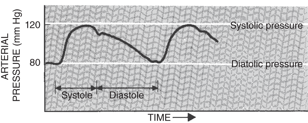
Figure 13.4 An EKG (electrocardiogram) tracing a heartbeat, measuring the systolic and diastolic pressure.
KEY TERMS
| agglutinogenic proteins | lymphocytes |
| anemia | matrix |
| anemic | monocytes |
| antibodies | neutrophils |
| antigen-antibody reaction | nodal tissue |
| antigens | nodes |
| atrioventricular node | pacemaker |
| A-V node | plasma |
| basophils | platelets |
| B cells | prothrombin |
| blood | pulse rate |
| blood pressure | pus |
| blood test | red blood cells |
| blood types | Rh system |
| bundle of His | S-A node |
| diastole | serum |
| differential count | sinoatrial node |
| eosinophils | sphygmomanometer |
| erythroblastosis fetalis | systole |
| erythrocytes | T cells |
| fibrin | thrombin |
| fibrinogen | thrombocytes |
| hematocrit value | thromboplastins |
| hemoglobin | tissue |
| hemolysis | tissue factor |
| histamine | universal donor |
| immune system | white blood cells |
| leukocytes |
SELF-TEST
Multiple-Choice Questions
Blood, Erythrocytes, Leukocytes, and Blood Types
- The fluid flowing through all the vessels of the body, excluding the lymph vessels, is the ___________.
- serum
- lymph
- blood
- plasma
- lumen
- Connective tissue includes the following types of tissues:
- nervous and connective
- epithelium and muscle
- muscle and nervous
- bone and epithelium
- lymph and cartilage
- ___________ is the fluid portion of both blood and lymph.
- plasma
- matrix
- ground substance
- water
- urea
- When the proteins involved in clotting are removed from blood plasma, the remaining liquid is called ___________.
- water
- urea
- ground substance
- serum
- lumen
- In addition to the plasma, the remaining 40–50% of the blood volume is composed of the following:
- thrombocytes and platelets
- erythrocytes and leukocytes
- white blood cells and red blood cells
- leukocytes
- all of the above
- The concentration of cells and particulate matter in the blood plasma is known as the ___________.
- erythrocyte count
- corpuscle count
- hemoglobin level
- anemia analysis
- hematocrit value
- When the blood's concentration of red blood cells or the red blood cell's concentration of hemoglobin drops below normal, a person becomes ___________.
- anemic
- weak
- pale
- all of the above
- none of the above
- The surfaces of erythrocytes contain genetically determined types of proteins that are responsible for the two major blood group classifications, which are:
- ABO and Rh
- Type A and Type B
- Type AB and O
- Rh− and Rh+
- Type O and Rh−
- Blood Type O will not agglutinate with any of the other blood types and therefore is known as the ___________.
- universal donor
- Blood Type A
- Blood Type B
- Blood Type AB
- Blood Type ABO
- Lymphocytes are involved in producing certain proteins known as ___________ inactivate foreign chemicals known as ___________ .
- antigens, antibodies
- antibodies, antigens
- basophils, eosinophils
- neutrophils, monocytes
- none of the above
Platelets, Ions, Salts, and Proteins in the Blood
- Both cut or damaged tissues as well as ruptured platelets release substances involved in clotting known as ___________.
- thrombins
- prothrombins
- fibrinogens
- calcium ions
- thromboplastins
- The presence of ___________ ions is needed for thromboplastins to initiate the blood clotting process.
- magnesium
- cadmium
- lead
- calcium
- chloride
- Once released, the thromboplastins convert the plasma protein ___________ into ___________ .
- fibrin, fibrinogen
- prothrombin, thrombin
- thrombinogen, thrombin
- thrombin, prothrombin
- profibrinogen, fibrinogen
- Thrombin converts another plasma protein into ___________ .
- fibrin
- histamine
- calcium ions
- prothrombin
- thromboplastin
- To aid the clotting process, damaged tissues release ___________, a chemical that expands the diameter of the capillaries and arterioles, thereby increasing the blood supply to the area, permitting more clotting proteins to leak out of the bloodstream into the damaged tissue.
- histamine
- fibrin
- calcium ions
- thrombin
- thromboplastin
- The most abundant positively charged inorganic ions found in the plasma include:
- sodium
- calcium
- potassium
- magnesium
- all of the above
- The primary negatively charged inorganic ions found in the human blood plasma include the following:
- chloride
- bicarbonate
- phosphate
- sulfate
- all of the above
Heartbeat and Blood Pressure
- The heartbeat begins in the ___________.
- sinoatrial node
- S-A node
- pacemaker
- all of the above
- none of the above
- A wave of contraction spreads from the S-A node across the walls of the atria to the ___________, which transmits impulses to the ventricles via ___________.
- atrioventricular node, a bundle of branching fibers
- A-V node, bundle of His
- atrioventricular node, bundle of His
- A-V node, a bundle of branching fibers
- all of the above
- The blood pressure exerted on the wall of any blood vessel when the ventricle is contracted is the ___________, and the pressure exerted on the wall of any vessel when the heart is relaxed is the ___________.
- systolic pressure, diastolic pressure
- diastolic pressure, systolic pressure
- sphygmomanomic pressure, blood pressure
- ventricular pressure, aventricular pressure
- ventricular pressure, auricular pressure
ANSWERS
- c
- e
- a
- d
- e
- e
- d
- a
- a
- b
- e
- d
- b
- b
- a
- e
- e
- d
- e
- a
Questions to Think About
- Analyze, compare, and contrast the contents of the blood of a vertebrate and an insect.
- How does blood clot?
- How does a heart beat?
- What is blood pressure?