Bronchopulmonary dysplasia (BPD) is one of the most significant medical complications in preterm infants, and pulmonary microvascular injury is associated with the pathogenesis of BPD [1]. Furthermore, impairments of developing pulmonary vasculature may cause secondary pulmonary hypertension (PH), which contributes significantly to morbidity and mortality among preterm infants [2]. To characterize the mechanisms of pulmonary vascular disease resulting from BPD, we studied the ultrastructural changes affecting pulmonary microvasculature.
Newborn ICR mice were exposed to 85% hyperoxia or normoxia for 14 days, and then normal air replacement conditions for the following 7 days. At postnatal day (P)14 and P21, lungs were harvested for ultrastructural examination and assessment of PH. To this end, we studied ultrastructural changes in pulmonary microvasculature, such as endothelial cells (ECs) and blood-air barriers (BABs), and examined the structure of pulmonary arteries associated with terminal and respiratory bronchioles as well as right ventricular hypertrophy (RVH).
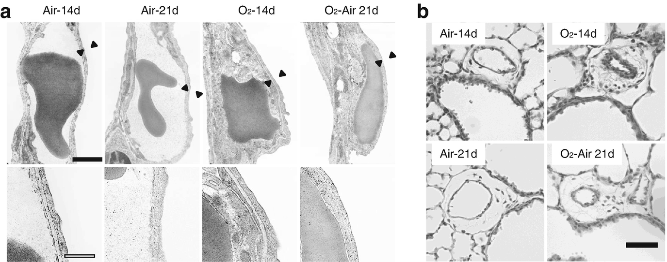
(a) Pulmonary capillaries and blood-air barriers in Air-14d, Air-21d, O2-14d, and normal air replacement conditions after hyperoxia (O2-Air-21d). Scale bar black, 1.0 μm; white, 0.35 μm. Black arrowheads denote the parts of BABs magnified in the lower panels. (b) H&E-stained lung sections indicating the thickness of pulmonary arteries associated with terminal and respiratory bronchioles. Scale bar (black), 50 μm
Pulmonary capillaries in the injured newborn mouse lung presented thick BABs consisting of ECs with abnormal morphology, which persisted following subsequent exposure to normal air replacement conditions. These ultrastructural changes might cause abnormal pulmonary microcirculation and finally lead to secondary PH in BPD.
This study was supported by the Ministry of Education, Culture, Sports, Science and Technology (MEXT) KAKENHI (Grant Number 16K10111).

Open Access This chapter is licensed under the terms of the Creative Commons Attribution 4.0 International License (http://creativecommons.org/licenses/by/4.0/), which permits use, sharing, adaptation, distribution and reproduction in any medium or format, as long as you give appropriate credit to the original author(s) and the source, provide a link to the Creative Commons license and indicate if changes were made.
The images or other third party material in this chapter are included in the chapter's Creative Commons license, unless indicated otherwise in a credit line to the material. If material is not included in the chapter's Creative Commons license and your intended use is not permitted by statutory regulation or exceeds the permitted use, you will need to obtain permission directly from the copyright holder.