Inositol triphosphate receptor (IP3R) is an intracellular Ca2+ release channel located on the membrane of the sarco/endoplasmic reticulum (SR/ER), a major intracellular storage site for Ca2+. There are three subtypes of IP3R (IP3R1, 2, and 3), each of which is known to have versatile roles in the pathophysiology of many organs [1].
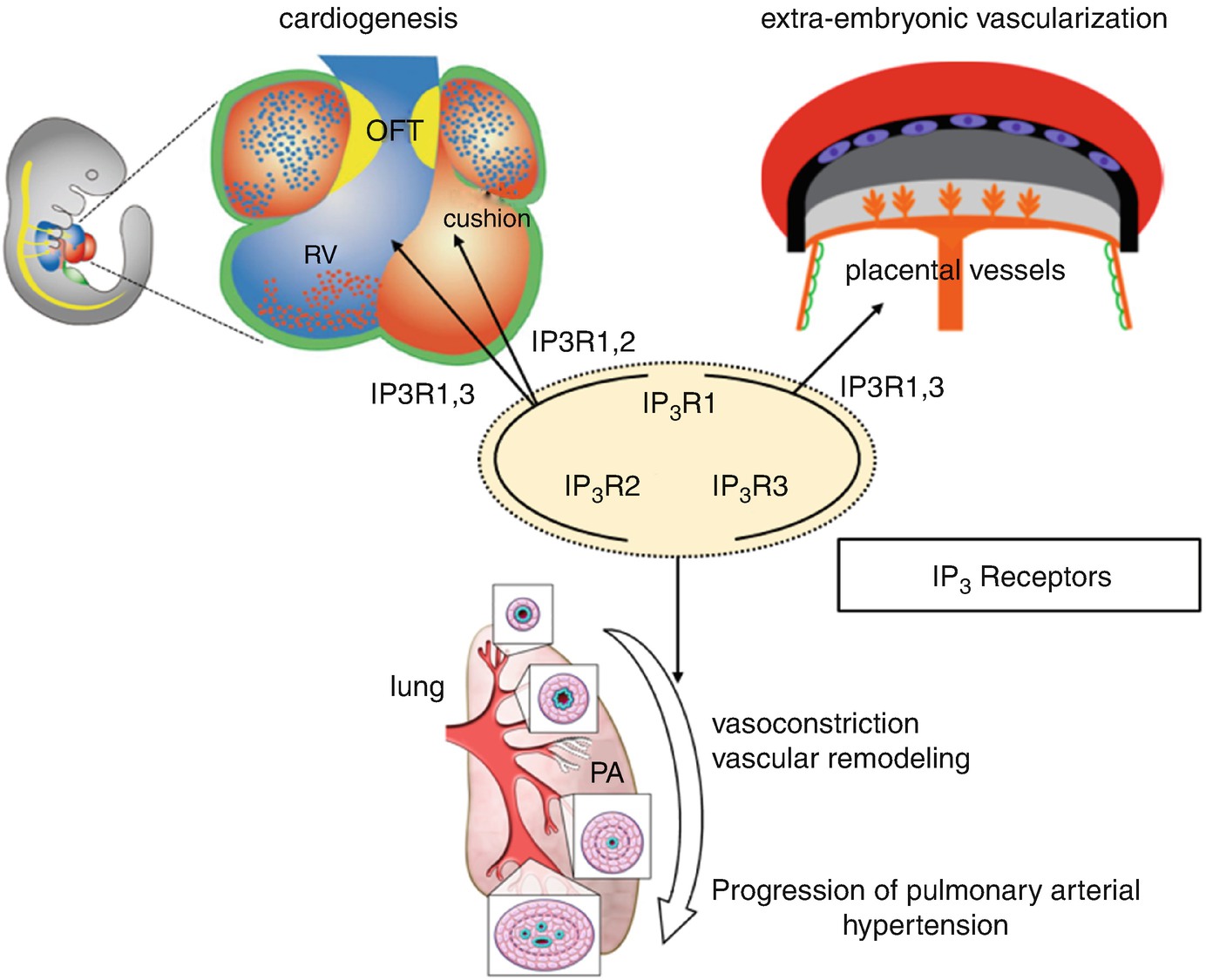
The role of IP3Rs for cardiogenesis, extra-embryonic vascularization, and suppression of pulmonary arterial hypertension. RV right ventricle, OFT outflow tract, PA pulmonary artery
Recently, we found the specific expression of IP3R in the pulmonary artery smooth muscle cells (PASMCs) of murine lungs. Although there are some reports that implicated IP3R1 in vasoconstriction or remodeling in vascular smooth muscle cells [5], the precise role of IP3Rs in PASMCs remains unknown. Since intracellular Ca2+ signaling in PASMCs is a major regulator for pulmonary vasoconstriction and remodeling, its dysregulation leads to the progression of pulmonary arterial hypertension (PAH). It was reported that store-operated Ca2+ (SOC) entry was upregulated in PASMCs with PAH patients. Our results suggest that intracellular Ca2+ depletion through Ca2+ release by IP3R from the SR/ER may lead to the opening of SOC channels, a mechanism potentially involved in the pathophysiology of PAH (Fig. 13.1) [6].
This work was supported by Acterion Academia Prize 2015 to A.S. and JSPS KAKENHI Grant Numbers JP19390288 to H.Y. and JP26461619 to K.U.

Open Access This chapter is licensed under the terms of the Creative Commons Attribution 4.0 International License (http://creativecommons.org/licenses/by/4.0/), which permits use, sharing, adaptation, distribution and reproduction in any medium or format, as long as you give appropriate credit to the original author(s) and the source, provide a link to the Creative Commons license and indicate if changes were made.
The images or other third party material in this chapter are included in the chapter's Creative Commons license, unless indicated otherwise in a credit line to the material. If material is not included in the chapter's Creative Commons license and your intended use is not permitted by statutory regulation or exceeds the permitted use, you will need to obtain permission directly from the copyright holder.