GFRA2 facilitates the isolation of CPs by fluorescence-activated cell sorting from differentiating mouse and human pluripotent stem cells. If utilizing pan-mesoderm marker PDGFRA and hemangioblastic marker KDR in conjunction with GFRA2, multipotent CPs giving rise to cardiomyocytes, smooth muscle cells and endothelial cells are identified as GFRA2+/PDGFRA+/KDR+ whereas CPs already committed only to cardiomyocytes are identified as GFRA2+/PDGFRA+/KDRneg.
In loss-of-function of Gfra2, the functional redundancy of GFRA1 compensated for the cardiac GFRA2 signal in the mutant mouse embryos. Compound mutant of Gfra1/2 showed non-compaction cardiomyopathy due to the down-regulation of NOTCH signal in the developing heart. Cardiac GFRA2 signal is distinct from the canonical pathway that depends on the RET tyrosine kinase and its established ligands.
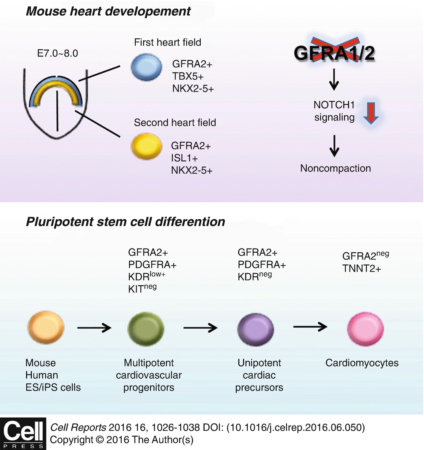
GFRA2 is a specific marker for CPs which governs myocardial compaction. Upper case: A signal transduction via GFRA1/2 is upstream of NOTCH signal that is indispensable for the myocardial compaction process. Lower case: GFRA2 is a specific surface antigen to identify CPs from differentiating pluripotent stem cells in mice and humans. This illustration is cited from Ishida, H et al. Cell Rep. 2016 Jul 26;16(4):1026–38, and is licensed under the Creative Commons Attribution 4.0 International License. To view a copy of this license, visit http://creativecommons.org/licenses/by/4.0/ or send a letter to Creative Commons, PO Box 1866, Mountain View, CA 94042, USA
Collectively our findings establish a platform for investigating the biology of CPs as a foundation for the future development of CP transplantation therapies for treating heart failure.
This work was supported by MRC New Investigator Research Grant G0900105 and MRC Research Grant MR/J007625/1 to K.Y., Japan Heart Foundation/Bayer Yakuhin Research Grant Abroad and JSPS (Japan Society for the Promotion of Science) Postdoctoral Fellowship for Research Abroad to H.I. We thank Ruchaya PJ at Kings College London for the support in writing.

Open Access This chapter is licensed under the terms of the Creative Commons Attribution 4.0 International License (http://creativecommons.org/licenses/by/4.0/), which permits use, sharing, adaptation, distribution and reproduction in any medium or format, as long as you give appropriate credit to the original author(s) and the source, provide a link to the Creative Commons license and indicate if changes were made.
The images or other third party material in this chapter are included in the chapter's Creative Commons license, unless indicated otherwise in a credit line to the material. If material is not included in the chapter's Creative Commons license and your intended use is not permitted by statutory regulation or exceeds the permitted use, you will need to obtain permission directly from the copyright holder.