2
Germs
What They Are and How They Interact with Humans
Germs are microbes—living creatures of extremely small size that, like all living organisms, eat, breathe, excrete, and multiply. When we speak of “germs,” we generally are referring to unicellular microbes that cause disease in humans and other creatures. There is one exception to this: the virus, which is only a portion of a cell and, as such, is technically not considered a living organism at all.
Germs can be broken down into four basic categories:
- Parasites
- Bacteria
- Fungi and yeasts
- Viruses
We’ll talk about each of them in turn later in this chapter. For now, let’s look at the way in which germs interact with the world and, most particularly, with human beings.
TINY BEINGS
Germs are so small that they are invisible to the naked eye. We need a microscope with great magnifying power to see them. To give an approximate idea of their size, let’s compare them to a hair. Parasites (the largest germs) are on average ten times smaller than the thickness of a hair, bacteria and molds are one hundred times smaller, and viruses are between one thousand and ten thousand times smaller. In other words, on a field that is 1 millimeter in size, you can lay out, side by side, ten hairs, one hundred parasites, one thousand bacteria or molds, and between ten thousand and one hundred thousand viruses.
Human cells on average have a diameter similar to that of parasites. Our cell wall thickness is smaller than any germ except for the smallest viruses. Even smaller in size are organic molecules like sucrose, for example (which is ten times smaller than the thickness of our cell walls and the smallest viruses), and atoms, of course. The hydrogen atom is ten times smaller than the sucrose molecule.

Figure 1. Germ dimensions (from largest to smallest)
When we talk of “germs,” we mean those microbes that negatively affect us. However, these represent only a very small minority of all microbes. For example, there are thousands of different kinds of bacteria, but only fifty-five kinds are known to make us sick. The same holds true for parasites, fungi, and viruses: thousands of different kinds exist, but only a small proportion of them have a pathogenic effect on human beings.
Microbes inhabit our entire environment. They live in the earth of the fields, meadows, and forest undergrowth. They are in the water of the streams, rivers, lakes, and swamps. They have even been discovered in the ice of the ice pack and in the depths of the sea. They are in the air we breathe. They can be found everywhere; no place seems to be spared their presence. They live even inside plants and animals. We ourselves, as human beings, have numerous nonpathogenic microbes on our skin and in our intestines (our intestinal flora), mouth, nasal passages, respiratory tract, and more.
THE ROLE OF MICROBES
Why are microorganisms so numerous and omnipresent in nature? The reason is that these microbes play a fundamental role in the manufacture and decomposition of living material.
All matter is constructed from the fundamental elements of the universe, as depicted on Mendeleev’s periodic table of elements. These basic substances have characteristics that are too crude to form plant and animal tissues, however. A dynamizing and vivifying labor must first be performed so that they can combine with other elements in order to acquire the qualities necessary to make the transition into constructing living matter. In other words, they must go from the inorganic state to the organic state. This labor is performed by germs.
The bacteria that produce nitrates (azotobacter), for example, transform the nitrogen in the air into a more sophisticated substance that plants can assimilate. These bacteria can be found in a free state in the soil (which the nitrogen penetrates when it rains), but some plants offer them a haven. The bumpy nodes on the roots of leguminous plants like alfalfa and beans are full of these nitrogen-fixing bacteria.
Similar processes take place for the preparation and manufacture of all the other mineral substances necessary for organic life. In addition to the nitrogen cycle, there is one for sulfur, one for phosphorus, and so on. For each of these cycles, other bacteria—or specialized microorganisms—are active. In this respect, microbes could be compared to tiny, almost invisible workers whose activity is essential for preparing inert matter so that plant and animal bodies can take form from it. These bodies in fact entirely depend on the microbial world for their existence.
In natural cycles, the manufacture, formation, and multiplication of matter is followed by the stage of decomposition. Dead plants and animals contain in their tissues a host of substances that can be reutilized if they are freed from the organic structures that hold them. Here again, it is the job of microbes to break down the plant and animal tissues so they can be brought down to their basic elements.
The transformational powers of microorganisms are well known to human beings, who use some of them to make certain food products. It is, in fact, bacteria and fungi that transform milk into yogurt or cheese, grape juice into wine, wine into vinegar, flour into bread dough, malt and hops into beer, and so forth.
There are thousands of different kinds of microbes that can be divided into subcategories, each containing a countless number of individuals. This enormous populace inhabits the entire surface of the earth and everything that is found on it. Stepping in at different stages of the manufacturing and decomposing processes of matter, extremely different types of germs are close neighbors in a single environment.
For example, there are more than four hundred different kinds of microbes in the intestinal environment of the human being. Their relations with one another are most often harmonious, especially when they form crews of “workers” on the same line of work. There are times, though, when this harmony is broken. The struggle for territory and food—the same kind of struggle that can be seen among plants and animals—also takes place among these infinitely tiny creatures. At such times some microbial populations will be decimated by other ones, which will then take their place.
At other times a change in external conditions—humidity, temperature, and so forth—can have an adverse impact on the living environment of one kind of germ, and another kind can then take advantage of this and move in.
THE MICROBE–HUMAN RELATIONSHIP
The relationship between microbes and higher life-forms—animals and human beings, for example—can be beneficial or harmful.
The relationship is beneficial when the microbes and the organism that houses them can help each other mutually. This is the case with resident microbes, which colonize a body for the long term, as opposed to those that enter it by accident. The microbes of the intestinal flora are permanent residents. In this association, they provide a service to the body by participating in various physiological processes: digestion of cellulose, synthesis of various vitamins and amino acids, and so on. And the body offers microbes an environment that is favorable for their development, as the hot, moist intestinal milieu has a wealth of nutritious substances that they can eat. This relationship is described as symbiotic, and the microbes that participate in it are called saprophytes.
In a harmful relationship, there are no exchanges, support, or aid between the microbes and the body that houses them. To the contrary, the microbes colonize the organism and exploit the resources it offers them without giving it anything in return. In addition, the body suffers from all the negative consequences of their presence: injury to the tissues they inhabit and poisoning of the body from the toxins the microbes secrete.
This kind of relationship, in which one of the protagonists lives entirely at the expense of the other, is a parasitical relationship. A parasitical relationship cannot last long without endangering the survival of the host. The microbes involved therefore do not take up permanent residence and are present only temporarily. All the germs that make us sick fall into this category.
THE HISTORY OF THE DISCOVERY OF GERMS
Today, even children know that a germ is an extremely tiny pathological organism that makes us sick when it enters our body. But this knowledge is quite recent. For most of history, humans knew nothing about the existence of germs. Of course, they noted the contagious nature of some diseases and suspected that a “something” was being transmitted, but as they were unable to see this “something” with the naked eye, they had no idea what it could be.
The notions our ancestors had of this “something” were vague and sometimes far-fetched, but when put back in the context of their times, they reveal concepts that are surprisingly close to reality. In antiquity, infectious diseases and epidemics were sometimes attributed to miasma, meaning impurities (dirty things) that came from putrefying plant and animal wastes or from stagnant water—in which there are teeming populations of many kinds of germs, actually. People also spoke of putrid gases emanating from this putrefaction, or of “corrupted air,” to explain the contagious nature of certain diseases—which is actually a fair description of airborne microbes.
“Little winged worms” were mentioned as a probable cause of infections by Ulrich von Hutten around 1519, whereas the Swiss doctor Paracelsus (1493–1441) mentioned “little living germs.” Other physicians hypothesized about animalcules (“tiny animals”) or “vermicular germs.” But these concepts were too novel and revolutionary for the times and were therefore rejected by the majority of physicians.
With the invention of the microscope in the seventeenth century, what had been until then only a vague hypothesis, though adhering fairly closely to reality, would be confirmed. The first direct observation of a germ most likely took place in 1680, which is to say a little more than 330 years ago. Using a microscope with several lenses, Antonie van Leeuwenhoek saw protozoa (the largest germs) as well as bacteria, which he described as “very little living animalcules, very prettily a-moving” and called infusoria.
The manner in which germs reproduce would be discovered forty years later. In 1720 Italian scientist Lazzaro Spallanzani isolated a germ in a drop of water and watched it divide in its environment to give birth to new germs.
The construction of increasingly sophisticated microscopes and the perfection of techniques for cultivating germs in a liquid environment would make it possible to obtain many more precise observations. Proof that certain germs were the source of different diseases was established in 1865 by two researchers pursuing their efforts separately: the Frenchman Louis Pasteur (1822–1895) and the German Robert Koch (1843–1910). They extracted germs from the blood of diseased animals and cultivated them in the laboratory. Once they had isolated several specimens, they used them to inoculate healthy animals. These animals then contracted the disease. These studies demonstrated the causeand-effect relationship between the presence of germs and the infection.
Systematic studies of the germs responsible for infectious diseases were then performed. In this manner staphylococci were discovered in 1878, streptococci in 1879, and the tuberculosis and cholera bacilli in 1882, followed by the diphtheria bacterium, the rabies virus, and so on.
PATHOLOGICAL GERMS
What are the germs responsible for illnesses in humans beings, and how are they different from other microbes? To explore these questions, we will successively examine the four major groups they form: the parasites, the bacteria, the fungi, and the viruses.
Parasites
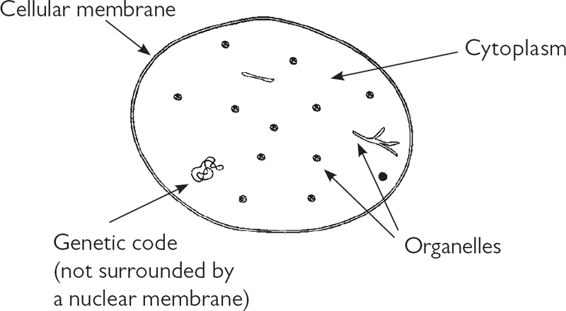
When we speak of parasites in this book, we are referring primarily to protozoans, unicellular organisms of the protist family (they should not be confused with the multicellular creatures like ticks, fleas, and intestinal worms that are also called parasites). They form the largest group of the germs that attack human beings. They consist of a protective envelope (cellular membrane) containing a fluid (cytoplasm) in which all the organs (organelles) responsible for breathing, digestion, energy production, and so on are immersed, as well as a command center (the nucleus of the cell).

Figure 2. The protozoan cell
To develop and, more importantly, multiply, some protozoans temporarily or permanently need a higher organism to house them; hence the name parasites. These higher organisms offer them a favorable environment and a generous source of food. Unfortunately for the host organism, “food” for a protozoan is not just the food its host consumes but also the host’s own tissues and secretions.
Parasites will not colonize just any animal. Each variety attacks a specific host species that offers the specific living conditions the invader requires. This means that a parasite that is extremely dangerous for one animal species will not be dangerous for another species, or it might attack human beings but not animals. Similarly, parasites will colonize not all the tissues in the organs of the host body but only a select few (known as target organs).
The protozoan Trichomonas vaginalis, for example, lives in vaginal secretions and causes vaginitis. Trypanosomes, which are transmitted by the tsetse fly, poison the human nervous system with their metabolic wastes and cause sleeping sickness.
Among the protozoans that are implicated in human disorders we also find the plasmodia and certain amoebas. Plasmodium, the agent responsible for malaria, is a parasite of red blood cells. It multiplies a thousandfold within each blood cell, causing them to explode. When this phenomenon takes place in many red blood cells at the same time, the bloodstream is suddenly overtaken by their decomposing cadavers and wastes. This causes fever, characterized by spikes of extremely high temperatures that correspond to the parasites’ reproductive cycle. The parasites that are freed during the crisis state then colonize more red blood cells, which explode in turn, starting the entire process over again. Moreover, the destruction of a large number of red blood cells is not inconsequential for the composition of the blood: the sharp drop in the proportion of red blood cells will cause serious anemia.
Amoebas are protozoans that live in both fresh and salt water. The majority are harmless to humans, but one kind, Entamoeba histolytica, is responsible for a form of dysentery characterized by painful and bloody diarrhea. E. histolytica is primarily found in the world’s hotter regions.
| PARASITICAL DISEASES | |
| PROTOZOAN | DISEASE |
| Entamoeba histolytica amoeba | Amebiasis (amebic dysentery) |
| Plasmodium | Malaria |
| Trichomonad | Vaginitis, urethritis |
| Trypanosome | Sleeping sickness |
Bacteria
In structure, bacteria are even less complex than protozoans. While they are constructed on the same basic model as all cells, bacteria still do not possess as many organelles as the parasites. In addition, the nucleus of the bacterial cell is not surrounded by a protective membrane, and its genetic code consists of only one DNA chromosome.

Figure 3. The bacterial cell
The thousands of various kinds of bacteria in existence can be divided into major families depending on their shape. The bacteria with a long rod shape are called bacilli, those that are spherical are called cocci, and those that have a spiral, or helical, shape are called spiral. Within the bacillus group we find bacilli arranged in pairs and in chains as well as bacteria that are curved and have tails. The coccus group appears in the shape of a single seed or as pairs, strings, or clusters. Spiral bacteria can be subclassified by the number of twists, cell flexibility, and motility.
| BACTERIA SHAPES | ||
| COCCI (SPHERICAL SHAPE) | BACILLI (ELONGATED ROD SHAPE) | SPIRAL (HELICAL SHAPE) |
|
Micrococci (in seeds) Pneumococci (in pairs) Streptococci (in strings) Staphylococci (in clusters) |
Filamentous bacteria (with a tail) Vibrio (curved) |
Spirilla (thick, rigid spirals) Spirochetes (thin, flexible spirals) |
Another classification is based on the degree to which bacteria are receptive to a violet dye known as a Gram stain (named for the inventor of the test, Danish scientist Hans Gram). Some bacteria take on an intense violet color when exposed to the stain; they are called Gram positive. Other bacteria turn pink; they are called Gram negative. Which reaction a bacterium has provides useful information about the structure of its outer membrane and, therefore, whether it will have more or less receptivity to various kinds of antibiotics.
A bacterial cell’s membrane has the distinctive feature of being extremely extensive in proportion to its volume. In fact, the membrane is nine million times larger than the cell’s volume. In comparison, the skin surface of a human being is only twenty times larger than the volume of the body. This large expanse is both to the advantage and to the detriment of bacteria. The advantage comes from the fact that bacteria root themselves into the environment they intend to exploit by surface contact, and with a greater surface area, they have a greater ability to take root. The drawback is that having a larger surface area makes it harder to defend themselves from attack—from antibiotics, for example.
As was the case for the protozoans, different bacteria have a preference for certain species, tissues, and organs. Consequently it is these organs and tissues that they will colonize and damage. But many bacteria produce toxins that are transported through the body by the bloodstream or the lymph flow, and these toxins can poison tissue that is quite distant from the area the bacteria have colonized.
The bacillus Bordetella pertussis, for example, colonizes the lungs and triggers violent coughing fits and whooping cough. The Helicobacter pylori bacterium establishes itself in the stomach and causes gastritis and gastroduodenal ulcers. Escherichia coli is a regular inhabitant of the lower intestine; it becomes virulent when it moves out of this organ and is responsible for 80 percent of all urinary tract infections. Vibrio cholerae is not a regular inhabitant of the intestine; however, when it gains entrance it will cause copious watery diarrhea that can lead to dangerous levels of dehydration and mineral deficiencies. Brucella melitensis, which is responsible for Malta fever (also known as brucellosis, Bang’s disease, or Gibraltar fever), does not develop within an organ cavity but inside the very cells themselves, which makes it harder to attack and eradicate with antibiotics.
The bacillus for botulism, Clostridium botulinum, poses a threat because of the toxins it produces. The starting point for this disease is the digestive tract, but the poisoning rapidly spreads into the central nervous system, causing paralysis. The same holds true for the bacillus for tetanus, Clostridium tetani. It multiplies over the surface of the body, but when it enters the body through wounds, the toxins it manufactures attack the central nervous system and then the muscles, causing them to contract painfully. In the same way, the damage that betahemolytic streptococci cause to the kidneys and heart valves (also known as hemolysis) is due not to the presence of the bacteria in these organs but to the toxins they secrete, which are transported to the organs by the bloodstream.
While some bacteria are responsible for only one disease—for example, Koch’s bacillus, Mycobacterium tuberculosis, is responsible for tuberculosis—others can be the source of a wide variety of diseases depending on which part of the body they attack, either by their direct presence or with their toxins. This is why staphylococci and streptococci are responsible not only for skin disorders (boils, abscesses, impetigo) and throat problems (angina/throat inflammation, rhinopharyngitis) but also ear problems (otitis) and disorders affecting the small intestine (enteritis), lungs (pneumonia), bladder (cystitis), kidneys (pyelitis), and brain (encephalitis), among other things.
It is also possible for one disease to be caused by different bacteria. Cystitis, for example, can originate in an infection caused by enterococci, streptococci, Escherichia coli, Proteus mirabilis, klebsiella, or Pseudomonas aeruginosa.
| BACTERIAL DISEASES | |
| BACTERIA | DISEASE |
| Bacillus anthracis | Anthrax |
| Bordetella pertussis | Whooping cough |
| Brucella melitensis | Malta fever (brucellosis) |
| Campylobacter jejuni | Various forms of gastroenteritis |
| Clostridium botulinum | Botulism |
| Clostridium perfringens | Gas gangrene |
| Clostridium tetani | Tetanus |
| Corynebacterium diphtheriae | Diphtheria |
| Escherichia coli | Cystitis |
| Haemophilus ducreyi | Chancroid |
| Haemophilus influenzae | Meningitis, otitis |
| Helicobacter pylori | Gastroduodenal ulcer |
| Legionella pneumophila | Legionnaire’s disease |
| Listeria monocytogenes | Listeriosis |
| Mycobacterium leprae | Leprosy |
| Mycobacterium tuberculosis | Tuberculosis |
| Neisseria gonorrhoeae | Gonorrhea |
| Neisseria meningitidis | Meningitis |
| Pseudomonas aeruginosa | Pneumonia, septicemia |
| Salmonella typhi | Typhoid fever |
| Shigella dysenteriae | Dysentery |
| Staphyloccus and Streptococcus | Boils, abscess, impetigo, sinusitis, angina/throat inflammation, rhinopharyngitis, otitis, septicemia, enteritis, pneumonia, osteomyelitis, arthritis, endocarditis, meningitis, cystitis, pyelonephritis, whitlow, breast abscess |
| Streptococcus (beta-hemolytic group A) | Scarlet fever |
| Treponema palladum | Syphilis |
| Vibrio cholerae | Cholera |
| Yersinia pestis | Plague |
Fungal and Yeast Infections
The agents for these infections are microscopic fungi. Like bacteria, they are formed from a single cell, and they feed primarily on sugars and starches. However, they also require proteins, vitamins, and minerals. They can be found everywhere the environment makes such nutrients available and in which a certain degree of moistness can be found: the ground, caves, cellars, dairy products, and, in human beings, the digestive tract. The most favorable environment for the development of yeasts and fungi is an acidic milieu (one with a pH of around 4). Heat is generally favorable for their growth, but there are those that live in ice. They are extremely adaptable life-forms.
The best-known yeast is most likely brewer’s yeast. As suggested by its name, it is used for crafting beer. It multiplies in a blend of water, malt (germinated barley), and hops—a milieu that is moist and sweet.
Among the 350 known species of yeasts and fungi, two are especially pathogenic for human beings. The first, for which the infection is serious but luckily rare, is Cryptococcus neoformans. Transmitted primarily by the guano of pigeons and other birds, it causes fungal meningitis. The second is Candida albicans, whose rate of infection has grown exponentially since the appearance of conventional antibiotics—and especially since the increase in their inappropriate use.
Candida albicans is a normal inhabitant of the human intestines. Working with the hundreds of other microbial species that make up the intestinal flora, it helps the body break down nutrients and other materials.
Occasionally this fungus can develop in another region of the human body, outside the intestines, such as the corner of the lips (causing perleche or angular cheilitis), in the mouth (thrush), on the toes (athlete’s foot), in the nails (ingrown toenails), in the skin creases of the groin, the armpits, or the back of the knees (intertrigo), and in the genital organs (vulvovaginitis and balanitis).
In all these afflictions, white deposits can be found on the affected tissues. These white areas are colonies of Candida albicans (albicans means “white”).
Fungal infections tend to be more tenacious than those caused by other germs because fungi are quite undemanding when it comes to living conditions. They establish themselves easily, and it is difficult to get rid of them once they have settled in.
| FUNGAL INFECTIONS | |
| YEAST OR FUNGUS | DISEASE |
| Candida albicans | Athlete’s foot (toe fungus), balanitis, chronic candidiasis, intertrigo (skin rash), ingrown toenail, perionyxis, perleche, thrush, various fungal infections, vulvovaginitis |
| Cryptococcus neoformans | Meningitis |
Viruses
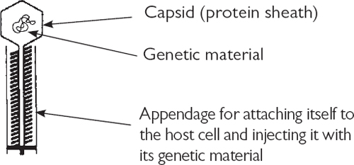
Viruses form their own separate category among germs. They are not cellular organisms—they have no cellular membrane, cytoplasm, or organelles but only genetic material. This may consist of either DNA or RNA rather than the two together, as is generally the case in a cell.
The genetic material of a virus is surrounded by a protective membrane, called the capsid, on which appendages (or tails) can be found. Those appendages permit a virus to attach itself and perforate the cell that it will inhabit as a parasite.
A virus is therefore nothing more than a piece of genetic information surrounded by a protective shell. It would be more accurately described as a molecule rather than a living organism. This makes it easy to grasp why viruses are the smallest germs. The viral molecule is most often spherical in shape, but some are elongated. Every kind of virus—there are at least a good thousand different types—possesses its own unique genetic material.
Once viruses were called “filterable viruses.” When scientists first begin to conduct studies on bacteria, they realized that the filters they were using allowed the passage of something that was smaller than bacteria—something that was invisible to the microscope (of that era) but still had the ability to cause disease. This mysterious entity was given the name filterable (can travel through a filter) virus (poison). As time passed, only the term virus was retained.

Figure 4. The virus
Because they lack organelles and enzymes, viruses are not capable of metabolizing substances. They do not breathe, nor do they eat. As long as they are not dwelling inside a cellular host, they remain inert. To multiply, they are entirely dependent on the metabolic activity of the cells they have invaded. They are therefore unavoidably cellular parasites.
To penetrate the cell with which it has come into contact, the virus bores a hole in the cell’s membrane with its appendage. Its genetic material is then injected into the cell, where it replaces the genetic material of the cell. The virus will then use the organs of the host cell, not to manufacture the substances it needs to survive but to produce up to several hundred new viruses. When the number of these newly created viruses exceeds the capacity of the cell, the cell explodes and loses its cytoplasm, causing it to die. The viruses freed by the death of the cell now spread and infect other cells.
A virus cannot enter any cell it chooses but only those with which it shares an affinity. It is necessary for the cell to offer it suitable living conditions; this cell is known as a target cell. This is why, for example, the mumps virus finds its target cells in the salivary glands, the herpes virus targets the skin, the rabies virus targets certain cells of the brain, and the polio virus targets the front cells of the spinal cord. The targets sought by flu virus (for which there are several hundred varieties) are the cells of the upper respiratory tract: the nose, pharynx, and larynx. The HIV virus, which causes AIDS, targets the white blood cells of the immune system.
| VIRAL DISEASES | |
| VIRUS FAMILY | DISEASE |
| Arbovirus | Yellow fever, encephalitis |
| Arenavirus, Coxsackie virus | Meningitis |
| Echovirus, adenovirus | Respiratory infections |
| Enterovirus | Poliomyelitis |
| Herpesvirus | Herpes, chickenpox, shingles |
| Human immunodeficiency virus (HIV) | AIDS |
| Myxovirus | Flu, measles, mumps |
| Poxvirus | Smallpox |
| Rhabdovirus | Rabies |
THE MULTIPLICATION OF MICROBES
Because of its tiny size, a single germ is incapable of causing harm to the human organism. If physical damage is present, it is because an entire colony of germs is present.
Even if we accept the fact of a single germ entering the human body—which is never the case, as it is always accompanied by cohorts—it will not remain alone for long. Germs are able to multiply with great speed. After a very short period of time, an entire population will be found at the spot where only a few had been present.
The rapidity with which unicellular germs are able to multiply comes from their specific fashion of reproduction. A mother cell will divide into two identical cells, each possessing all the attributes of the mother cell: cellular membrane, cytoplasm, organelles, and a nucleus. The two cells that have been created this way are the daughter cells. The mother cell ceases to exist at the “birth” of its descendants.
When the two daughter cells divide in turn, they also vanish, but only after creating two daughter cells each, for a total of four separate cells. In other words, the number of descendants doubles with each succeeding generation. And contrary to human beings, for whom a new generation appears every twenty years, a new generation of germs can come into being every fifteen minutes to an hour. Consequently, the speed with which a population of germs increases is extremely quick.
Let’s take as an example the bacteria Escherichia coli (called a colibacillus, or colon bacillus), which is a habitual and harmless inhabitant of the intestine but can become virulent under certain circumstances. This bacteria gives birth to a new generation every twenty minutes. Because it doubles in number every time it divides, it will have eight descendants after one hour and sixty-four one hour after that. One hour and twenty minutes after this, it will have attained a population of one thousand descendants. The growth rate accelerates with each new generation: one hour and thirty minutes after this, there will be ten thousand descendants. A million individuals will be reached a little over two hours after this stage, and one billion after three hours and thirty minutes.
So, in one night of eleven hours, we can go from one individual to a population of ten billion. All of this from just one single microbe!
Is there anything that could put a stop to the dizzying pace of such growth? Wouldn’t the body be irremediably condemned to succumb to an invasion like this and find itself completely overrun every time it is infected? No! On the one hand, the body does not remain passive when confronted by invasion but defends itself by killing some of the invaders with the help of its immune system. On the other hand, living conditions in the target cells become more or less quickly unfavorable to the germs because of their number. The lack of space, oxygen, and food makes itself felt; additionally, the metabolic wastes excreted by the germs damage the environment and make it toxic. The microbial growth rate inevitably slows and then stagnates after a certain time because each tissue and organ has limits to just how many microbes it can “welcome.”
However, the number of germs is not the only factor in the severity of an infection. We cannot say that the greater the number of germs, the more dangerous the disease. The virulence of germs—that is, their ability to sicken the organism—varies from one species to the next. A small number of extremely virulent germs can cause more damage than a large number of mildly aggressive germs. The level of damage they can cause therefore depends more on their intrinsic nature than on the size of their population.
Among the factors that restrict the growth of germ populations, temperature plays an important role. Like all other living things, germs require a favorable temperature within a certain range. The ideal temperature for the germs that infect human beings is generally around 37° Celsius/98.6° Fahrenheit. While low temperatures slow their metabolisms and eventually paralyze them, high temperatures will kill them. At 45° Celsius/113° Fahrenheit the majority of germs will die. It so happens that during an infection, the body accelerates all its metabolisms (oxygenation, blood circulation, and so on), which results in a rise in the body’s temperature—that is, fever—of up to 40° Celsius/104° Fahrenheit. If this warming does not kill the germs, it does make their living conditions extremely adverse and thereby contributes to arresting their multiplication. This is why it is therapeutically valuable to not indiscriminately bring fevers down.*1