CHAPTER 2
The Amazing History of Modern Neuroscience The Search for a Winning Formula
Modern neuroscience is built upon a strong foundation of individual discoveries, each
of which has played its part in unraveling the mysteries of the brain and how it supports
our thoughts and behavior. This foundation is critical for understanding how the brain
works and identifying the specific factors involved in developing a Winner’s Brain.
THE CHICAGO WORLD’S FAIR opened on May 27, 1933, forty years after the previous Chicago
World’s Fair, the Columbia Exposition of 1893, which had celebrated the 400th anniversary
of Columbus’s discovery of America. It was very much a science fair, and its lights
and electricity were activated by rays from the star Arcturus, 40 light-years away.
Visitors were awed by the towering dream city that rose up from what had been landfill
and now stretched across the 427 acres of Burnham Park, on the shores of Lake Michigan.
Though the fair was held in the depths of the Great Depression, more than 48 million
people passed through its gates, gladly paying the 50 cents daily admission to witness
the uncharted limits of human imagination and to feast upon all the information and
experience the Great Fair had to offer.
The event was conceived as a tribute to science and technology, hence its moniker,
“A Century of Progress.” Its 400,000-square-foot Hall of Science included a miniature
oil refinery, an exhibit on evolution, and a ten-foot-tall robot that explained digestion.
On public display for one of the first times ever was a dazzling new invention called
television.
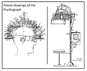
And then there was the psychograph, which for just ten cents scanned a fairgoer’s
brain. This brain-scanning machine resembled an old-fashioned beauty salon hair dryer
with dozens of long metal probes jutting from its helmet. The subject sat in a chair
and the headpiece was lowered and adjusted, then the operator pulled back a lever to activate the belt-driven
motor, which sent out low-voltage signals meant to map the various regions, or what
the psychograph read as “organs,” of the brain. Once the examination was complete,
an enormous dot matrix printer whirred into action, providing each client with a personal
“neuroanalysis” titled “The Guide o’ Life.”

The psychograph was the mechanical counterpart of a then century-old practice known
as phrenology, a mixture of brain science, psychology, and philosophy. Phrenologists
believed that certain traits literally shaped the brain, and the brain responded by
pushing and shifting against the skull, leaving telltale lumps and divots. To phrenologists,
an individual’s talent and character were easily readable if you knew how to correctly
interpret the skull’s topography; for example, the phrenological brain spot for musicality
was located at the base of the temple, just above the left eye. The justification
for this was found in paintings of Mozart, who was often depicted with his finger
placed on that very spot when composing music.
Before the invention of the psychograph, phrenologists relied on a series of charts
superimposed upon ink drawings and plaster models of skulls to make their case. But
by the turn of the twentieth century, many of their theories had been largely discredited and were beginning to be supplanted
by the burgeoning fields of psychology and neuroscience. By the time the machine made
its appearance at the Chicago fair, both professionals and the general public alike
had come to regard phrenology as more novelty act than science (this may be why the
psychograph was banished from the Hall of Science and placed among the circus and
carnival attractions on the Midway; the Temple of Phrenology exhibit lay sandwiched
between Midget Village and the Ripley’s Believe It or Not tent, just around the corner
from the flea circus).
Yet phrenologists played an important, if misguided, role in the early advancement
of modern neuroscience. They were attempting to unlock the secrets of personality
and behavior by linking specific mind functions to size differences across the different
areas of the brain. The phrenologists’ notion that the brain was the seat of thought,
emotion, and behavior was revolutionary compared to some historical viewpoints. The
ancient Egyptians, for example, thought the heart was responsible for thought and
higher functioning; they carefully preserved the heart and discarded the brain during
the embalming process. And while subsequent scholars, such as the ancient Greek physician
Alcmaeon, advanced the possibility that the brain was important for thought and perception,
even notable thinkers such as Aristotle continued to believe that the heart was the
most critical thought-related organ—indeed, he thought the brain acted primarily as
a radiating device for cooling blood coming through the heart. These examples illustrate
how mankind has been striving to understand the seat of intellect for millennia. The
various and shifting points of view found along the way eventually settled squarely
on the brain. We have been fascinated ever since by what makes this critical organ
tick and by pondering how its power might be harnessed.
In this chapter, we highlight some of the critical points in the recent history of
psychology and neuroscience, instances over the past 200 years that have helped advance
our knowledge of the brain and how it works. Many of the cases from earlier days depended
upon unhappy events where patients literally (and often unintentionally) gave their lives for
the betterment of neuroscience when their brains were examined after accident or death.
These sacrifices gave investigators a starting point for gaining knowledge about where
memories are formed, which structures hold the key to moral behavior, how we process
language, and so on. Every example we describe provides supporting evidence of how
changing the brain changes how you think, feel or behave and how these changes can
lead you to success.
In our search for what makes a Winner’s Brain operate so effectively, our goals were
not so different from those of the phrenologists. They invented the psychograph in
an attempt to scan the brain. To them, the landscape of the skull was a window into
the function of the specialized areas of the brain that lay beneath. With this book,
we are pursuing the same goals, but with the knowledge that the skull is too hard
and the brain too soft to give us answers in a topographical way. What you do with
your brain causes it to grow and change—and this has been proven by examining the
brain with the incredible tools we have available to us today. It is also demonstrated
in the real world by people who do the things they want in life.
Brain Science with a Bang
The phrenologists’ contention that specific structures in the brain might be specialized
for different aspects of thought, emotion, and behavior was radical at the time and,
to some extent, on the right track. Their influence can be seen front and center in
one of the most pivotal cases from the history of brain study.
In the summer of 1848, Phineas P. Gage, a 25-year-old railroad construction foreman
from Vermont, accidentally set off an explosion that sent an iron bar rocketing through
the air. The bar entered his left cheek, pierced the base of his skull, traversed
through the front of his brain, and exited at high speed through the top of his head before landing at least 100 feet
away, covered with blood and bits of brain. Although Phineas recovered his senses
shortly after the accident and witnesses reported he spoke rationally, they soon realized
that “Gage was no longer Gage.”
Previously he had been described as having a well-balanced mind and as someone who
was persistent about executing all of his plans into action. Now he was rude, profane,
and wildly inconsiderate, someone totally incapable of making good choices. Gage spent
the rest of his days wandering aimlessly from one menial farm job to the next, even
at one point displaying himself as an oddity in P. T. Barnum’s Museum in New York
City alongside bearded ladies and dwarfs. He carried the five-foot-long bar around
with him until his death at the age of 38. Recently we were able to see the bar, along
with Phineas Gage’s skull, at the Warren Anatomical Museum, part of Harvard’s Countway
Library of Medicine in Boston.
Prominent phrenologists who reflected on Gage’s case accurately attributed his changed
personality to brain damage. Regardless of phrenology’s shortcomings, its basic premise
that different parts of the brain contribute to different traits and abilities correctly
supported the conclusion that the blast had spared Gage’s language and motor centers
but not the parts of the brain responsible for character and reason. Given the location
of the injury, it was one of the first solid clues that the prefrontal cortex, part
of the frontal lobe of the brain, might actually contribute to basic traits like good
judgment and social manners.
This fundamental truth about how such faculties are localized within the brain would
take decades to prove. French scientist Paul Broca kept the ball rolling in 1861 by
tracing language difficulties of stroke patients to damage in their brains’ left frontal
lobes, eventually attributing the master center of speech production to one square
inch of grey matter rather than the lumpiness of a person’s head. In his honor, this
tiny patch of cerebral real estate was christened Broca’s area.
Twelve years later, another European, the psychiatrist Carl Wernicke, connected a
patient’s inability to comprehend speech to damage within another area, the left temporal lobe. The temporal lobes can be found on each side
of the bottom half of the brain, running from just behind the temples to just behind
the ears. Beyond advancing the understanding of how the brain processes language,
the possibility that the brain region involved in language comprehension was separate
from that for language production helped scientists understand that a basic ability
such as language was not confined to a single part of the brain. They were beginning
to realize that different aspects of these fundamental abilities could arise in different
regions of the brain. This meant that there had to be a lot of communication and coordinated
effort going on between these different brain structures to support something as effortless
as carrying on a conversation.
Living in the Moment
Not quite a century after Gage’s meeting with the metal rod, another ill-fated soul
unwittingly furthered the study of neuroscience. Henry Molaison, also known as Patient
H.M. until his recent death, could recall the stock market crash of 1929 and the Chicago
World’s Fair, but not a recent stroll through the woods or a person he had met the
day before. At the age of nine, H.M. fell off a bike and subsequently suffered from
epileptic seizures until age 27, when a surgeon, in 1953, removed sections of his
medial temporal lobes—parts closer to the middle of his brain—as a treatment. This
did the trick for eliminating H.M.’s seizures—but consequently wiped out his ability
to create new long-term memories. Thereafter he became the subject of intense scientific
study; experts from all over the world came to assess him. But H.M. never seemed to
mind. Actually, he never remembered any of it.
In the course of his epilepsy surgery, two-thirds of H.M.’s hippocampus—a curved structure
within the medial temporal lobes—had been removed. Considering H.M. was then only
able to recall experiences that happened before his operation, his surgeon, William Scoville, and neuropsychologist,
Brenda Milner, concluded that a key function of the hippocampus is to form declarative
memories, the type of memories you can actively reflect upon and talk about. But what
Milner and subsequent investigators also found interesting is that many of H.M.’s
other memory functions were preserved. In one experiment, H.M. was asked to trace
a line between two outlines of a five-point star, one inside the other, while watching
his hand and the star in a mirror, a difficult skill for anyone to master. Every time
H.M. picked up the pencil, he experienced tracing the star as if it was the first
time. Yet he gradually got very good at it.
This is because H.M.’s cerebellum, the fist-sized lump at the base and back of the
brain, and his basal ganglia, nearer to the center of the brain, remained intact.
These regions are key for what scientists call procedural memory, which enabled him
to acquire new motor skills like tracing the star or solving a puzzle that required
stacking disks in a specific order. However, lacking the ability to form conscious
memories of new events, he could not explicitly recall ever having learned these skills.
His short-term and working memory were also in good shape; he was able to memorize
lists of words and hold information in his head for short periods of time about as
well as the average person. But for the next 55 years, until his death in a Connecticut
nursing home, you could say H.M. truly lived in the moment.
Like Gage before him, H.M.’s bad luck stands as one of the great mile-stones in the
study of neuroscience. His selective memory function helped explain the link between
the brain’s structure and specific psychological processes. It was further proof of
both the localized and holistic nature of the brain; that is, the brain has separate
working parts that typically work together as a whole. The history of modern brain
study is riddled with brain damage cases such as those of Gage and H.M. that have
helped us, for example, link vision to the occipital lobes, attention and visual-spatial
processing to the parietal lobes, and emotion to structures such as the amygdala.
In these instances, things didn’t work out so well for the individuals, but each stroke, knock on the head, and rod through the
brain advanced our understanding of how this sophisticated organ works.
Moving Past Damage
Thankfully, we no longer have to wait for accidents to happen to examine the inner
workings of the human mind. In fact, it was a few years before the Chicago fair in
1929 that a German psychiatrist, Hans Berger, was among the first to come up with
a less invasive way to study the brain in action. He changed neuroscience forever
in 1929 by attaching electrodes to his subjects’ skulls to produce a graphic representation
of the electrical activity of the brain. Berger watched these brain waves, as he called
them, in a variety of different circumstances, charting how they shifted depending
upon what a patient was doing or even thinking. If a patient sat quietly in a chair
with his eyes closed, Berger noted slower alpha waves, and if the patient remained
in the chair and opened his eyes, the brain switched to higher-frequency beta waves.
In this way, Berger was able to assign the brain’s electrical response to different
types of attention and focus.
Refined forms of his electroencephalography (EEG) method are now used routinely in
every hospital and neurologist’s office. EEG and its cousin, MEG (magnetoencaphalography),
which measures magnetic fields produced by electrical activity, both have millisecond-or-better
temporal resolution. And so they are considered invaluable research tools for studying
the timing of different brain functions as well as the general location from which
they originate.
Since Berger, scientists have continued to devise ways of revealing the mysteries
of the brain without having to wait for a patient to die. By 1970, Phineas Gage could
have walked into any emergency room and undergone a simple battery of tests to reveal
the damage to his prefrontal cortex, the part of the brain now known to be associated with complex cognitive behaviors, personality expression, decision making, and social behavior—precisely
the traits that went south for Gage once the iron bar pierced his brain. Testing would
not change the nature of the brain damage in a case as severe as Gage’s, but at least
doctors are able to determine the extent and effect of such an injury and how best
to help the patient.
We now understand that the brain operates on many different levels. Besides plotting
brain waves, we also learn about it by monitoring changes in blood flow and chemical
interactions, and by observing how the different tissues and brain cells respond both
in the moment and to repeated experiences. Some of the most common tools used in cognitive
neuroscience research today are MRI (Magnetic Resonance Imaging) and the fMRI (functional
Magnetic Resonance Imaging.)
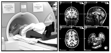
During an MRI scan, a patient lies on a bed that slides into a large cylinder. The
scanner’s strong magnetic field forces the protons within the brain to line up much
the same way metal filings line up under a magnet. Brief radio frequency pulses then
push the protons out of alignment, causing them to emit a signal as they move back
into place. How quickly they can move back into place, however, depends on the type
of tissue in which the protons occur. The scanner picks up these differences and translates
the contrasting signals into an anatomical map of the brain.
Liz Neporent inside an MRI scanner at the A.A. Martinos Center for Biomedical Imaging
(left) and the resulting anatomical images of her brain (right). Photos by Cristine
Lee.
An f MRI scan works much the same way, but instead of looking for difference in anatomy,
it detects brain activity through relative changes in levels of blood oxygen. As active
brain cells use up available oxygen, local increases in blood flow bring fresh red
blood cells with iron-rich, oxygen-carrying hemoglobin. After releasing their oxygen,
the iron atoms in hemoglobin produce small distortions in the surrounding magnetic
field. Changes in the relative concentration of oxygen-carrying vs. oxygen-free hemoglobin
therefore provide another set of contrasting signals that can be picked up by the
scanner, which can be associated with differences in brain activity. With its focus
on brain structure, MRI can measure differences in the thickness, density, and volume
of different parts of the brain, whereas fMRI focuses on brain function, measuring
levels of activity at thousands of points across the brain as it is actually happening.
While these are currently among the most widely used neuroimaging techniques, several
other methods also provide critical data. Metabolic activity in the brain, for example,
can be measured using a PET (positron emission tomography) scanner, while the brain’s
chemical makeup can be gauged using MRS (magnetic resonance spectroscopy). Meanwhile,
carefully placed electrodes provide single-cell recordings, which can pinpoint the
role of individual neurons in different brain functions, such as those involved in
identifying your spouse, your child—or Brad Pitt. Researchers also continue to examine
actual brain tissue, although they no longer have to rely on the unplanned sacrifice
of people like Gage and H.M. Instead, people now willingly donate their brains upon
their deaths to places like the Harvard Brain Tissue Resource Center at McLean Hospital
in Belmont, Massachusetts, the largest brain bank in the world. Because most studies
now require only a small sample of tissue, one brain can potentially support hundreds
of projects.
All of these incredible techniques are the fruit of many decades of innovation and
technological advances. Someday, the technology we describe in this chapter may allow
us to gauge the effectiveness of cognitive strategies in real time, isolate and make
changes at the cellular level, or even create new ways to maximize the brain. Right now, as we’ll see throughout this
book, these techniques can give us a glimpse of how a Winner’s Brain operates.
The promise of this book lies in our current ability to identify areas in the brain
associated with key cognitive elements connected to success, and to track new approaches
that can lead to changes in how the brain operates. Recent advances in neuroimaging
demonstrate that such approaches can lead to improvements in mental functions and
that these improvements can cause changes in the physical structure of the brain itself.
The extent of the positive changes made to the brain’s structure partly reflects the
durability of the corresponding improvements in the brain’s ability to function. Such
improvements can help you develop a Winner’s Brain, and in the pages that follow we
give examples of people who have improved themselves and done well in life using strategies
that we describe in this book.
As Scott L. Rauch, M.D., chair of Partners Psychiatry and Mental Health and president
and psychiatrist in chief of McLean Hospital, told us, “Modern brain imaging techniques
have revolutionized neuroscience. In particular, in psychiatry, these methods have
enabled investigators to safely explore the structure, function and chemical basis
of psychiatric diseases as well as their treatments. Coupled with genetics, brain
imaging approaches hold the greatest promise for [diagnosis and treatment].”
Scanning the Winner’s Brain
In writing this book—and aiming to show how brain function relates to success—we’ve
taken considerable care to avoid passing along erroneous or incomplete information.
Each study we quote has been carefully considered, and in many cases we’ve spoken
at length to the researchers who carried it out. We spoke to top thinkers in the fields
of neuroscience and psychology, too, to avoid interpreting single studies in a vacuum and to ensure we
have presented a comprehensive, cutting-edge view of how the brain works. We’ve also
reinforced the findings by talking to people who personify a Winner’s Brain. In almost
every instance, we’ve been pleased to discover that what works in the lab often seems
to lead to success in the real world, too. The people we interviewed throughout this
book are living examples of winning ways of thinking, feeling, and behaving. They
represent different professions, nationalities, and worldviews. With each study and
interview, we have aimed to provide a better understanding of which traits—we call
them Win Factors—make the greatest contributions to bettering the brain, along with
deliberate strategies that are most likely to lead to positive changes in both brain
and behavioral functioning.
Instead of simply reporting measurements from studies looking at the capacity of the
brain, we’ve worked hard to uncover how that power helps someone become successful,
and how you can harness it too. It makes sense that preserving and enhancing brain
power by consciously trying to strengthen qualities like Memory and Emotional Balance
(to name just two Win Factors) will increase your chances of achieving what’s important
to you.
As it turns out, optimizing your brain function is the key to feeling more satisfied,
becoming more engaged in life, reaching your potential, and realizing your dreams.
And the best part is, just about anyone can do it.


