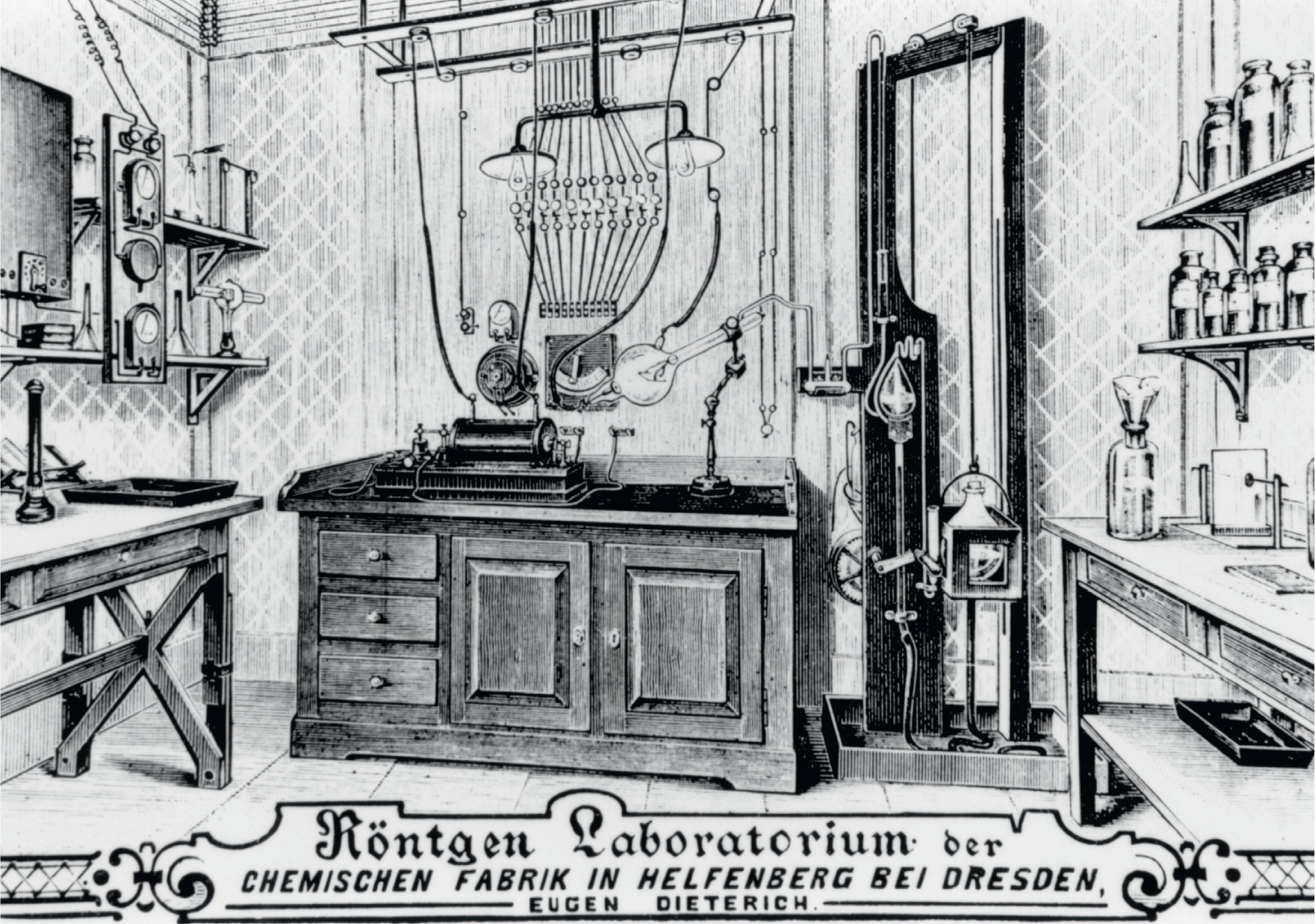
Laboratory of Wilhelm Konrad Röntgen (1845–1923).
The close link between experimental science and technology is particularly powerfully demonstrated by the development of physics at the end of the nineteenth century. One key piece of technology, the vacuum (or Crookes) tube, produced a revolution in the understanding of the world of the very small, and led to the birth of atomic physics.
In 1894, Philipp Lenard, working in Germany and following up experiments carried out by Heinrich Hertz, studied the way cathode rays could pass through a thin sheet of metal foil inside a vacuum tube. Because the rays passed through the metal without leaving any detectable holes, he thought that they must be waves, not particles. This idea would soon be proved wrong (see here), but in a separate development Wilhelm Röntgen, the professor of physics at the University of Würzburg, decided to follow up Lenard’s work to find out if the cathode rays could penetrate the glass of the Crookes Tube itself. Röntgen was already 50 years old when he carried out his key experiments in November 1895 – an established scientist with a reputation as a skilled experimenter, but still open to new ideas.

To see if cathode rays were passing through the glass of the tube and out into the laboratory, Röntgen set his apparatus up in a darkened room and covered the entire tube with a thin layer of black cardboard to stop any light from the glow inside escaping. He wanted to be sure his eyes were adapted to the dark and could pick up any flashes from the simple cathode-ray detector he had prepared. It was already known that a sheet of paper painted with barium platinocyanide would fluoresce when struck by cathode rays, so Röntgen set up such a screen in the dark lab to look out for the fluorescence. After carrying out some initial tests, Röntgen decided to repeat the experiment using a vacuum tube with a thicker glass wall. On 8 November 1895, after he had set up the new tube, and covered it with black cardboard, he put the barium platinocyanide screen to one side while he turned the vacuum tube on and the lab lights off, simply to make certain no light was escaping from the tube. To his surprise, he saw a faint shimmering light from the screen, which was several feet away from the tube and off to one side, not in the ‘line of fire’ of the cathode rays. Something else – something previously unknown – was making the screen glow when the tube was turned on.
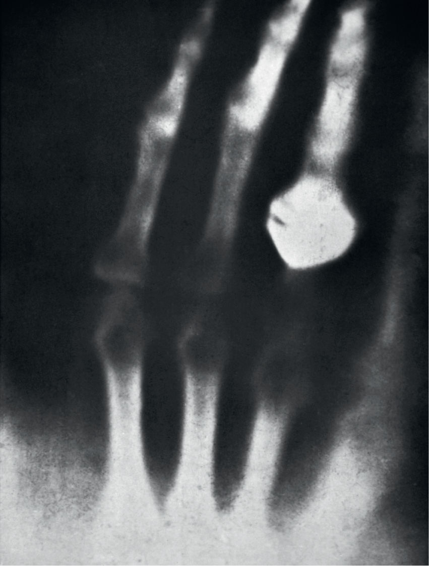
Over the next few weeks, Röntgen made a careful study of what he called ‘X-rays’ (‘X’ is traditionally the unknown quantity; in some countries they became known, to his embarrassment, as Röntgen-rays). He found that they were produced where the cathode rays hit the glass wall of the Crookes tube, and spread out in all directions. They travelled in straight lines, and were not deflected by electric or magnetic fields. But the most dramatic discovery was that they would penetrate many materials, including human flesh. Just two weeks after the discovery of X-rays, Röntgen used them to make the first X-ray photograph, of his wife’s hand. It showed the bones of her fingers and her wedding ring, and it was published in the scientific paper in which he announced the discovery. Titled ‘On a new kind of ray’ (‘Über eine neue Art von Strahlen’), it was written on 28 December 1895, and published early in 1896.

The discovery was sensational news, not least because of the publication of the X-ray photograph of Anna Röntgen’s hand (on seeing the image, apparently she exclaimed ‘I have seen my death!’). On 13 January 1896, Röntgen demonstrated the phenomenon to Emperor Wilhelm II, in Berlin. English translations of the paper were published in the journals Nature (on 23 January) and Science (on 14 February). X-rays were soon recognized as part of the electromagnetic spectrum as light, but with higher frequencies (shorter wavelengths).
The medical implications of the discovery were obvious, and X-rays soon became an important diagnostic tool, enabling doctors to see inside the human body without cutting it open. Before the end of 1896, the first radiology department opened in a Glasgow hospital, where they obtained pictures of a kidney stone and of a penny lodged in a child’s throat. Less than two years after the discovery, X-rays were used for the first time in a battlefield context, to find bullets and broken bones inside patients during the Balkan War of 1897.
The first Nobel Prize in physics was awarded to Röntgen in 1901, ‘in recognition of the extraordinary services he has rendered by the discovery of the remarkable rays subsequently named after him.’