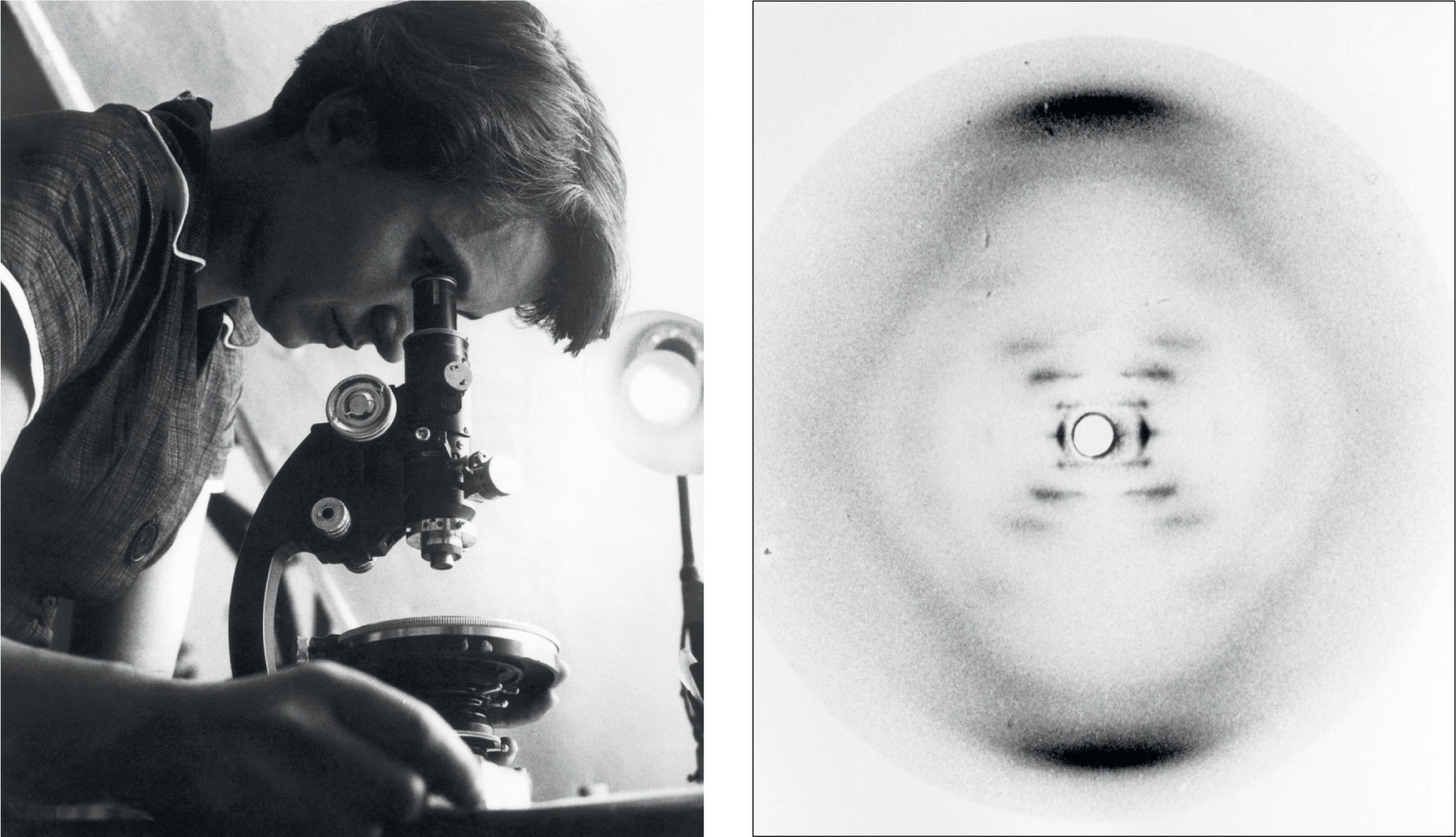
The structure of DNA, the molecule of life, was revealed by experiments carried out by a team working at the Medical Research Council’s Biophysics Research Unit at King’s College, London. The head of the unit, John Randall, was one of the first people to accept the evidence that genetic information is carried by DNA, and in 1950 he assigned a research student, Raymond Gosling, to work with Maurice Wilkins to obtain X-ray diffraction images of DNA.
Working in a basement at King’s, Gosling was able to crystallize DNA – the first person to crystallize genes. He knew that it was then just a matter of time before the structure of the molecules would be revealed. Under Wilkins’ supervision, Gosling obtained the first X-ray diffraction images of crystalline DNA. These were presented by Wilkins at a meeting in Naples, where the American James Watson was in the audience and realized their significance.
Back at King’s, the team were joined by Rosalind Franklin, who already had considerable expertise at X-ray diffraction work. Randall was particularly eager to bring her on board, because he doubted that Wilkins and Gosling had the expertise to solve the problem of working out the structure of DNA from the spotty diffraction images. Armed also with new equipment, Franklin and Gosling obtained better images, and discovered that there are two forms of crystalline DNA. When it is wet, it forms a long, thin fibre, but when it is dry, it is short and fat. These became known as the ‘B type’ and ‘A type’, respectively. Because of the humidity inside cells, the B type was expected to be more like the DNA found in living things.
The X-ray diffraction photographs hinted at the structure of the molecules of DNA, but a great deal of analysis was required to work out the positions of the atoms in the molecules. Franklin, with Gosling, concentrated on solving this puzzle for the A type, while Wilkins focused on the B type. There was some crossover between their work, but not as much as there should have been, because of a personality clash between Franklin and Wilkins. By the beginning of 1953, it was clear that both forms of DNA were based on a helical structure, and Franklin prepared two scientific papers suggesting a double-helical structure of the A type DNA, which were submitted to the journal Acta Crystallographica in March that year. Just at that time, she moved from King’s to Birkbeck College, also in London. Also just at that time, the structure of the B type DNA had been determined by another team, in Cambridge.
In January 1953, on a visit to King’s, Watson, who was now based in Cambridge, had been shown a print of the best X-ray diffraction image of the B type DNA. It had actually been taken in May 1952, by Franklin and Gosling, but was passed on without their knowledge. The image, known as Photo 51, showed the X-ray diffraction pattern of DNA in the highest quality available at the time and clearly shows a cross-shaped pattern that could only result from a helical structure. If one experiment could be said to have unlocked the secret of DNA, it was the one that produced this image.
Back in Cambridge, Watson and his colleague, Francis Crick, who had together been puzzling about the structure of DNA for some time, tried building models of the molecule that would fit the image, using Pauling’s bottom-up approach (see here). This quickly led them to the discovery that everything fitted together if a DNA molecule consisted of two strands twined around each other in a double helix, with the bases on the inside, so that the bases on one strand linked with the bases on the other strand like the steps on a spiral staircase. Adenine always links with thymine, and cytosine always links with guanine. So the strands are like mirror images of each other, and if they unravel each lone strand can build a new double helix by adding the appropriate units to build up the other strand.

LEFT: © Science Source/Science Photo Library
Rosalind Franklin (1920–1958).
RIGHT: © King’s College London Archives/Science Photo Library
‘Photo 51.’ X-ray diffraction photograph of DNA (deoxyribonucleic acid), obtained in May 1952 by King’s College London researchers Rosalind Franklin and Raymond Gosling (1926–2015).

© A. Barrington Brown, Gonville & Caius College/Science Photo Library
James Watson (b.1928), left, and Francis Crick (1916–2004), with their model of part of a DNA molecule in 1953.
The structure also carries much more information than a boring repetition of a four-letter word (see here). The A, T, C and G can occur along the strand in any order, such as AATCAGTCAGGCATT …, like a message in a four-letter code. This can carry a great deal of information, just as the two-letter Morse code or binary computer code can convey a great deal of information. Crick and Watson completed their model building on 7 March 1953, and sent a paper off to Nature.
In 1962, Crick, Watson and Wilkins shared the Nobel Prize in Physiology or Medicine for this work. Franklin, who had died of cancer in 1958, could not share the honour as Nobels are never given posthumously.

