7
Integrated Plasmonic Detectors
7.1 INTRODUCTION
Surface plasmons are known to have the potential to guide, concentrate, and scatter light at the nanoscale. These properties are particularly interesting in emerging fields that rely on either small-footprint and fast devices like in on-chip optical communication, optical absorption in small volumes such as cost-effective solar cells, or biosensing, where the extent of the field is similar to the size of the molecules that are probed. In all these cases, surface plasmon-based devices have been investigated extensively to improve the performance or to create new possibilities. In one important application, surface plasmons can directly enhance the absorption in (thin film) solar cells. In optical communication schemes and biosensing, surface plasmons have been studied extensively using optical techniques, while it is highly desirable to detect them locally using integrated plasmon detectors.
To understand how surface plasmons can be applied for enhancing the response of semiconductor detectors, the detection of biochemical reactions, or signal transmission, one has to gain insight into the nature of surface plasmons. Surface plasmons are collective electron oscillations at the interface between a metal and a dielectric. As the reader will have discovered while wandering through this book, one of the most amazing properties of surface plasmons is the way in which they can focus and channel light using subwavelength-sized metallic structures. For this reason, surface plasmons have the potential of merging the dimensions of fast photonics with nowadays electronic components.
For the development of plasmon-enhanced photodetectors, small-area photodetectors are combined with a plasmonic antenna structure, which can collect light over a large area and focus it on the much smaller semiconductor area. Also metal particle resonances can be applied to maximize the overlap of the plasmonic mode and the small detector active area. In this way it is possible to combine the advantages of small photodetectors, like a low noise level and high-speed operation, with the advantages of large-area photodetectors, such as the high detector photoresponse.
In the study of optical communication on chip, plasmonic waveguides have been studied extensively in the last decade [1–7]. The delay caused by electrical interconnects increases when their dimensions are scaled down, opposite to the transistors, for which the performance increases with scaling. Researchers today are investigating if the use of light as the information carrier can supply an integrated circuit with a better performance as their copper counterparts. At optical frequencies, it is possible to communicate with a high bandwidth, and as a consequence an optical waveguide can carry digital signals with a capacity exceeding that of its electrical counterpart with a factor of more than one thousand. Unlike diffraction-limited dielectric waveguides, metallic waveguides can be fabricated with subwavelength dimensions and benefit from the same properties of optical signal transmission and can be used for both optical and electrical signal transmission. For this reason, they have been suggested to function as optical interconnects on chip. In order to integrate these metallic waveguides on chip, electro-optical transducers need to be developed to realize a fast and efficient coupling between electrical and optical signals, leading to the design of plasmon sources and detectors.
In the field of solar cell research, the properties of surface plasmons are applied to increase the conversion efficiency. Material cost is the main factor determining the price of solar cells today. As a consequence, great interest has arisen in both solid state and organic thin film solar cells, where thin semiconductor layers are deposited on a cheap substrate. A lower material cost asides, thin semiconductor junctions offer an increased collection efficiency compared to wafer-based solar cells because the minority carrier diffusion length is larger than the film thickness. However, the absorption length of the incident radiation can exceed the semiconductor film thickness, leading to only a partial conversion of the optical power in the semiconductor junction. Both localized surface plasmons on metallic nanoparticles and propagating surface plasmon polaritons (SPPs) on metallic films can offer a way to increase the absorption of the incident light by channeling and concentrating the electromagnetic fields inside the thin semiconductor layer.
SPPs are intrinsically very sensitive to small changes in the refractive index near the metal/dielectric interface in a confined region whose spatial extent is dictated by the evanescent tail of the SPP. At visible wavelengths, this is in the order of a few hundred nanometers or below. Therefore SPPs provide excellent means to probe biochemical events occurring at the metal surface. Bio-recognition is achieved by using localized surface plasmon resonance (LSPR) biosensors through the detection of LSPR changes caused by adsorbate-induced changes in local dielectric constant. Usually the LSPR spectrum of a nanoparticle is studied with an optical detector in the far field. In order to develop lab-on-chip plasmonic biosensors, the integration of plasmonic sources and detectors is of paramount importance.
In general we can divide the plasmon detection mechanisms into three large groups. The underlying physical principle is presented in Figure 7.1. More than two decades ago, the study of surface plasmon detection by means of tunnel junction detectors started. In Figure 7.1a the detection process is visualized. If plasmons are focused inside the tunnel junction, it will bias the junction and induce tunneling of electrons back and forth at the plasmon frequency, even when no external bias voltage is applied. Due to the rectification property caused by the nonlinear and asymmetric nature of the IV characteristics of the tunnel junction, a DC bias develops across the junction under illumination and the system will behave as a detector. Such bias voltage can be detected either as a light-induced DC current or as an additional bias if the tunnel junction is placed under an externally applied bias voltage. The second detection mechanism is the integration of standard semiconductor detectors with plasmonic nanostructures, shown in Figure 7.1b. Among these detectors we can find photoconductors, MSM photodetectors, and photodiodes. MSM photodetectors are mainly studied for plasmonic enhancement of ultrafast photodetectors while photodiodes mainly find their applications in plasmon-enhanced solar cells. A third detection method is the application of Schottky photodiodes, depicted in Figure 7.1c. In Schottky photodetectors, two detection modes are possible. For plasmon energies above the bandgap energy of the semiconductor, electron–hole pair generation across the bandgap is the main contributor to the photocurrent. For energies below the bandgap but larger than the work function of the junction, direct excitation of electrons to the conduction band is impossible, but emission of carriers can occur from the metal to the semiconductor over the Schottky barrier, leading to a measurable photocurrent. In this chapter we will discuss the combination of these three types of detectors with plasmonic waveguides and nanostructures.
FIGURE 7.1 Plasmon detection mechanisms. (a) A tunnel junction detector. (b) Standard semiconductor photodetectors. (c) A Schottky diode photodetector.

7.2 ELECTRICAL DETECTION OF SURFACE PLASMONS
7.2.1 Plasmon Detection with Tunnel Junctions
The first demonstrations of electrical detection of surface plasmons were performed using electrical tunnel junctions. Indeed, as shown in Figure 7.1a, the rectifying nature of an asymmetric tunnel barrier results in a DC component upon AC excitation, similar to the rectification in a pn-diode. The particular advantage of a tunnel junction compared to a pn-diode is the intrinsic speed of the tunnel junction. As the speed is determined by the tunnel time, which can go down to 1 fs or below, bandwidths up to the NIR and visible are feasible [8]. However, these types of detectors have very low efficiencies, which can be strongly enhanced using plasmon excitations. Substantial experimental evidence for this has been shown already in the 1980s, although care has to be taken to rule out temperature effects, as surface plasmon excitations usually involve sample heating, which is reflected as well in the tunnel junction conductance. More specifically, plasmons in tunnel junctions were excited in the Kretschmann geometry [9] or using metal–insulator–metal (MIM) junctions deposited on gratings [10] for visible wavelengths, and clear plasmon-related enhancements of the photoconductivity were found. Similar effects were shown to exist in scanning tunneling microscopy (STM) on a metal film, where a clear light-induced tunneling current was observed upon the excitation of surface plasmons using the Kretschmann geometry [11].
Tunnel junctions are particularly interesting for the ultrafast detection of terahertz or IR radiation as little fast alternatives are available. In order to decrease the response time, and hence increase the bandwidth, the junctions should exhibit a relatively low resistance-area product. A well-established tunnel junction is the Ni/NiO/Ni junction, which has a high-quality NiO barrier, but exhibits a very small barrier height. Its properties have been investigated thoroughly by Hobbs et al., who optimized the fabrication technique and completely characterized the electrical properties [12]. The same group at IBM reported later on a waveguide-integrated version of their detector, which is depicted in Figure 7.2 [13].
FIGURE 7.2 Schematic representation of the waveguided integrated detector from Hobbs et al. On top of the silicon waveguide there is a Au antenna, connected to metal connection pads. In the gap between the two leads, the Ni/NiO tunnel junction is deposited.

In this work a Au antenna is fabricated on top of a Si photonic waveguide. The Au antenna captures and concentrates the light into the Ni/NiO tunnel junction that is deposited on top of and in-between the Au antenna (that serves as an electrical contact simultaneously). The device operates at 1.6 μm and exhibits a quantum efficiency of 6%, which is a remarkably high number compared to older results.
Next to tunnel junctions, gold quantum point contacts also exhibit photoconductivity through a process that is called photo-assisted transport (PAT). This was shown earlier by Ittah et al. by direct laser irradiation of a 1G0 Au point contact, where photo-assisted processes were observed and attributed to interactions between the photons and the ballistic electrons in the point contact [14]. The point contacts were fabricated in a double process. First, two Au electrodes with a nanometer-sized gap were deposited using a shadow masking technique. Subsequently, by applying a relatively large voltage across the gap, the mobile Au atoms restructure in the zone of high field until a contact is established. This proved to be a rather reliable method for fabricating quantum point contacts. In a next paper, the same group integrated this sensor in a plasmonic waveguide, where free-space radiation was converted to running SPPs using a grating on a Au SPP waveguide strip. A polarization and distance-dependent analysis demonstrated the electrical conversion of the optically excited SPPs [15].
7.2.2 Plasmon-Enhanced Solar Cells
Conventional photovoltaic solar cells presently employ a range of optical techniques to couple incident sunlight into the solar cell. For example, random surface textures are used in commercial mono- and multi-crystalline silicon solar cells and highly ordered inverted pyramid designs used in the highest efficiency silicon solar cells. These textured surfaces play two roles: One is to reduce the overall surface reflection and the second is to scatter the light internally within the solar cell where it becomes trapped to a significant extent. These textured surface light-trapping techniques can be highly effective in practice [16] and approach the ergodic limit for optical path length extension [17]. The technology is mature and is commonly used in commercial solar cell devices. However, the texturing process, to be efficient, consumes typically 10 μm of active material and becomes therefore practically unusable for solar cells with thickness much smaller than 100 μm. Plasmonic structures have the ability to enhance the electric field locally, making them suitable for ultrathin or nanostructured active regions.
The consequences of plasmonics in photovoltaics are profound, not only in terms of fundamental physics but also for enabling the manufacture of new, highly efficient solar cells. The ability to greatly increase the effective absorption of semiconductors by several orders of magnitude will enable conventional solar cells to be made with the minimum of semiconductor material, both reducing costs and manufacturing time. It also enables nanostructured absorbers to collect appreciable quantities of light, leading to great flexibility in cell structure and design.
Next to the scattering or near-field response of local surface plasmons in metal nanoparticles, the excitations of waveguided SPPs at the interface between a metal and an absorbing photo-active film can be utilized to enhance the photoabsorption in that film. A paper from Caltech theoretically examines possibilities to include such mechanisms in thin film solar cells and proposes to embed nanocorrugations in the back metal contact [18]. The corrugations can be chosen such that an efficient coupling is achieved between the incoming light and SPPs on the metal film.
Local surface plasmons are collective charge oscillations that occur when light is incident upon subwavelength metallic structures [19]. Unlike the surface textures, which offer only a randomizing diffractive path for scattering light, plasmonic structures behave as nanoantennas, effectively channeling light into different internal modes within a high refractive index material. Depending on the particle size and geometry, four possible effects can be obtained and are depicted in Figure 7.3: (a) preferential scattering of light inside a solar cell, (b) electric field enhancement in the vicinity of the plasmonic structure via far- to near-field conversion, (c) efficient conversion of light reaching the rear of the solar cell to surface plasmons, and (d) efficient guiding of light from normally incident (TE) to laterally propagating (TM) SPP modes or optically guided (TE or TM) modes.
FIGURE 7.3 Four possible configurations for a plasmonic structure: (a) scattering of light, (b) local field enhancement, (c) (back) scattering of light that reaches the rear into surface plasmons, and (d) conversion of light into surface plasmon polariton resonance.

Currently 90% of the solar cell market is based on crystalline Si wafers. The general trend in the sector, hence, goes toward thinner cells. Unfortunately, Si is a weak absorber, especially near the indirect bandgap and substantial thicknesses (>200 μm) are needed for complete absorption of light. Therefore, techniques that can enhance light trapping in Si solar cells are certainly of interest in Si solar cells. Moreover, in the limit of extremely thin Si layers ( 100 μm) the common texturing of the top surface cannot be used. Plasmonic enhancement strategies based on nanoparticle scattering have been applied by a number of groups both theoretically and experimentally, and in particular, photocurrent enhancements up to 19% for planar wafers have been achieved [20, 21]. These experiments, partially inspired by a seminal paper by Stuart and Hall [22], indicated that the most promising enhancement mechanism based on plasmonic nanoparticles is given by their large scatter cross sections. Recent papers suggest possible geometries for optimized scatter cross sections [20, 23]. There are now also strong indications that particle scattering is probably most effective at the rear of the solar cell, as instead of broadband scattering, the absorption enhancement can be engineered in a narrow spectra range around the semiconductor bandgap, where the absorption coefficient drops significantly [24]. Although some experiments have made use of Au nanoparticles, cost and contamination issues typically favor Ag for the plasmonic metal. Ag also suffers less from absorption losses in the metal itself.
Next to silicon cells, a lot of effort goes into thin film solar cells, such as dye-sensitized, CIGS, or organic cells. In the case of organic cells, most of the light is already absorbed in a very thin (tens to a few hundred nanometers) film compared to conventional silicon cells. However, the required exciton splitting and the limited exciton diffusion lengths in these materials favor thinner cells. Also issues related to the series resistance require relatively thin cells. Due to this inherent trade-off that exists between optical and electrical properties, the ability to enhance the absorption of organic thin films could provide a significant boost to the efficiencies of solar cells. Electrical properties can be maintained, such that devices have a high fill factor, while absorption is increased, resulting in a direct increase of the short circuit current. The use of surface plasmons seems particularly well suited to fill this role. In an early work, it was shown that near-field plasmon-enhanced absorption was responsible for a 15% increase in photocurrent for a series-connected tandem cell compared with that of a single cell [25]. There, it was also shown that the near-field enhancement extended for distances up to ∼10 nm and covered a broad range of wavelengths, even those significantly red-shifted from the plasmon energy.
Since then, there have been numerous reports in the literature where metal NPs have been incorporated into bulk heterojunction solar cells [26, 27]. In both cases, the Ag NPs were not embedded within the organic semiconductor layer, but either underneath [26] or within [27] the hole-injecting layer that is positioned between the anode indium tin oxide layer and the bulk heterojunction. In each of these reports, a photocurrent enhancement was seen, but the origin of the effect could not be explicitly identified, particularly due to the fact that the metal NPs were separated from the organic chromophores by distances too large such that near-field effects could play a significant role. Other studies focused on investigating the possibility of exploiting SPPs, via either nanohole arrays or grating structures [28–31].
In these studied SPPs enhanced effects were clearly shown. However, as usually a relatively simple model system was chosen, the reached efficiencies were moderate. An experiment where the efficiency of an already optimized cell can still be improved using plasmonic effects has yet to be demonstrated.
7.2.3 Plasmon-Enhanced Photodetectors
Next to solar cells, a lot of work has been published on the enhancement of photodetectors. While the main objective of plasmon-enhanced solar cells is to reduce the cost per cell and increase the conversion efficiency, the goal of plasmon-enhanced detectors is determined by its application. We will categorize the different plasmon-enhanced detectors by the method used to focus the incident light in the active region of the detector. Different geometries are presented in Figure 7.4: (a) grating couplers to convert incident light to SPPs which are focused inside a small-scale detector, (b) particle antenna on a small-scale detector, and (c) metallic photonic crystal structures to enhance the photoresponse. The inclusion of an antenna or resonator can be used to enhance the photoresponse or to make the detector wavelength- and polarization-specific.
FIGURE 7.4 Geometries for plasmon-enhanced detectors. (a) Grating couplers to focus the generated SPPs inside a small-scale detector. (b) Particle antenna on a small-scale detector. (c) Metallic photonic crystal structures.

In the first geometry, a nanoscale semiconductor photodetector is discussed. Small-area photodetectors benefit from low noise levels, a low junction capacitance, and a possible high-speed operation. However, under the same optical power density, a lower output is obtained due to the decrease of the active area of the semiconductor detector. In order to increase the responsivity, and maintaining the advantages of nanoscale photodetectors, metallic antenna structures are fabricated on top of the small active semiconductor region. When the correct geometrical parameters are chosen, the plasmonic antennas convert the incident optical radiation into SPPs propagating in the sample plane, which are guided toward the active region of the photodetector, generating a photocurrent. This way, one can realize a small detector with a large collection area.
One of the first designs realizing a large photocurrent enhancement by means of an SPP antenna structure was realized by Ishi et al. [32]. They studied the photocurrent enhancement of a 300 nm diameter silicon Schottky photodiode equipped with a silver surface plasmon antenna. A schematic presentation of this so-called bull's-eye antenna detector is shown in Figure 7.5a. When the grating geometrical parameters are designed correctly so the corrugations can provide the correct momentum difference, coupling between incident photons and propagating surface plasmons on the silver surface is achieved. Under illumination of 840 nm laser light, a photocurrent enhancement of several 10-folds was measured compared with an unpatterned silver film. As the authors mention in Reference 32, at this wavelength the absorption length in silicon is approximately 10 μm and therefore propagating light cannot generate carriers efficiently within the thin absorption layer, leading to very low quantum efficiency. In future work, a great improvement could be made by resonantly coupling the incident SPPs to a resonant cavity in which the resonant mode strongly overlaps with the nanoscale-active region of the photodetector. The limiting cutoff frequency due to the transit time of the carriers across the 200 nm thick depletion layer was estimated to be 80 GHz. This strongly suggests that plasmonic antennas can effectively increase the responsivity of a nanoscale photodetector while conserving the high-speed operation. The largest enhancements can be achieved with resonant systems, indicating that this approach only works at one specific excitation wavelength for which the metallic antenna is optimized. Later Laux et al., however, experimentally demonstrated that it was possible to let multiple antennas of this type overlap in space in order to separate light according to its wavelength and polarization [33].
FIGURE 7.5 Plasmonic SPP antennas. (a) Circular bull's-eye antenna enhancing the photocurrent in a nanoscale Si Schottky diode detector. (b) Linear Bragg grating on a planar MSM detector.

In several designs, linear Bragg gratings were used to excite, guide, and focus plasmons on metal surfaces [34–36]. Shackleford et al. reported the experimental investigation of a metal–semiconductor–metal (MSM) photodetector where the metallic cathode and anode are each modified by the deposition of a series of 10 parallel linear corrugations [37]. These corrugations are designed to selectively couple 830 nm light to propagating SPPs, which in turn are guided to and concentrated in a slit MSM detector with an aperture of approximately 1 μm. A colored SEM picture of the device can be seen in Figure 7.5b. The planar structure of this device makes it very simple to fabricate and integrate in photonic circuitry. By performing current transient measurements for wavelengths between 800 and 870 nm, a maximum enhancement of approximately 1.9 was found for a wavelength of 830 nm. The FWHM of the transient response for both the MSM detector with and without corrugations was measured to be 15 ps. Also the decay time constant was found to be approximately equal, indicating that the integration of the plasmonic lens provides an increase of about 90% in responsivity without negatively affecting the speed of the detector. Also here, a larger increase in photocurrent enhancement could be obtained by designing the detector active area inside a plasmonic resonant cavity to which the incident SPPs can efficiently couple. White et al. calculated that a 10-fold enhancement of the optical absorption can be obtained by fabricating a detector inside a single slit by tuning the geometry in such a way that the resonant slit mode almost completely overlaps with the active semiconductor material, both spatially and spectrally [38]. A resonant slit with antenna geometry was theoretically proposed by Yu et al. [39] for the enhanced detection of mid-infrared radiation. They proposed a metallic slit filled with MCT (HgCdTe) surrounded by a linear grating structure, entirely placed on top of an insulating oxide. With the appropriate choice of geometry, the detection slit forms a Fabry–Perot resonator and light in the slit is resonantly enhanced, while the grating slits improve the absorption of the light in the detector slit by converting incident radiation into surface plasmons on the metal surface which are channeled to the detection slit. By modeling a detector with 20 grooves on either side, they achieved an enhancement factor of about 250, compared with the absorption in the same detector volume without plasmonic antenna. Above 20 grooves the absorption cross section saturates because of SPP propagation losses which are dominated by reradiation to free space.
As depicted in Figure 7.4b, the second way we presented in the introduction to enhance the photocurrent response is integrating a nanoscale structure on the detector that has a local plasmon resonance. Resonant antennas can confine strong optical fields inside a subwavelength volume. This was previously shown for dipole antennas [40] and bow-tie antennas [41]. By designing the structure in such a way that the region with highly confined optical fields overlaps with the active region of the photodetector, a strong enhancement of the photocurrent can be achieved. Both LSPR and local SPP resonances are used for this purpose. De Vlaminck et al. developed a technique for the local electrical transduction of a plasmon resonance in a single metal nanostructure [42]. A gold strip with nanoscale-transverse dimensions ranging between 200 and 500 nm was deposited on a free-standing GaAs photoconductor with a thickness of 100 nm. A SEM picture of the device can be found in Figure 7.6a. The combination of the Au strip on top of a thin GaAs layer results in a Fabry–Perot resonator for SPPs at the Au/GaAs interface where the edges of the particle act as reflective mirrors. Through comparison of the photoresponse of this detector with a reference detector without Au strip, they were able to study the effect of the resonance on the response of the photoconductor. The GaAs layer was explicitly chosen such that it probed the near field of the Au strip. It was found that the resonance wavelength of the metal structures scales linearly with the metal–semiconductor cavity size, which offers wide range tuneability. The signal enhancement for devices with widths between 230 and 400 nm was experimentally measured and varied between 1 and 3.2. If the devices and measurement setup discussed by De Vlaminck et al. could be optimized, devices like these photoconductors can be very promising for applications like integrated biosensors for refractive index sensing. Later, Barnard et al. also studied similar resonances in a tapered metal nanostrip antenna [43]. On a silicon-on-insulator (SOI) wafer, they fabricated an MSM detector, where the plasmonic nanoantenna was inserted in-between the two electrodes. A schematic overview of the sample is given in Figure 7.6b. Also in this case the (top) Si layer is very thin (40 nm), enabling to electrically probe the near field of the tapered gold nanostrip in function of its width. By scanning a monochromatic focused laser beam across the antenna, a photocurrent enhancement was observed each time the resonance condition for the Fabry–Perot resonator was satisfied. By performing photocurrent scans along the lengths of the tapered gold nanostrip, it was possible to match the experimental photocurrent enhancement map with the theoretical predictions based on a simple Fabry–Perot model. The work performed by Barnard et al. clearly showed that large micron-scale photodetectors are excellent tools to investigate the optical near-field properties of nanoscale plasmonic particles.
FIGURE 7.6 Photodetector enhancement by local surface plasmon resonances. (a) GaAs photoconductor with gold nanostrip. (b) Nanostrip resonance measurement by silicon MSM diode. (c) Half-wave antenna for photocurrent enhancement of a nanoscale Ge detector. (d) Nanoparticle plasmon-enhanced Schottky detector.
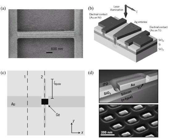
While the two previous detectors were mainly fabricated to perform an electrical near-field study of the properties of metal nanoparticles, nanoparticle resonators were—just like SPP antennas—also used to increase the photoresponse of small photodetectors. In 2008, Tang et al. demonstrated a very small germanium MSM detector with an active region of 150 × 60 × 80 nm which was equipped with a half-wave Hertz dipole antenna [44]. A schematic presentation is depicted in Figure 7.6c. The half-wave antenna was designed to concentrate the electric field inside the high-index germanium of the detector in the antenna gap. Polarization-dependent photocurrent measurements confirmed the half-wave behavior of the antenna. For a given device, the authors measured the photocurrent for the polarization along the antenna axis to be 20 times that for the perpendicular polarization for a very low bias voltage. Also spectral measurements showed a clear dipole peak at 1390 nm which was characteristic of the half-wavelength antenna. A rough approximation of the cutoff frequency was found to be over 100 GHz, suggesting the possibility of high-speed operation. Also Knight et al. reported on a nanoparticle antenna-based photodetector [45]. In this design, illustrated in Figure 7.6d, a nanorod antenna is fabricated on top of a silicon layer, forming a Schottky contact. At the resonance frequency of the nanorod antenna, the decay of the excited surface plasmons results in hot electrons that can cross the metal/semiconductor barrier and hence lead to a photocurrent. Arrays of these antenna-equipped Schottky detectors were studied in function of the particle geometry and light polarization. The expected dependency of the resonance wavelength on the particle size was found and showed good agreement to calculated absorption spectra. Furthermore Collin et al. demonstrated resonant cavity-enhanced MSM detectors [46, 47]. A large photocurrent enhancement was found for TE polarization, showing a good correspondence with calculations. However, the plasmon-enhanced mode in TM polarization exhibited a much weaker resonance than expected, due to a thin titanium layer at the metal–semiconductor interface and to the morphology of the deposited metal layers. 1D grating structures were also integrated on top of graphene photodetectors. Efficient conversion of light to a local plasmon resonance in the nanostrips leads to dramatic increase in the local electric field at the electrode–graphene interface, causing a photocurrent enhancement of more than 20 at the resonance frequency [48]. Like in previous detector designs, here also the polarization and wavelength sensitivity were demonstrated. Also Liu et al. demonstrated a dramatic increase in external quantum efficiency of plasmon-enhanced graphene photodetectors [49].
The third way presented of enhancing the photoresponse of a photodetector is the inclusion of a metallic photonic crystal on the detector area or arranging the detector structures in a periodic way, forming a photonic crystal structure. Like other approaches, the idea of integrating a resonant structure on the detector is increasing the interaction length between the incoming light and the active semiconductor region. This is interesting for thin film semiconductor detectors with a large absorption length like, for example, silicon. Instead of passing only once through the absorbing layer, at resonance the light passes multiple times before being absorbed in the semiconductor and the metal or being reradiated, increasing the probability of being absorbed in the active layer. One way of making use of metallic photonic crystal structures is introducing defect structures inside a 1D or 2D metallic photonic crystal, creating a resonant cavity with a high field enhancement. A more common approach is to use photonic crystal structures to efficiently couple incident light to an in-plane confined resonant mode which overlaps with the underlying active area of the photodetector. When designing a photonic crystal-enhanced photodetector, several aspects have to be considered: The resonance wavelength has to match the maximum absorption region of the active semiconductor material, the crystal needs both excellent incident light coupling and in-plane SPP confinement properties and the plasmonic fields at resonance need to spatially match with the active region of the photodetector. A thorough investigation of these concepts was theoretically described by Rosenberg et al. [50] and also realized experimentally [51]. The calculated band structure of the square crystal lattice of square holes in the metal layer revealed no band gaps but several flat-band regions. A SEM picture of the device under study can be seen in Figure 7.7a. These band-edge modes have a group velocity close to zero and consequently the light propagates very slowly and is confined within the patterned region. The authors realized the developed concept on a dots-in-a-well photodetector material [51]. FDTD simulations revealed a fundamental plasmonic dipole-like mode at 7.8 μm and higher-order modes of the in-plane square lattice photonic crystal around 5.5 μm. By varying the lattice constant between 1.83 and 2.38 μm, the peak wavelength could be shifted from 5.5 to 7.2 μm. The sensor could be made polarization-dependent by stretching the square holes in one dimension, splitting the resonance in two well-defined peaks. By comparing the responsivity of the photonic crystal photodetector with an unpatterned detector, an enhancement of 5 was achieved at T = 77 K. A similar detector was developed by Chang et al. [52]. In this publication a hexagonal lattice of etched circles was processed in a 50 nm thick gold film on an InGaAs quantum well detector. The detector was optimized for dual band detection at 5 and 9 μm wavelengths. The resonance wavelength was found to increase linearly with the lattice constant, while the size of the holes determines the light transmission via evanescent tunneling through the thin metallic 2D holes. A maximum photoresponse enhancement of 2.3 was found at 8.8 μm for a lattice constant of 3.2 μm and a hole diameter of 1.6 μm (T = 77 K). Several other research groups also studied metallic photonic crystal-enhanced photodetectors based on InAs/InGaAs quantum wells or quantum dots [53–55].
FIGURE 7.7 Photonic crystal-based enhanced photodetectors. Top row: the entire device, bottom row: a zoom of the photonic crystal lattice. (a) Square lattice photonic crystal for MIR quantum dot detector. (b) Hexagonal lattice for MIR quantum dot detector. (c) Periodically ordered array of nanopillar photodetectors with self-aligned hole array.
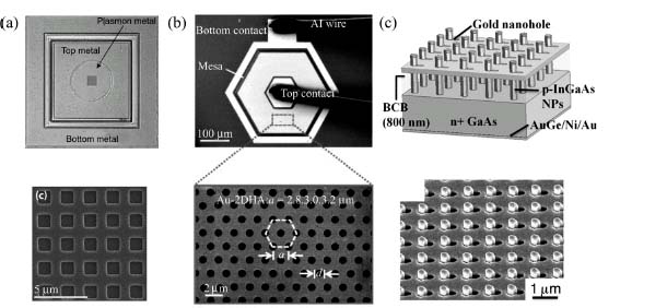
In the previous papers, a large-scale metallic photonic crystal was fabricated on top of a large-volume detector for mid-infrared light detection. Another way to use a photonic crystal structure is to fabricate small-scale detectors and position them in a periodic pattern. Senanayake et al. demonstrated a nanopillar-based plasmon-enhanced photodetector array operating in the NIR spectral range [56]. The authors were able to fabricate elongated nanoholes in a metal surface, self-aligned to a periodic array of nanopillar photodetectors (see Fig. 7.7c). SPPs can be excited by incident light on the nanohole array. The periodicity of the nanohole array supports Bloch wave resonances which cause a high electric field enhancement inside the nanopillars, leading to an enhanced photoresponse. However, when comparing the responsivity with a reference detector with ITO contacts, no enhancement was measured. The lower responsivity was attributed to the small spatial overlap between the nanopillar–substrate depletion region and the hot spots from the photonic crystal mode. The authors demonstrated that this nanopillar detector could be made wavelength- and polarization-dependent. The wavelength where the detector responsivity reaches a maximum can be tuned by changing the lattice constant of the nanohole array and the polarization sensitivity is determined by the stretching of the nanoholes in one dimension.
Finally, far-field integrated electrical detection of plasmonic effects was also realized. Dunbar et al., for example, fabricated slit and groove structures on top of a standard CMOS camera and achieved an eight times enhancement in the optical transmission [57]. Also several other applications electrically detected plasmons in the far field; for example, in biosensor applications, Jonsson et al. studied supported lipid bilayers by detecting LSPR changes [58].
7.2.4 Waveguide-Integrated Surface Plasmon Polariton Detectors
Plasmonic waveguides have been studied for a variety of applications, ranging from detection of biological properties to optical communication. For most applications, it would be useful to be able to excite and detect SPPs locally in the metallic waveguide by means of an integrated plasmon source and detector, respectively. Depending on the application, the important aspects of these electro-optical transducers are the efficiency, speed, and scalability. Here we will present various ways of detecting SPPs on chip. Direct electrical detection of SPPs propagating on metallic waveguides has been demonstrated with several types of detectors and waveguides. First, we will discuss SPP detection with both organic and solid-state semiconductors. Later we will present SPP detection with a superconducting detector and last the detection of waveguide modes in dielectric waveguides with plasmon-assisted photodetectors.
The first demonstration of direct electrical detection of waveguided SPPs was by Ditlbacher et al. [59]. The authors fabricated a 150 × 500 μm2 large organic photodiode on top of a 100 nm thick silver film that serves as both the plasmonic waveguide and the bottom contact of the diode. A schematic overview can be seen in Figure 7.8a. In the silver film, a grating structure and a slit were etched as SPP excitation structures. SPPs are generated by coupling incident laser light to SPPs at the slit or grating. They propagate along the silver waveguide and excite excitons in the organic materials when entering the diode, giving rise to an electric current that can be assessed directly. By scanning the focused laser beam across the device while measuring the photocurrent, photocurrent maps were generated, showing a strongly enhanced photocurrent when the laser spot scans across the SPP excitation structures, due to the excitation, propagation, and subsequent electrical detection of guided SPPs. Blocking regions were inserted between the excitation and detection region to exclude that the photocurrent generation was due to scattered light. Organic photodiodes offer a large processing flexibility and can be deposited on cheap substrates. However, in terms of speed, efficiency, and scalability, they cannot compete with solid-state photodetectors at this time.
FIGURE 7.8 Integrated SPP detection methods. (a) Organic photodiode SPP detector. (b) GaAs MSM detector inside MIM waveguide. (c) Ge nanowire detector for plasmons propagating on silver nanowires.

In 2009, several research groups started working on integrating solid-state photodetectors inside plasmonic waveguides. We demonstrated the electrical detection of SPPs in MIM waveguides by means of a nanoslit GaAs MSM detector [60]. MIM waveguides offer the prospects of combining a high confinement of the plasmonic mode with a reasonably long propagation distance [7]. A schematic representation of the device under study can be seen in Figure 7.8b. In the bottom layer of the MIM waveguide, a subwavelength detection slit is processed, forming the two contacts of the nanoscale MSM detector. SPPs are generated by focusing a laser beam on the subwavelength slit in the top metal layer; they propagate through the MIM waveguide and couple to a local mode at the detection slit, which is strongly absorbed by the GaAs layer. 2D photocurrent scans were performed for both TE and TM polarization, revealing only a high photocurrent for TM polarization, consistent with theory. Photocurrent line scans were acquired for different distances between the excitation slit and the detection slit in order to study the SPP decay along the waveguide. For a gold waveguide with 100 nm thick cured HSQ insulating layer, 1/e decay lengths of 3.5–9.5 μm were found for, respectively, 660–870 nm excitation wavelengths. Because of their extremely low capacitance and short transit times, these detectors are very suitable for high-speed integrated electrical detection of SPPs.
Next to flat film waveguides and MIM waveguides, metallic nanowire waveguides also were extensively studied in literature [6]. It was shown that nanowire waveguides too can have a high confinement of the electromagnetic fields and maintain a long propagation length. The challenge with nanowire waveguides remains the controlled fabrication of horizontally positioned nanowires on chip. Falk et al. proposed the electrical detection of nanowire waveguide plasmons by means of a germanium nanowire field-effect transistor [61]. The detection scheme is depicted in Figure 7.8c and consists of a silver nanowire crossing a germanium field-effect transistor. The silver nanowire guides the SPPs toward the crossing point, where they are converted to electron–hole pairs, giving rise to a photocurrent through the germanium nanowire. Also here the photocurrent was mapped in function of the laser spot position, revealing that SPPs are launched only when the laser is focused on the silver nanowire ends. A gating voltage on the silver nanowire was applied in order to realize a 300-fold increase in the plasmon-to-charge conversion efficiency. For some devices this conversion efficiency exceeded 50 electrons per plasmon at 1 V bias voltage. The authors demonstrated the utility of this SPP detector concept by electrically detecting the emission from a CdSe quantum dot that acts as a single-plasmon source. Photon correlation measurements of the far-field fluorescence revealed an antibunching signature, proving that the observed spot corresponds to an individual quantum dot. Together with an integrated SPP source [62, 63], the previous two approaches could lead to a plasmonic optoelectronic circuit. Also for plasmon biosensing, for example, like described by Dostalek et al. [64], integrated SPP sources and detectors would be of great interest in order to achieve a fully integrated plasmon biosensor.
The last direct semiconductor SPP detector which will be discussed in this chapter was demonstrated by Akbari et al. [65, 66]. The authors propose detection of SPPs with a Schottky photodetector. A gold stripe is deposited on an n-type silicon layer, functioning simultaneously as the waveguide and a Schottky contact. An ohmic contact is fabricated at the bottom of the wafer. The sample was cleaved perpendicular to the stripe axis and the asb0 SPP mode at the gold–silicon interface is excited by end-fire coupling with a polarization maintaining tapered optical fiber at the side of the sample. The excitation energy is chosen to be smaller than the bandgap energy so no direct electron–hole pair generation across the bandgap occurs. Absorption of the SPPs in the metal waveguide causes hot electrons which can cross the Schottky barrier if the excitation energy is large enough. These electrons can be collected, giving rise to a photocurrent. To prove the asb0 SPP mode was excited, photocurrent measurements were performed for TE and TM polarization, revealing that only a high photocurrent was obtained for TM-polarized light. This type of detector, however, is not confined to a small area, since a distance of several times the propagation length is necessary if one wants to achieve complete absorption of the optical power in the injected plasmon pulse.
Also in 2009, a completely different approach to integrated detection of SPPs was published by Heeres et al. [67]. The authors coupled a plasmon waveguide to a superconducting single-photon detector, thereby demonstrating on-chip electrical detection of single plasmons. The photodetector consisted out of a meandering NbN wire. The wire enters the superconductive state below approximately 9 K. If a bias current close to the critical current is applied, a single photon can create a local region in the normal resistive state, which is detected as a voltage pulse at the terminals of the wire. A flat polycrystalline gold waveguide was deposited on top of the detectors, separated by a thin dielectric, and again grating structures were used for achieving SPP excitation. Measuring the pulse frequency in function of the laser spot position confirmed plasmon excitation, propagation, and subsequent electrical detection by the superconductor detector. Time correlation measurements using a single-photon source proved the single-plasmon sensitivity of this type of detector. This publication demonstrates a very sensitive plasmon detector with an excellent time resolution which can be widely applied for research purposes. However, for commercial applications, the low-temperature operation still forms a large barrier.
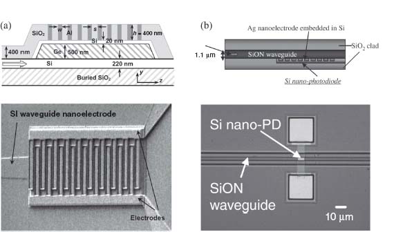
Not only are plasmon detectors useful for the detection of free-space radiation or waveguided SPPs, but they can also be employed for integrating dielectric waveguides with small, high-speed detectors, as plasmonic detectors can considerably shorten the length over which the full optical power of the light pulse is absorbed. Ren et al. proposed a Ge-on-SOI MSM photodetector with interdigitated electrodes to obtain a large TM enhancement at a wavelength of 1550 nm [68]. In Figure 7.9a both a schematic presentation of the device and a SEM picture are shown. A TE/TM-polarized optical mode propagates through the silicon core layer (220 nm thick). By calculating the correct grating pitch, the detector can be optimized for 1550 nm light detection. Under TE and TM illumination, the quantum efficiencies are found to be 14.1% and 86.7%, revealing a very high responsivity for TM polarization. FDTD simulations were performed to confirm that the interdigitated electrodes function as a 1D grating coupler above the detector's depletion layer. Also the time response of the detector was studied by the authors. A 3 dB bandwidth of 11.4 and 15.6 GHz was found for the TM and TE modes, respectively, meaning there is a trade-off between the enhancement in responsivity and the response speed. Also Fujikata et al. presented a similar detection scheme [69]. A low-loss SiON waveguide in-between SiO2 cladding layers was studied. On the silicon-active photodetector surface, silver grating MSM detector-interdigitated electrodes were processed. The resulting device can be seen in Figure 7.9b. An 850 nm laser light was introduced in the waveguide via butt-coupling with an optical fiber. A 10% quantum efficiency was achieved for TM polarization, which was two to three times larger than for TE polarization due to the surface plasmon resonance-enhanced near-field optical coupling between the waveguide and the Si layer. Also here the time response of the detector was measured, reaching a 17 ps FWHM of the generated electrical pulse following a 780 nm optical pulse smaller than 2 ps. The authors applied their design to a prototype of a large-scale integration on-chip optical clock system. They demonstrated a 5 GHz optical clock circuit operation with a four-branching H-tree structure, validating that plasmon-enhanced integrated photodetection for dielectric waveguide has very promising prospects for future on-chip optical circuits based on photonics and plasmonics.
FIGURE 7.9 Plasmon-enhanced detectors for detection of dielectric waveguide optical modes. Top row: device cross-sections. Bottom row: microscope pictures. (a) Plasmon-enhanced germanium detector fabricated on a silicon waveguide. (b) Plasmon-enhanced silicon detector on a SiON waveguide.
7.3 OUTLOOK
The unique properties of plasmonic nanostructures to guide and focus light into deep-subwavelength volumes have led to a number of demonstrations where plasmonic nanoantennas strongly enhance the absorption in photovoltaic cells and photodetectors for free-space radiation or integrated in plasmonic or photonic waveguide. Most of the demonstrations so far, however, are proof-of-principle demonstrations on model systems. To embed them in current or future applications, future research efforts should be focused to integrate plasmonic nanostructures in a more industrial setting.
These are clearly very different applications, as in solar cells broadband absorption is required while in photodetectors a narrow-band response is often desirable. Plasmonic resonances are relatively narrow band in nature and hence the resulting absorption enhancement is intrinsically more favorable in the case of a photodetector. Nevertheless, if plasmonic resonances can be employed to enhance the absorption of specific parts of the solar spectrum, where absorption is weak, such as near the band edge of (indirect) semiconductors, without disturbing or shadowing the other part of the spectrum, they can still be considered very useful in the quest for thinner and cheaper cells. The field of plasmonic solar cells has been very active recently, with a number of companies also active in the field. In order to be successfully embedded in a solar cell flow, issues related to contamination and cost-effective plasmonic patterning will need to be tackled.
Plasmon (or antenna)-enhanced photodetectors have a bright future in ultrafast, multi- or hyperspectral imaging in a wide range of wavelengths. The wavelength and polarization selectivity that has been demonstrated will with high probability find its way in several applications. Moreover, recent results on plasmonic-enhanced absorption in graphene show a route toward highly confined and atomically thin photodetectors that can be supported by an arbitrary substrate. More problematic is the integration of plasmonic effects in conventional silicon photodetectors, where contamination issues and the limited temperature budget are a potential road block.
Integrating plasmonic detectors with plasmonic and photonic waveguides is a very promising application, as the waveguided light can be captured in extremely small volumes, leading to potentially very fast, sensitive, and dense photodetectors. This is interesting for a number of applications, such as highly multiplexed integrated surface plasmon resonance-based biosensing and fast detection in integrated optical communication schemes. In the latter application, however, the lack of electrically excitable coherent plasmon sources and the large losses present in plasmonic waveguides prevent a pure plasmonic information routing scheme. However, when exploiting the nearly loss-free propagation in silicon or silicon nitride photonic waveguides, the ability of plasmonic nanostructures to reduce the interaction length between a photodetector and the waveguide will lead to faster detectors. However, in this case also, issues related to integration in an industrial fabrication platform need to be considered.
REFERENCES
1. Boltasseva A, Nikolajsen T, Leosson K, Kjaer K, Larsen MS, Bozhevolnyi SI (2005) Integrated optical components utilizing long-range surface plasmon polaritons. J. Lightw. Technol. 23(1): 413–422.
2. Berini P, Charbonneau R, Lahoud N, Mattiussi G (2005) Characterization of long-range surface-plasmon-polariton waveguides. J. Appl. Phys. 98: 043109.
3. Bozhevolnyi SI, Volkov VS, Devaux E, Ebbesen T (2005) Channel plasmon-polariton guiding by subwavelength metal grooves. Phys. Rev. Lett. 95: 046802.
4. Moreno E, Rodrigo SG, Bozhevolnyi SI, Martin-Moreno L, Garcia-Vidal FJ (2008) Guiding and focusing of electromagnetic fields with wedge plasmon polaritons. Phys. Rev. Lett. 100: 023901.
5. Maier SA, Kik PG, Atwater HA, Meltzer S, Harel E, Koel BE, Requicha AG (2003) Local detection of electromagnetic energy transport below the diffraction limit in metal nanoparticle plasmon waveguides. Nature 2: 229–232.
6. Oulton RF, Sorger VJ, Genov DA, Pile DFP, Zhang X (2008) A hybrid plasmonic waveguide for subwavelength confinement and long-range propagation. Nat. Photonics 2: 496–500.
7. Dionne JA, Lezec HJ, Atwater HA (2006) Highly confined photon transport in subwavelength metallic slot waveguides. Nano Lett. 6: 1928–1932.
8. Nagae M (1972) Response time of metal-insulator-metal tunnel junctions. Jpn. J. Appl. Phys. 11: 1611–1621.
9. Soole JBD, Lamb RN, Hughes HP, Apsley N (1986) Surface plasmon enhanced photoconductivity in planar metal-oxide-metal tunnel junctions. Solid State Commun. 59: 607–611.
10. Berthold K, Höpfel RA, Gornik E (1985) Surface plasmon polariton enhanced photoconductivity of tunnel junctions in the visible. Appl. Phys. Lett. 46(7): 626–628.
11. Möller R, Albrecht U, Boneberg J, Koslowski B, Leiderer P, Dransfeld K (1991) Detection of surface plasmons by scanning tunneling microscopy. J. Vac. Sci. Technol. B 9: 506.
12. Hobbs PCD, Laibowitz RB, Libsch FR (2005) Ni-NiO-Ni tunnel junctions for terahertz and infrared detection. Appl. Opt. 44: 6813.
13. Hobbs PCD, Laibowitz RB, Libsch FR, LaBianca NC, Chiniwalla PP (2007) Efficient waveguide-integrated tunnel junction detectors at 1.6 μm. Opt. Express 15: 16376.
14. Ittah N, Gilad N, Yutsis I, Selzer Y (2009) Measurement of electronic transport through 1G0 gold contacts under laser irradiation. Nano Lett. 2009: 1615.
15. Ittah N, Selzer Y (2011) Electrical detection of surface plasmon polaritons by 1G0 gold quantum point contacts. Nano Lett. 11: 529.
16. Campbell P, Green MA (1987) Light trapping properties of pyramidally textured surfaces. J. Appl. Phys. 62(1): 243–249.
17. Yablonovitch E (1982) Statistical ray optics. J. Opt. Soc. Am. 72(7): 899–907.
18. Ferry VE, Sweatlock LA, Pacifici D, Atwater HA (2008) Plasmonic nanostructure design for efficient light coupling into solar cells. Nano Lett. 8(12): 4391–4397.
19. Maier SA (2007) Plasmonics: Fundamentals and Applications. New York: Springer.
20. Hägglund C, Zäch M, Petersson G, Kasemo B (2008) Electromagnetic coupling of light into a silicon solar cell by nanodisk plasmons. Appl. Phys. Lett. 92: 053110.
21. Rockstuhl C, Fahr S, Lederer F (2008) Absorption enhancement in solar cells by localized plasmon polaritons. J. Appl. Phys. 104: 123102.
22. Stuart HR, Hall DG (1998) Island size effects in nanoparticle-enhanced photodetectors. Appl. Phys. Lett. 73: 3815.
23. Catchpole K, Polman A (2008) Design principles for particle enhanced solar cells. Appl. Phys. Lett. 93(19): 191113.
24. Ouyang Z, Pillai S, Beck F, Kunz O, Varlamov S, Catchpole KR, Campbell P, Green MA (2010) Effective light trapping in polycrystalline silicon thin-film solar cells by means of rear localized surface plasmons. Appl. Phys. Lett. 96(26): 261109.
25. Rand BP, Peumans P, Forrest SR (2004) Long-range absorption enhancement in organic tandem thin-film solar cells containing silver nanoclusters. J. Appl. Phys. 96(12): 7519.
26. Morfa AJ, Rowlen KL, Reilly TH, Romero MJ, van de Lagemaat J (2008) Plasmon-enhanced solar energy conversion in organic bulk heterojunction photovoltaics. Appl. Phys. Lett. 92(1): 013504.
27. Kim S, Na S, Jo J, Kim D, Nah Y (2008) Plasmon enhanced performance of organic solar cells using electrodeposited Ag nanoparticles. Appl. Phys. Lett. 93(7): 073307.
28. Mapel JK, Singh M, Baldo MA, Celebi K (2007) Plasmonic excitation of organic double heterostructure solar cells. Appl. Phys. Lett. 90(12): 121102.
29. Tvingstedt K, Persson N, Inganas O, Rahachou A, Zozoulenko IV (2007) Surface plasmon increase absorption in polymer photovoltaic cells. Appl. Phys. Lett. 91(11): 113514.
30. Reilly TH, van de Lagemaat J, Tenent RC, Morfa AJ, Rowlen KL (2008) Surface-plasmon enhanced transparent electrodes in organic photovoltaics. Appl. Phys. Lett. 92(24): 243304.
31. Lindquist NC, Luhman WA, Oh S, Holmes RJ (2008) Plasmonic nanocavity arrays for enhanced efficiency in organic photovoltaic cells. Appl. Phys. Lett. 93(12): 123308.
32. Ishi T, Fujikata J, Makita K, Baba T, Ohashi K (2005) Si nano-photodiode with a surface plasmon antenna. Jpn. J. Appl. Phys. 44(12): 364–366.
33. Laux E, Genet C, Skauli T, Ebbesen TW (2008) Plasmonic photon sorters for spectral and polarimetric imaging. Nat. Photonics 2: 161–164.
34. Ropers C, Neacsu CC, Elsaesser T, Albrecht M, Raschke MB, Lienau C (2007) Grating-coupling of surface plasmon onto metallic tips: a nanoconfined light source. Nano Lett. 7(9): 2784–2788.
35. Weeber J-C, Gonzalez MU, Baudrion A-L, Dereux A (2005) Surface plasmon routing along right angle bent metal strips. Appl. Phys. Lett. 87: 221101.
36. Chen C, Verellen N, Lodewijks K, Lagae L, Maes G, Borghs G, Van Dorpe P (2010) Groove-gratings to optimize the electric field enhancement in a plasmonic nanoslit-cavity. Appl. Phys. Lett. 108: 034319.
37. Shackleford JA, Grote R, Currie M, Spanier JE, Nabet B (2009) Integrated plasmonic lens photodetector. Appl. Phys. Lett. 94: 083501.
38. White JS, Veronis G, Yu Z, Barnard ES, Chandran A, Fan S, Brongersma ML (2009) Extraordinary optical absorption through subwavelength slits. Opt. Lett. 34(5): 686–688.
39. Yu Z, Veronis G, Fan S, Brongersma ML (2006) Design of midinfrared photodetectors enhanced by surface plasmons on grating structures. Appl. Phys. Lett. 89: 151116.
40. Muhlschlegel P, Eisler H-J, Martin OJF, Hecht B, Pohl DW (2005) Resonant optical antennas. Science 308(5728): 1607–1609.
41. Kinkhabwala A, Yu Z, Fan S, Avlasevich Y, Mullen K, Moerner WE (2009) Large single-molecule fluorescence enhancements produced by a bowtie nanoantenna. Nat. Photonics 3: 654–657.
42. De Vlaminck I, Van Dorpe P, Lagae L, Borghs G (2007) Local electrical detection of single nanoparticle plasmon resonance. Nano Lett. 7(3): 703–706.
43. Barnard ES, Pala RA, Brongersma ML (2011) Photocurrent mapping of near-field optical antenna resonances. Nat. Nanotechnol. 6: 588–593.
44. Tang L, Kocabas SE, Latif S, Okyay AK, Ly-Gagnon D-S, Saraswat KC, Miller DAB (2008) Nanometre-scale germanium photodetector enhanced by a near-infrared dipole antenna. Nat. Photonics 2: 226–229.
45. Knight MW, Sobhani H, Nordlander P, Halas NJ (2011) Photodetection with active optical antennas. Science 332: 702–704.
46. Collin S, Pardo F, Teissier R, Pelouard J-L (2004) Efficient light absorption in metal-semiconductor-metal nanostructures. Appl. Phys. Lett. 85(2): 194–196.
47. Collin S, Pardo F, Pelouard J-L (2003) Resonant-cavity-enhanced subwavelength metal-semiconductor-metal photodetector. Appl. Phys. Lett. 83(8): 1521–1523.
48. Echtermeyer TJ, Britnell L, Jasnos PK, Lombardo A, Gorbachev RV, Grigorenko AN, Geim AK, Ferrari AC, Novoselov KS (2011) Strong plasmonic enhancement of photovoltage in graphene. Nat. Commun. 2(458): 1–5.
49. Liu Y, Cheng R, Liao L, Zhou H, Bai J, Liu G, Liu L, Huang Y, Duan X (2011) Plasmon resonance enhanced multicolour photodetection by graphene. Nat. Commun. 2: 579.
50. Rosenberg J, Shenoi RV, Krishna S, Painter O (2010) Design of plasmonic photonic crystal resonant cavities for polarization sensitive infrared photodetectors. Opt. Express 18(4): 3672–3686.
51. Rosenberg J, Shenoi RV, Vandervelde TE, Krishna S, Painter O (2009) A multispectral and polarization-selective surface-plasmon resonant midinfrared detector. Appl. Phys. Lett. 95: 161101.
52. Chang C-C, Sharma YD, Kim Y-S, Bur JA, Shenoi RB, Krishna S, Huang D, Lin S-Y (2010) A surface plasmon enhanced infrared photodetector based on InAs quantum dots. Nano Lett. 10(5): 1704–1709.
53. Lee SC, Krishna S, Brueck SRJ (2011) Plasmonic-enhanced photodetectors for focal plane arrays. IEEE Photonics Technol. Lett. 23(14): 935–937.
54. Lee SC, Krishna S, Brueck SRJ (2009) Quantum dot infrared photodetector enhanced by surface plasma wave excitation. Opt. Express 17(25): 23160–23168.
55. Wu W, Bonakdar A, Mohseni H (2010) Plasmonic enhance quantum well infrared photodetector with high detectivity. Appl. Phys. Lett. 96: 161107.
56. Senanayake P, Hung C-H, Shapiro J, Lin A, Liang B, Williams BS, Huffacker DL (2011) Surface plasmon-enhanced nanopillar photodetectors. Nano Lett. 11(12): 5279–5283.
57. Dunbar LA, Guillaumee M, De Leon-Perez F, Santschi C, Grenet E, Eckert R, Lopez-Tejeira F, Garcia-Vidal FJ, Martin-Moreno L, Stanley RP (2009) Enhanced transmission from a single subwavelength slit aperture surrounded by grooves on a standard detector. Appl. Phys. Lett. 95: 011113.
58. Jonsson MP, Jonsson P, Dahlin AB, Hook F (2007) Supported lipid bilayer formation and lipid-membrane-mediated biorecognition reactions studied with a new nanoplasmonic sensor template. Nano Lett. 7(11): 3462–3468.
59. Ditlbacher H, Aussenegg FR, Krenn JR, Lamprecht B, Jakopix B, Leising G (2006) Organic diodes as monolithically integrated surface plasmon polariton detectors. Appl. Phys. Lett. 89: 161101.
60. Neutens P, Van Dorpe P, De Vlaminck I, Lagae L, Borghs G (2009) Electrical detection of confined gap plasmons in metal–insulator–metal waveguides. Nat. Photonics 3: 283–286.
61. Falk AL, Koppens FHL, Yu CL, Kang K, de Leon Snapp N, Akimov AV, Jo M-H, Lukin MD, Park H (2009) Near-field electrical detection of optical plasmons and single-plasmon sources. Nat. Phys. 5: 475–479.
62. Neutens P, Lagae L, Borghs G, Van Dorpe P (2010) Electrical excitation of confined surface plasmon polaritons in metallic slot waveguides. Nano Lett. 10(4): 1429–1432.
63. Walters RJ, van Loon RVA, Brunets I, Schmitz J, Polman A (2009) A silicon-based electrical source of surface plasmon polaritons. Nat. Mater. 9: 21–25.
64. Dostalek J, Ctyroky J, Homola J, Brynda E, Skalsky M, Nekvindova P, Spirkova J, Skvor J, Schrofel J (2001) Surface plasmon resonance biosensor based on integrated optical waveguide. Sens. Act. B 76: 8–12.
65. Akbari A, Berini P (2009) Schottky contact surface-plasmon detector integrated with an asymmetric metal stripe waveguide. Appl. Phys. Lett. 95: 021104.
66. Akbari A, Tait RN, Berini P (2010) Surface plasmon waveguide Schottky detector. Opt. Express 18(8): 8505–8514.
67. Heeres RW, Dorenbos SN, Koene B, Solomon GS, Kouwenhoven LP, Zwiller V (2010) On-chip single plasmon detection. Nano Lett. 10: 661–664.
68. Ren F-F, Ang K-W, Song J, Yu M, Lo G-Q, Kwong D-L (2010) Surface plasmon enhanced responsivity in a waveguided germanium metal-semiconductor-metal photodetector. Appl. Phys. Lett. 97: 091102.
69. Fujikata J, Nose K, Ushida J, Nishi K, Kinoshita M, Shimizu T, Ueno T, Okamoto D, Gomyo A, Mizuno M, Tsuchizawa T, Watanabe T, Yamada K, Itabashi S, Ohashi K (2008) Waveguide-integrated Si nano-photodiode with surface-plasmon antenna and its applications to on-chip optical clock distribution. Appl. Phys. Express 1: 022001.