Appendix A
Properties of Biological Fluids
This appendix represents a compilation of information relevant to biomaterials scientists regarding the properties and composition of several body fluids, i.e., blood, plasma (serum), cerebrospinal fluid, synovial fluid, saliva, tear fluid, and lymph. Where possible, ranges of values are provided, but the reader should recognize that significant variations are possible, particularly in states of disease. Further, the data reported here reflect adult measurements and may be substantially different in a pediatric population. Values for cerebrospinal fluid refer to the lumbar region, those for synovial fluid refer to the knee joint, and those for lymph refer to the thoracic duct, unless otherwise specified. Table A1 lists the physico-chemical properties of these fluids. Table A2 provides the typical cellular composition of human blood. Table A3 shows the normal volumes of these fluids in males and females, and presents equations whereby such volumes can be estimated from the mass of the individual. Next, Table A4 lists the approximate concentrations of the major proteins present in various biological fluids. Tables A5 and A6 present the concentrations of inorganic and organic species, respectively, in these fluids. Table A7 provides data on the major plasma proteins, i.e., concentration, molecular weight, isoelectric point (pI), sedimentation coefficient (S), diffusion coefficient (D), extinction coefficient (E280), partial specific volume (V20), carbohydrate content (C), and half-life. Finally, Tables A8 and A9 present information on the proteins involved in the complement pathway and blood coagulation pathway, respectively. Some of the information contained in this appendix have been previously published in Black, J., & Hastings, G. (eds.) (1998). Handbook of Biomaterial Properties. Chapman & Hall: New York, NY, pp. 114–124.
TABLE A1 Physico-chemical Properties of Several Biological Fluids
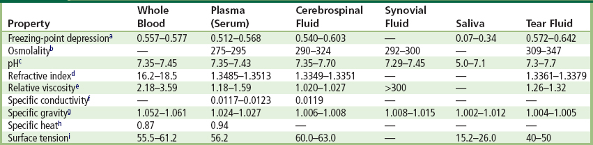
bUnits are mosm/kg H2O. Calculated from freezing-point depression.
cpH measured from arterial blood and plasma, and from cisternal portion of CSF.
eMeasured in vitro at 37°C for whole blood, plasma, and synovial fluid, and at 38°C for cerebrospinal fluid. The viscosity of serum is slightly less than plasma due to the absence of fibrinogen.
fUnits are S/cm. Measured at 25°C for plasma, 18°C for CSF.
iUnits are dyn/cm. Measured at 20°C.
TABLE A2 Cellular Composition of Blood
| Cell Type | Cells/μl | Half-Life in Circulation |
| Erythrocytes | 4.6–6.2 × 10⁶ (M)4.2–5.2 × 10⁶ (F) | 25 ± 2 days |
| Leukocytes | ||
| Neutrophils | 3000–5800 | 6–8 hours |
| Eosinophils | 50–250 | 8–12 hours |
| Basophils | 15–50 | ? |
| Monocytes | 300–500 | 1–3 days |
| Lymphocytes | 1500–3000 | Variable |
| Platelets | 1.5–3.5 × 10⁵ | 3.2–5.2 days |
| Reticulocytes | 2.3–9.3 × 10⁴ | — |
TABLE A3 Volumes of Various Biological Fluidsa

Males (M)Females (F)
BV = 41.0 × b + 1530BV = 47.16 × b + 864
PV = 19.6 × b + 1050PV = 28.89 × b + 455
EV = 21.4 × b + 490EV = 18.26 × b + 409
aThe following equations can be used to estimate blood volume (BV), erythrocyte volume (EV), and plasma volume (PV) from the known body mass (b, kg) with a coefficient of variation of approximately 10%:
TABLE A4 Protein Concentrations (mg/dl) in Various Biological Fluids

TABLE A5 Concentrations of Major Inorganic Substances (mmol/L) in Various Biological Fluids

TABLE A6 Concentrations of Organic Compounds (mg/dl) in Various Biological Fluids

TABLE A7 Properties of the Major Plasma Proteins
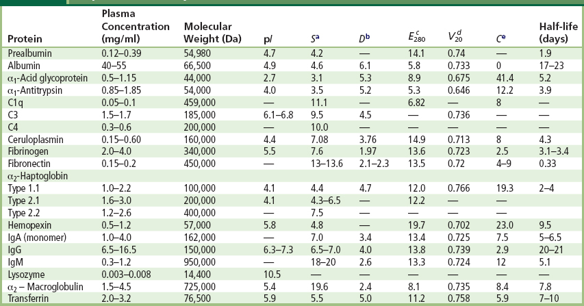
aSedimentation constant in water at 20°C, expressed in Svedberg units.
bDiffusion coefficient in water at 20°C, expressed in 10−7 cm2/sec.
cExtinction coefficient for light of wavelength 280 nm traveling 1 cm through a 10 mg/ml protein solution.
dPartial specific volume of the protein at 20°C, expressed as mlg−1.
eCarbohydrate content of the protein, expressed as the percentage by mass.
TABLE A8 Properties of Proteins Involved in the Complement System

TABLE A9 Properties of Proteins Involved in Blood Coagulation

∗Dr. Steven M. Slack died on April 29, 2010. He was Professor in the Department of Biomedical Engineering and Associate Dean of Graduate Studies in the Herff College of Engineering, at the University of Memphis in Tennessee. He will be sorely missed by the four editors of this textbook, by his colleagues around the world, and especially by his many students at the University of Memphis.