Penetrating foreign bodies
Appearances on plain radiographs
Glass
All glass is radio-opaque. Visibility of glass is not dependent on its lead content1,2.
The radiographic density of the different types of glass does vary. Imaging technique is important. A soft tissue exposure is essential.
Zooming on a digital image is often necessary, otherwise very small fragments are easily overlooked.
Metal
Most metals are radio-opaque. A notable exception is aluminium.
Wood or plastic
Only occasionally will wood be visualised3–5. A splinter might be well defined on a radiograph if the fragment has paint on its surface.
Why is wood almost non-opaque on a radiograph?
▪ Explanation: the detection of a foreign body is dependent on the atomic numbers of its constituent atoms in contrast to those of human soft tissue. Wood and soft tissue are both largely composed of carbon and other atoms with similar, low, atomic numbers. Therefore they are not readily differentiated by the X-ray beam.
In clinical practice it is best to assume that all splinters, thorns, and fragments of plastic will be non-opaque on a radiograph.
This splinter of wood was only visible because it had a thick coat of paint on its surface. It is usually very difficult to visualise wooden splinters or thorns on a radiograph.
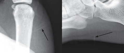
Wood splinters are often invisible, or barely visible, on a radiograph. On the left, a wood splinter (arrow); on the right, a toothpick (arrow).
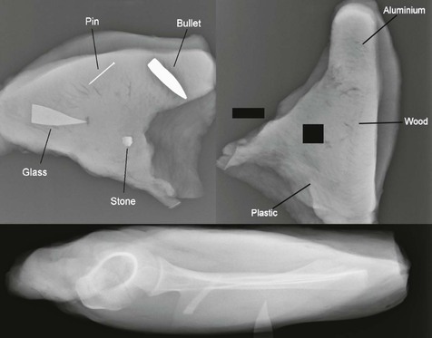
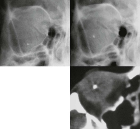
Visibility of penetrating foreign bodies. The top left image shows four different foreign bodies covered by soft tissue (turkey meat). The glass shard is from a broken picture frame. The top right image shows three other foreign bodies covered by turkey meat. All are invisible. The bottom image shows two pieces of glass buried in the soft tissues of a shoulder of lamb. Note that bone overlies part of one piece of glass and it is that portion of the fragment that is most difficult to visualise.
Suspected foreign bodies
Soft tissue laceration6–8
Foreign body detection
First choice in the Emergency Department: plain radiography.
Back up options in specific cases:
□ Glass, metal, wood and plastic well visualised.
□ But… operator dependent, and will detect superficial foreign bodies only.
□ Most foreign bodies are well visualised.
Imaging pitfalls: Glass and metallic foreign bodies2,3,8,9
▪ Pitfall (1): Overlying bone on a radiograph will hide/obscure glass fragments including very dense shards. Project the site of injury away from bone. Two or more projections are required.
▪ Pitfall (2): If there has been high energy trauma, such as a fall from a height, a glass foreign body may be driven deep into the tissues and well away from the laceration. The precise history and circumstance in relation to the glass injury will determine if the radiographic field needs to be extended well beyond the laceration.
▪ Pitfall (3): With gun shot wounds the bullet (if there is no exit wound) can be situated far from the entry point. A wide radiographic field may need to be covered. On occasion, fluoroscopy can be helpful when searching for a missing bullet that has not been located on the original radiographs.
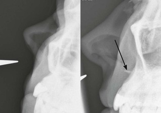
Glass wound to the face.
The left hand image shows the pointer indicating the site of the wound—but there is no evidence of a glass foreign body. The second image was taken with the maxilla rotated away from the site of injury, revealing a glass foreign body (arrow). Be careful: overlying bone will obscure or hide a glass foreign body.
Foreign body removal
Prior to surgical exploration it will sometimes be helpful if the precise position of a glass fragment or wooden splinter is shown beneath the skin. In these instances sonography can provide guidance at the time of removal. If a foreign body is situated deep in the soft tissues and sonography cannot provide the required information, then CT or MRI will often provide excellent localisation.
Orbital injury
Foreign body detection10,11
Most foreign bodies will be detected with slit lamp ophthalmoscopy.
Plain radiography, ultrasound, or CT will be of assistance in selected cases.
▪ Glass or metal fragments
Plain film radiography is recommended.
▪ Wood or plastic fragments
Sonography is recommended. The accuracy of detection is dependent on an experienced operator and the quality of the equipment. CT is an alternative to ultrasound and is the preferred investigation in some centres. CT is sensitive, shows the retrobulbar space better than ultrasound, and is less operator-dependent10. MRI is also available. It can be utilised when CT findings are uncertain11. A history of a ferromagnetic foreign body injury to the orbit is a contraindication to a MRI examination.
Orbital radiography.
Two frontal projections: downward gaze (top left) and then upward gaze (top right). Movement (or lack of movement) of a fragment on upward and downward gaze will indicate whether it is situated within or outside the globe.
CT is available should there be any lack of certainty as to the position of a foreign body after the plain films have been scrutinised. The CT image shown on the right is of a different patient.
Foreign body removal
Ultrasound or CT can provide accurate localisation prior to exploration of the orbit10.