20
PART 1 Organization of the Human Body


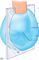
Outer balloon wall
Inner balloon wall
Cavity
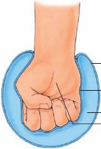
Fist (organ)
Outer balloon wall(parietal serousmembrane)Inner balloon wall(visceral serousmembrane)
Cavity
Fist (organ)
(b)
(c)
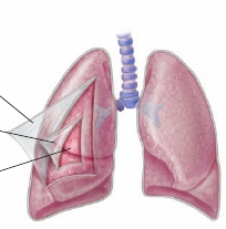
FIGURE 1.15
Serous Membranes
(
a
) A fist pushing into a balloon. A “glass” sheet indicates the location of a section through the balloon. (
b
) Interior view produced by the section in (
a
). Thefist represents an organ, and the walls of the balloon represent the serous membranes. The inner wall of the balloon represents a visceral serous membranein contact with the fist (organ). The outer wall of the balloon represents a parietal serous membrane. (
c
) The relationship of the parietal and serous mem-branes to the lungs. Figure 1.16 shows the relationship of the parietal and visceral membranes to the heart.
esophagus, and other structures, such as blood vessels and nerves.The two lungs are located on each side of the mediastinum.Abdominal muscles primarily enclose the
abdominal cavity,
which contains the stomach, the intestines, the liver, the spleen,the pancreas, and the kidneys. Pelvic bones encase the small spaceknown as the
pelvic cavity,
where the urinary bladder, part of thelarge intestine, and the internal reproductive organs are housed.The abdominal and pelvic cavities are not physically separatedand sometimes are called the
abdominopelvic cavity.
Serous Membranes
Serous
(s ē r′ ŭ s)
membranes
line the trunk cavities and coverthe organs within these cavities.
Parietal
membranes are foundagainst the outer wall of a body cavity, and
visceral
membranesare found covering the organs in a body cavity. Imagine pushingyour fist into an inflated balloon (figure 1.15). Your fist representsan organ; the inner balloon wall in contact with your fist representsthe
visceral
(vis′er- ă l; organ)
serous membrane
covering theorgan; and the outer part of the balloon wall represents the
pari-etal
(p ă -r ī ′ ĕ -t ă l; wall)
serous membrane.
The cavity, or space,between the visceral and parietal serous membranes is normallyfilled with a thin, lubricating film of serous fluid produced bythe membranes. As organs rub against the body wall or againstanother organ, the combination of serous fluid and smooth serousmembranes reduces friction.The thoracic cavity contains three serous membrane–lined cav-ities: a cavity for the heart called the
pericardial
(per-i-kar′d ē - ă l;around the heart)
cavity
and two lung cavities called the
pleural
(ploor′ ă l; associated with the ribs)
cavities.
The pericardial cav-ity surrounds the heart (figure 1.16
a
). The visceral pericardiumcovers the heart, which is contained within a connective tissue saclined with the parietal pericardium. The pericardial cavity, which
contains pericardial fluid, is located between the visceral pericar-dium and the parietal pericardium.Each lung is covered by visceral pleura and surrounded by apleural cavity (figure 1.16
b
). Parietal pleura line the inner surfaceof the thoracic wall, the outer surface of the parietal pericardium,and the superior surface of the diaphragm. The pleural cavity liesbetween the visceral pleura and the parietal pleura and containspleural fluid.The abdominopelvic cavity contains a serous membrane–lined cavity called the
peritoneal
(per′i-t ō -n ē ′ ă l; to stretch over)
cavity
(figure 1.16
c
).
Visceral peritoneum
covers many of theorgans of the abdominopelvic cavity.
Parietal peritoneum
linesthe wall of the abdominopelvic cavity and the inferior surfaceof the diaphragm. The peritoneal cavity is located between thevisceral peritoneum and the parietal peritoneum and containsperitoneal fluid.The serous membranes can become inflamed, usually as a resultof an infection.
Pericarditis
(per′i-kar-d ī ′tis;
-itis,
inflammation) isinflammation of the pericardium,
pleurisy
(ploor′i-s ē ) is inflamma-tion of the pleura, and
peritonitis
(per′i-t ō -n ī ′tis) is inflammation ofthe peritoneum.The abdominopelvic cavity also has other specialized mem-branes in addition to the parietal and visceral membranes, called
mesenteries
(mes′en-ter- ē z). The mesenteries anchor the organsto the body wall and provide a pathway for nerves and blood ves-sels to reach the organs. The mesenteries consist of two layersof peritoneum fused together (figure 1.16
d
). They connect thevisceral peritoneum of some abdominopelvic organs to the pari-etal peritoneum on the body wall. The mesenteries also connectcertain organs’ visceral peritoneum to the visceral peritoneumof other abdominopelvic organs. Other abdominopelvic organsare more closely attached to the body wall and do not have mes-enteries. Parietal peritoneum covers these other organs, which