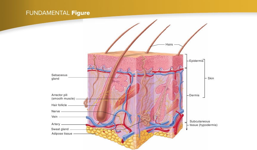
FIGURE 5.1
Skin and Subcutaneous Tissue
The skin, consisting of the epidermis and the dermis, is connected by the subcutaneous tissue to underlying structures. Note the accessory structures (hairs,glands, and arrector pili), some of which project into the subcutaneous tissue, as well as the large amount of adipose tissue in the subcutaneous tissue.

basale, stratum spinosum, stratum granulosum, stratum lucidum,and stratum corneum. The number of cell layers in each stratumand even the number of strata in the skin vary, depending on theirlocation in the body.
Stratum Basale
The deepest portion of the epidermis is a single layer of cuboidal orcolumnar cells called the
stratum basale
(b ā ′s ă -l ē ), or
stratumgerminativum
(jer′mi-n ă -t ī v′um; figure 5.3,
stratum 1
). The epider-mis is anchored to the basement membrane by hemidesmosomes. Inaddition, desmosomes hold the keratinocytes together (seechapter 4). The connections formed by the hemidesmosomes anddesmosomes provide structural strength to the epidermis. Keratino-cytes are strengthened internally by keratin fibers (intermediatefilaments) that insert into the desmosomes. Keratinocyte stem cellsof the stratum basale undergo mitotic divisions approximately every19 days. One daughter cell remains a stem cell in the stratum basaleand divides again, but the other daughter cell is pushed toward thesurface and becomes keratinized. It takes approximately 40–56 daysfor the cell to reach the epidermal surface and slough off.
(figure 5.3,
stratum 2
). As the cells in this stratum are pushed tothe surface, they flatten; desmosomes break apart, and newdesmosomes form. During preparation for microscopic observa-tion, the cells usually shrink from one another, except where theyare attached by desmosomes, causing the cells to appear spiny—hence the name stratum spinosum. Additional keratin fibers andlipid-filled, membrane-bound organelles called
lamellar
(lam′ ĕ -l ă r, l ă -mel′ar)
bodies
form inside the keratinocytes.
Stratum Granulosum
The
stratum granulosum
(gran- ū -l ō ′s ŭ m) consists of two to fivelayers of somewhat flattened, diamond-shaped cells. The longaxes of these cells are oriented parallel to the surface of the skin(figure 5.3,
stratum 3
). This stratum derives its name from thepresence of protein granules of
keratohyalin
(ker′ ă -t ō -h ī ′ ă -lin),which accumulate in the cytoplasm of the cell. The lamellar bod-ies, formed as the cells pass through the stratum spinosum, moveto the plasma membrane and release their lipid contents intothe extracellular space. Inside the cell, a protein envelope formsbeneath the plasma membrane. In the most superficial layers ofthe stratum granulosum, the nucleus and other organelles degener-ate, and the cell dies. Unlike the other organelles and the nucleus,however, the keratin fibers and keratohyalin granules within thecytoplasm do not degenerate.
143
Stratum Spinosum
Superficial to the stratum basale is the
stratum spinosum
(sp ī -n ō ′s ŭ m), consisting of 8–10 layers of many-sided cells