CHAPTER 7 Skeletal System: Gross Anatomy
199

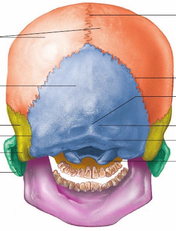
Sagittal suture
Parietal bones
Lambdoid suture
Occipital bone
External occipitalprotuberance
Temporal bone
Occipitomastoidsuture
Mastoid process
Superiornuchal line
Inferiornuchal line
Occipital condyle
Zygomatic arch
Posterior view
FIGURE 7.3
Posterior View of the Skull
(The names of the bones are in bold.)
Posterior View of the Skull
The parietal and occipital bones are the major structures visible inthe posterior view (figure 7.3). The parietal bones are joined to the
occipital bone
by the
lambdoid
(lam′doyd)
suture.
Occasionally,extra small bones called
sutural
(soo′choor- ă l)
bones
(
wormianbones
) form along the lambdoid suture.An
external occipital protuberance
is present on the posteriorsurface of the occipital bone (figure 7.3). It can be felt through thescalp at the base of the head and varies considerably in size fromperson to person. The external occipital protuberance is the site ofattachment of the
ligamentum nuchae
(noo′k ē ; nape of neck), anelastic ligament that extends down the neck and helps keep the headerect by pulling on the occipital region of the skull.
Nuchal lines,
aset of small ridges that extend laterally from the external occipitalprotuberance, are the points of attachment for several neck muscles.
Lateral View of the Skull
The parietal bone and the squamous part of the temporal bone forma major portion of the side of the head (figure 7.4). The term
temporal
means related to time, and the temporal bone is sonamed because the hair of the temples is often the first to turnwhite, indicating the passage of time. The
squamous suture
joins
these bones. A prominent feature of the temporal bone is a largehole, the
external auditory canal
(
external acoustic meatus
),which transmits sound waves toward the eardrum, or tympanicmembrane. The external ear, or auricle, surrounds the canal. Justposterior and inferior to the external auditory canal is a large infe-rior projection, the
mastoid
(mas′toyd)
process.
The process canbe seen and felt as a prominent lump just posterior to the ear. Theprocess is not solid bone but is filled with cavities called
mastoidair cells,
which are connected to the middle ear. Important neckmuscles involved in rotating the head attach to the mastoid pro-cess. The superior and inferior
temporal lines,
which are attach-ment points of the temporalis muscle, one of the major musclesof mastication, arch across the lateral surface of the parietal bone.The lateral surface of the
greater wing
of the
sphenoid
(sf ē ′noyd)
bone
is immediately anterior to the temporal bone(figure 7.4). Although appearing to be two bones, one on each sideof the skull, the sphenoid bone is actually a single bone thatextends completely across the skull. Anterior to the sphenoidbone is the
zygomatic
(z ī ′g ō -mat′ik)
bone,
or cheekbone, whichcan be easily seen and felt on the face (figure 7.5).The
zygomatic arch,
which consists of joined processes fromthe temporal and zygomatic bones, forms a bridge across the side