Mental protuberance
Anterior view
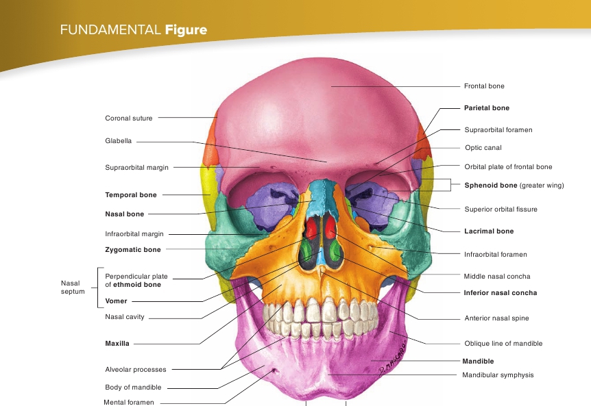
FIGURE 7.6
Anterior View of the Skull

(The names of the bones are in bold.)

Glabella
Supraorbitalmargin
Zygomaticbone
Frontal bone
Maxilla
Mandible
Mental protuberance
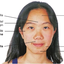
FIGURE 7.7
Anterior View of Bony Landmarks on the Face
(The names of the bones are in bold.)
fossa with its apex directed posteriorly (figure 7.8; see figure 7.6).They are called orbits because the eyes rotate within the fossae. Thebones that make up the orbits provide both protection for the eyesand attachment points for the muscles that move the eyes. Themajor portion of each eyeball is within the orbit, and the portion ofthe eye visible from the outside is relatively small. Each orbit con-tains blood vessels, nerves, and adipose tissue, as well as the eye-ball and the muscles that move it. The bones forming the orbit arelisted in table 7.4.The orbit has several openings through which structures com-municate between the orbit and other cavities. The nasolacrimal ductpasses from the orbit into the nasal cavity through the
nasolacrimalcanal,
carrying tears from the eyes to the nasal cavity. The opticnerve for vision passes from the eye through the
optic canal
at theposterior apex of the orbit and enters the cranial cavity. Superior andinferior fissures in the posterior region of the orbit provide open-ings through which nerves and blood vessels communicate withstructures in the orbit or pass to the face.
201