CHAPTER 8 Joints and Movement
259
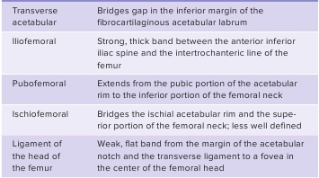
TABLE 8.3
Ligaments of the Hip Joint(see figure 8.23)
A number of bursae surround the knee (figure 8.24
f
). Thelargest is the
suprapatellar bursa,
a superior extension of thejoint capsule that allows the anterior thigh muscles to move overthe distal end of the femur. Other knee bursae include the subcutane-ous prepatellar bursa and the deep infrapatellar bursa, as well asthe popliteal bursa, the gastrocnemius bursa, and the subcutaneousinfrapatellar bursa (not shown in figure 8.24).


Ankle Joint and Arches of the Foot
The
ankle joint,
or
talocrural
(t ā ′l ō -kroo′r ă l) joint, is a highly mod-ified hinge joint formed by the distal tibia and fibula (figure 8.25).The medial and lateral malleoli of the tibia and fibula, which formthe medial and lateral margins of the ankle, are rather extensive,whereas the anterior and posterior margins are almost nonexistent.As a result, a hinge joint is created. A fibrous capsule surroundsthe joint, with the medial and lateral parts thickened to form liga-ments. Other ligaments also help stabilize the joint (table 8.5).Dorsiflexion, plantar flexion, and limited inversion and eversioncan occur at this joint.The arches (see figure 7.37) have ligaments that serve twomajor functions: to hold the bones in their proper relationship assegments of the arch and to provide ties across the arch somewhatlike a bowstring. As weight is transferred through the arch system,some of the ligaments are stretched, giving the foot more flexibil-ity and allowing it to adjust to uneven surfaces. When weight isremoved from the foot, the ligaments recoil and restore the archesto their unstressed shape.The arches of the foot normally form early in fetal life.Failure to form results in congenital
flat feet,
or fallen arches,in which the arches, primarily the medial longitudinal arch,are depressed or collapsed (see figure 7.37). This condition issometimes, but not always, painful. Flat feet may also occurwhen the muscles and ligaments supporting the arch fatigue andallow the arch, usually the medial longitudinal arch, to collapse.During prolonged standing, the plantar calcaneonavicular liga-ment may stretch, flattening the medial longitudinal arch. Thetransverse arch may also become flattened. The strained liga-ments can become painful. The plantar fascia is composed of thedeep connective tissue superficial to the ligaments in the centralplantar surface of the foot and the thinner fascia on the medialand lateral sides of the plantar surface (see figure 8.25).
Plantarfasciitis,
an inflammation of the plantar fascia, can be a problemfor distance runners.
anteriorly. This position is relaxing because the iliofemoralligament supports much of the body’s weight. The
ligament ofthe head of the femur
(round ligament of the femur) is locatedinside the hip joint between the femoral head and the acetabu-lum. This ligament does not contribute much toward strengthen-ing the hip joint; however, it does carry a small nutrient arteryto the head of the femur in about 80% of the population. Thedeepened acetabular labrum, ligaments of the hip, and surround-ing muscles make the hip joint much more stable but less mobilethan the shoulder joint.
Knee Joint
The
knee joint
is traditionally classified as a modified hinge jointlocated between the femur and the tibia (figure 8.24). Actually,it is a complex ellipsoid joint that allows flexion, extension, anda small amount of rotation of the leg. The distal end of the femurhas two large, ellipsoid surfaces with a deep fossa between them.The femur articulates with the proximal end of the tibia, which isflattened and smooth laterally, with a crest called the intercondy-lar eminence in the center (see figure 7.35). The margins of thetibia are built up by menisci—thick, articular disks of fibrocar-tilage (figure 8.24
b,d
), which deepen the articular surface. Thefibula articulates only with the lateral side of the tibia, not withthe femur.The knee joint is stabilized by a combination of ligaments andtendons. The major ligaments that provide knee joint stability arethe cruciate and collateral ligaments. Two
cruciate
(kroo′sh ē - ā t;crossed)
ligaments
extend between the intercondylar eminenceof the tibia and the fossa of the femur (figure 8.24
b,d,e
). The
anterior cruciate ligament
prevents anterior displacement of thetibia relative to the femur, and the
posterior cruciate ligament
prevents posterior displacement of the tibia. The
medial
(tibial)and
lateral
(fibular)
collateral ligaments
stabilize the medialand lateral sides, respectively, of the knee. Joint strength is alsoprovided by popliteal ligaments and tendons of the thigh musclesthat extend around the knee (table 8.4).
Predict 6
Ford Dent hurt his knee in an auto accident when his knee was rammedinto the dashboard. The doctor tested the knee for ligament damageby having Ford sit on the edge of a table with his knee flexed at a90-degree angle. The doctor attempted to pull the tibia in an anteriordirection (anterior drawer test) and then tried to push the tibia in aposterior direction (posterior drawer test). Results of the anterior drawertest were normal, but unusual movement did occur during the posteriordrawer test. Explain the purpose of each test, and describe whichligament was damaged.