CHAPTER 9 Muscular System: Histology and Physiology
303
Types of Smooth Muscle
There are two types of smooth muscle, visceral and multiunit.
Vis-ceral
(vis
′
er- ă l)
smooth muscle
is the more common of the twotypes. It occurs in sheets and includes the smooth muscle of thedigestive, reproductive, and urinary tracts. Visceral smooth musclehas numerous gap junctions (see chapter 4), which allow actionpotentials to pass directly from one cell to another. As a conse-quence, sheets of smooth muscle cells function as a unit, and awave of contraction traverses the entire smooth muscle sheet. Vis-ceral smooth muscle is often autorhythmic but in some areas itcontracts only when stimulated. For example, visceral smoothmuscle in the digestive tract contracts spontaneously and at relativelyregular intervals, whereas visceral smooth muscle in the urinarybladder contracts only when stimulated by the nervous system.
Multiunit smooth muscle
occurs in various configurations:sheets, as in the walls of blood vessels; small bundles, as in thearrector pili muscles and the iris of the eye; and single cells, as inthe capsule of the spleen. Multiunit smooth muscle has fewer gapjunctions than visceral smooth muscle, and cells or groups of cellsact as independent units. It normally contracts only when stimu-lated by nerves or hormones.In visceral smooth muscle tissue, the arrangement between neu-rons and smooth muscle fibers differs from that in skeletal muscletissue. The synapses are more diffuse than in skeletal muscle. Axonsof neurons terminate in a series of dilations along the branchingaxons within the connective tissue among the smooth muscle cells.These dilations have vesicles containing neurotransmitter moleculesthat, once released, diffuse among the smooth muscle cells and bindto receptors on their surfaces. Multiunit smooth muscle has synapsesmore like those found in skeletal muscle tissue.
Electrical Properties of Smooth Muscle
The resting membrane potential of smooth muscle cells is usuallynot as negative as that of skeletal muscle fibers. It normally rangesbetween –55 and –60 mV, compared with approximately –85 mVin skeletal muscle fibers. Furthermore, the resting membrane
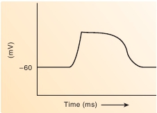
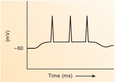
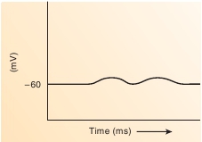
potential of many visceral smooth muscle cells fluctuates, withslow depolarization and repolarization phases. These slow wavesof depolarization and repolarization are propagated from cell to cellfor short distances (figure 9.27
a
). More “classic” action potentialscan be triggered by the slow waves of depolarization and usuallyare propagated for longer distances (figure 9.27
b
). In addition, somesmooth muscle types have action potentials with a plateau, or pro-longed depolarization (figure 9.27
c
). The slow waves in the restingmembrane potential may result from a spontaneous and progressiveincrease in the permeability of the plasma membrane to Na
+
and
+ +
Ca
2
, or they may be controlled by neurons. Sodium ions and Ca
2
diffuse into the cell through their respective channels and producethe depolarization.Smooth muscle does not respond in an all-or-none fashion toaction potentials. A series of action potentials in smooth musclecan result in a single, slow contraction followed by slow relax-ation instead of individual contractions in response to each actionpotential, as occurs in skeletal muscle. A slow wave of depo-larization that has one to several more classic-appearing actionpotentials superimposed on it is common in many types of smoothmuscle. After the wave of depolarization, the smooth musclecontracts. Action potentials with plateaus are common in smoothmuscle that exhibits periods of sustained contraction.Spontaneously generated action potentials that lead to con-tractions are characteristic of visceral smooth muscle in theuterus, the ureter, and the digestive tract. Certain smooth musclecells in these organs function as
pacemaker cells,
which tend todevelop action potentials more rapidly than other cells.The nervous system can regulate smooth muscle contractionsby increasing or decreasing action potentials carried by neuronaxons to smooth muscle. Responses of smooth muscle cells resultin depolarization and increased contraction or hyperpolarizationand decreased contraction. The nervous system can also regulatethe pacemaker cells.Hormones and ligands produced locally in tissues can bindto receptors on some smooth muscle plasma membranes. Thecombination of a hormone or other ligands with a receptor causes


(a)
Slow waves of depolarization
(c)
Action potential with prolongeddepolarization (plateau)
(b)
Action potentials in smooth musclesuperimposed on a slow wave ofdepolarization
FIGURE 9.27
Membrane Potentials in Smooth Muscle