370

PART 3 Integration and Control Systems
Dendrites
Neuroncellbody
Bipolar neurons
(
bi-,
two) have two processes: one dendriteand one axon (figure 11.5
b
). The dendrite is often specialized toreceive the stimulus, and the axon conducts action potentials tothe CNS. Bipolar neurons are located in some sensory organs,such as in the retina of the eye and in the nasal cavity.
Pseudo-unipolar neurons
(
pseudo-,
false +
uni-,
one) havea single process extending from the cell body, which divides intotwo branches a short distance from the cell body (figure 11.5
c
).The two branches function as a single axon. One branch extendsto the CNS, and the other extends to the periphery and has den-dritelike sensory receptors. The sensory receptors respond tostimuli, producing action potentials that are transmitted to theCNS. Most sensory neurons are pseudo-unipolar. According to astrictly functional definition of dendrite, the branch of pseudo-unipolar neurons that extends from the periphery to the neuron cellbody could be classified as a dendrite because it conducts actionpotentials toward the neuron cell body. However, this branch isusually referred to as an axon, for two reasons: It cannot be distin-guished from an axon on the basis of its structure, and it conductsaction potentials in the same fashion as an axon.
Glial Cells of the CNS
cell
Glial cells are the major supporting cells in the CNS. Glial cellshelp form a protective permeability barrier between the blood andthe brain and spinal cord, they phagocytize foreign substances,they produce cerebrospinal fluid, and they form myelin sheathsaround axons. There are four types of CNS glial cells, each withunique structural and functional characteristics. Refer to table 11.1for a description and an illustration of each glial cell type.
Astrocytes
of Ranvier
Presynaptic terminals
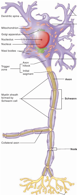
FIGURE 11.4
Neuron
The structural features of a neuron are a cell body and two types of cellprojections: dendrites and an axon.
Multipolar neurons
(
multi-,
many) have many dendrites anda single axon. The dendrites vary in number and in their degree ofbranching (figure 11.5
a
). Most of the neurons within the CNS andmotor neurons are multipolar.
Astrocytes
(as′tr ō -s ī tz;
aster,
star) are glial cells that are star-shaped because cytoplasmic processes extend from the cell body.These extensions widen and spread out to form foot processes,which cover the surfaces of blood vessels (table 11.1), neurons, andthe pia mater. (The pia mater is a membrane covering the outside ofthe brain and spinal cord.) Astrocytes have an extensive cytoskel-eton of microfilaments (see chapter 3), which enables them to forma supporting framework for blood vessels and neurons.Astrocytes help regulate the composition of extracellularbrain fluid. They do this by releasing chemicals that promote theformation of tight junctions (see chapter 4) between the endothelialcells of capillaries. The endothelial cells with their tight junctionsform the
blood-brain barrier,
which determines what substancescan pass from the blood into the nervous tissue of the brain andspinal cord. The blood-brain barrier protects neurons from toxicsubstances in the blood, allows the exchange of nutrients and wasteproducts between neurons and the blood, and prevents fluctuationsin blood composition from affecting brain functions.Astrocytes aid both beneficial and detrimental responsesto tissue damage in the CNS. Almost all injuries to CNS tissueinduce
reactive astrocytosis,
in which astrocytes wall off theinjury site and help limit the spread of inflammation to the sur-rounding healthy tissue. Reactive scar-forming astrocytes alsolimit the regeneration of the axons of injured neurons.