CHAPTER 11 Functional Organization of Nervous Tissue
371


Dendrites
Sensoryreceptors
Axonbranchesfunctionas asingleaxon.
body
body
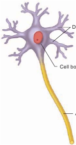
(a) A
multipolar neuron
hasmany dendrites and an axon.
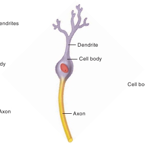
(b) A
bipolar neuron
has adendrite and an axon.
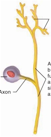
(c) A
pseudo-unipolar neuron
appearsto have an axon and no dendrites.
FIGURE 11.5
Structural Classes of Neurons
Neurons are classified structurally by the number of cellular projections extending from their cell bodies. Dendrites and sensory receptors are specialized toreceive stimuli, and axons are specialized to conduct action potentials.
Astrocytes also release chemicals that promote the develop-ment of synapses and help regulate synaptic activity by synthesiz-ing, absorbing, and recycling neurotransmitters.
many times around the axons, they form an insulating materialcalled a
myelin
(m ī ′ ĕ -lin)
sheath.
One oligodendrocyte can formmyelin sheaths around axons of multiple neurons (table 11.1).
Ependymal Cells
Ependymal
(ep-en′di-m ă l)
cells
line the ventricles (cavities) ofthe brain and the central canal of the spinal cord (table 11.1).Specialized ependymal cells and blood vessels form structurescalled
choroid plexuses
(ko′royd plek′s ŭ s-ez; table 11.1), whichare located within certain regions of the ventricles. The choroidplexuses secrete the cerebrospinal fluid that flows through the ven-tricles of the brain (see chapter 13). The ependymal cells frequentlyhave patches of cilia that help circulate cerebrospinal fluid throughthe brain cavities. Ependymal cells also have long processes attheir basal surfaces that extend deep into the brain and the spinalcord and seem, in some cases, to have astrocyte-like functions.
Glial Cells of the PNS
There are two types of glial cells in the PNS: Schwann cells andsatellite cells.
Schwann cells
form myelin sheaths. However,unlike oligodendrocytes, each Schwann cell forms a portion of themyelin sheath around only one axon (table 11.1).
Satellite cells
surround neuron cell bodies in sensory andautonomic ganglia (table 11.1). Besides providing support andnutrition to the neuron cell bodies, satellite cells protect neuronsfrom heavy-metal poisons, such as lead and mercury, by absorb-ing them and reducing their access to the neuron cell bodies.
Microglia
Microglia
(m ī -krog′l ē - ă ) are CNS-specific immune cells. Microg-lia become mobile and phagocytic in response to inflammation.They phagocytize necrotic tissue, microorganisms, and other for-eign substances that invade the CNS (table 11.1). Areas of the brainor spinal cord that have been damaged by infection, trauma, orstroke have more microglia than healthy areas. There the microgliaperform phagocytosis of dead cells and pathogens. A pathologistcan identify these damaged areas in the CNS during an autopsybecause large numbers of microglia are found there.
Myelinated and Unmyelinated Axons
Cytoplasmic extensions of the Schwann cells in the PNS and ofthe oligodendrocyte extensions in the CNS surround axons toform either myelinated or unmyelinated axons. Myelin protectsand electrically insulates axons from one another. In addition,action potentials travel along myelinated axons more rapidly thanalong unmyelinated axons (see “Propagation of Action Potentials”in section 11.5).In
myelinated axons,
Schwann cells or oligodendrocyteextensions repeatedly wrap around a segment of an axon to forma series of tightly wrapped membranes rich in phospholipids,with little cytoplasm sandwiched between the membrane layers(figure 11.6
a
). One way to picture the overlapping wrappings,especially for Schwann cells, is to imagine rolling up a hot dog(axon) inside a tortilla (Schwann cell). The tightly wrapped
Oligodendrocytes
Oligodendrocytes
(ol′i-g ō -den′dr ō -s ī tz) have cytoplasmic exten-sions that can surround axons. If the cytoplasmic extensions wrap