CHAPTER 16 Autonomic Nervous System


559
divisions were described. This section will detail the distribution ofthe autonomic nerve fibers to the target organs (figure 16.5). In somecases, the postganglionic axons extend directly through nerves to thetarget organ. In other cases, the postganglionic axons become part ofautonomic nerve plexuses.
Autonomic nerve plexuses
are complex,interconnected neural networks formed by neurons of the sympa-thetic and parasympathetic divisions. Axons of sensory neurons alsocontribute to these autonomic nerve plexuses. The autonomic nerveplexuses are typically named according to the organs they supply orthe blood vessels along which they are found. For example, the car-diac plexus supplies the heart, and the thoracic aortic plexus is foundalong the thoracic aorta. Plexuses following the route of blood ves-sels are a major means by which autonomic axons are distributedthroughout the body. Since they contain both sympathetic and para-sympathetic neurons, autonomic nerve plexuses are associated withboth the sympathetic and parasympathetic divisions.
Sympathetic Division Distribution
Sympathetic outflow is through spinal nerves, sympathetic nerves,and splanchnic nerves (see figure 16.3). Branches of these nerveseither extend directly to effectors or join autonomic nerve plexusesto be distributed to effectors. The major means by which sympa-thetic postganglionic axons reach effectors include the following:1.
Spinal nerves
. From all levels of the sympathetic chain, somepostganglionic axons project through gray rami communicatesto spinal nerves. The axons extend to a specific region innervatedby each pair of spinal nerves, regulating the activity of sweatglands in the skin, the smooth muscle in the blood vessels ofthe skin, and the smooth muscle of the arrector pili. Seefigure 12.14 for the distribution of spinal nerves to the skin.2.
Head and neck nerve plexuses
. Most of the sympathetic nervesupply to the head and neck is derived from the superior cervicalsympathetic chain ganglion (figure 16.5). Postganglionic axonsof sympathetic nerves form plexuses that extend superiorly tothe head and inferiorly to the neck. The plexuses associatedwith the head and neck give off branches to regulate the
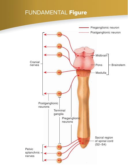
FIGURE 16.4
Parasympathetic Division
The location of parasympathetic preganglionic (
solid red
) and postganglionic(
dashed red
) neurons. The preganglionic neuron cell bodies are in the brain-stem and the lateral gray horns of the sacral part of the spinal cord, and thepostganglionic neuron cell bodies are within terminal ganglia.
TABLE 16.2
Comparison of the Sympathetic and Parasympathetic Divisions