678
PART 4 Regulation and Maintenance
are mainly flat, but the interior of both auricles and a part of theright atrial wall contain muscular ridges called
pectinate
(pek ′ ti-n ā t)
muscles.
The pectinate muscles of the right atrium are sepa-rated from the larger, smooth portions of the atrial wall by a ridgecalled the
crista terminalis
(kris ′ t ă ter ′ mi-nal ′ is; terminal crest).The interior walls of the ventricles contain larger, muscular ridgesand columns called
trabeculae
(tr ă -bek ′ ū -l ē ; beams)
carneae
(kar ′ n ē - ē ; flesh).
External Anatomy and CoronaryCirculation
The heart consists of four chambers: two
atria
( ā ′ tr ē - ă ; sing.
atrium
) and two
ventricles
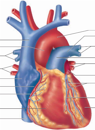
(ven ′ tri-klz). The thin-walled atria formthe superior and posterior parts of the heart, and the thick-walledventricles form the anterior and inferior portions (figure 20.5).
Auricles
(aw ′ ri-klz; ears) are flaplike extensions of the atria thatcan be seen anteriorly between each atrium and ventricle. Theentire atrium used to be called the auricle, and some medical per-sonnel still refer to it as such.Blood enters the atria of the heart through several large veins.The
superior vena cava
(v ē ′ n ă k ā ′ v ă ) and the
inferior vena cava
carry blood from the body to the right atrium. In addition, the
smaller coronary sinus carries blood from the walls of the heartto the right atrium. Four
pulmonary veins
carry blood from thelungs to the left atrium.Blood leaves the ventricles of the heart through two arteries:the
pulmonary trunk
and the
aorta.
The pulmonary trunk carriesblood from the right ventricle to the lungs. The aorta carries bloodfrom the left ventricle to the body. Because of their large size, thepulmonary trunk and aorta are often called the great arteries.The coronary circulation consists of blood vessels that carryblood to and from the tissues of the heart wall. The major vesselsof the coronary circulation lie in several grooves, or sulci, on thesurface of the heart. A large
coronary
(k ō r ′ o-n ā r- ē ; circling likea crown)
sulcus
(sool ′ k ŭ s; ditch) runs obliquely around the heart,separating the atria from the ventricles. Two more sulci extendinferiorly from the coronary sulcus, indicating the division betweenthe right and left ventricles. The
anterior interventricularsulcus
(
groove
) is on the anterior surface of the heart, extend-ing from the coronary sulcus toward the apex of the heart(figure 20.5
a,b
). The
posterior interventricular sulcus
(
groove
)is on the posterior surface of the heart, extending from thecoronary sulcus toward the apex of the heart (figure 20.5
c
). In ahealthy, intact heart, the sulci are covered by adipose tissue, andonly after this tissue is removed can they be seen.

Aortic arch
Left pulmonary artery
Superior vena cava
Aorta
Branches of rightpulmonary artery
Right pulmonary veins
Left atrium
Right atrium
Coronary sulcus
Right coronary artery
Great cardiac vein(in anterior interventricular sulcus)
Anterior interventricular artery(in anterior interventricular sulcus)
Left ventricle
Branches of leftpulmonary artery
Pulmonary trunk
Left pulmonary veins
Right ventricle
Inferior vena cava
(a)
Anterior view
FIGURE 20.5
Surface View of the Heart
(
a
) The two atria (right and left) are located superiorly, and the two ventricles (right and left) are located inferiorly. The superior and inferior venae cavae enterthe right atrium. The pulmonary veins enter the left atrium. The pulmonary trunk exits the right ventricle, and the aorta exits the left ventricle.