CHAPTER 24 Digestive System
877

ASSESS
YOUR PROGRESS
12.
Where are the visceral peritoneumperitoneum found? Defineretroperitoneal organs.
13.
What is the function of the
14.
What are the mesenteries?location of the mesenteries
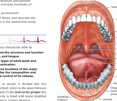
Upper lip (labium)
Labial frenulum of upper lip
Vestibule (superior)
Gingiva covering themaxillary alveolarprocess
Hard palate
Soft palate

24.6
Oral Cavity
LEARNING OUTCOMES
Fauces
Uvula
Cheek
Tongue
Lingual frenulum
Molars
Openings of thesubmandibular ducts
Premolars
Gingiva covering themandibular alveolarprocess
Labial frenulumof lower lip Lower lip (labium)
Anterior view
Incisors
After reading this section, you
A.
Describe the oral cavity andof the lips, cheeks, palate,
B.
Outline the structure anddescribe the process of mastication.
C.
Compare the structures andsalivary glands and describefunctions of saliva and the
The
oral cavity
(figure 24.6),regions: (1) The
vestibule
(ves′ti-bool;the lips or cheeks and the teeth,medial to the teeth. The oral cavitysquamous epithelium, which protects
Lips, Cheeks, and Palate
The
lips,
or
labia
(l ā ′b ē - ă ; figure 24.6), are muscular structuresformed mostly by the
orbicularis oris
( ō r-bik′ ū -l ā ′ris ō r′is)
muscle
(see figure 10.9) and connective tissue. The outer surfaces of the lipsare covered by skin. The keratinized stratified epithelium of the skinis thin at the margin of the lips and is not as highly keratinized as theepithelium of the surrounding skin (see chapter 5); consequently, itis more transparent than the epithelium over the rest of the body. Thecolor from the underlying blood vessels shows through the relativelytransparent epithelium, giving the lips a reddish-pink to dark redappearance, depending on the overlying pigment. At the internalmargin of the lips, the epithelium is continuous with the moist strati-fied squamous epithelium of the mucosa in the oral cavity.One or more
labial frenula
(fren′ ū -l ă ; sing.
frenulum
), whichare mucosal folds, extend from the alveolar process of the maxillato the upper lip and from the alveolar process of the mandible tothe lower lip.The
cheeks
form the lateral walls of the oral cavity. Theyconsist of an interior lining of moist stratified squamous epithe-lium and an exterior covering of skin. The substance of the cheekincludes the
buccinator muscle
(see chapter 10), which flattensthe cheek against the teeth, and the
buccal fat pad,
which roundsout the profile on the side of the face.The lips and cheeks are important in mastication and speech.They help manipulate food within the oral cavity and hold it inplace while the teeth crush or tear it. They also help form wordswhen we speak. A large number of the muscles of facial expres-sion are involved in moving the cheeks and lips (see chapter 10).

FIGURE 24.6
Oral Cavity
The roof of the oral cavity is called the
palate.
The palate sepa-rates the oral and nasal cavities and prevents food from passing intothe nasal cavity during chewing and swallowing. The palate consistsof two parts (figure 24.6). The anterior, bony part is the
hard palate
(see chapter 7). The posterior, nonbony part is the
soft palate,
which consists of skeletal muscle and connective tissue. The
uvula
( ū ′v ū -l ă ; a grape) is a posterior projection from the soft palate. Theposterior boundary of the oral cavity is the
fauces
(faw′s ē z), whichis the opening into the pharynx, or
throat
. The
palatine tonsils
arein the lateral wall of the fauces (see chapter 22).
Tongue
The
tongue
is a large, muscular organ that occupies most of the oralcavity proper when the mouth is closed. Its major attachment in theoral cavity is through its posterior part. The anterior part of thetongue is relatively free, except for attachment to the floor of themouth by a thin fold of tissue called the
lingual
(tongue)
frenulum.
The muscles associated with the tongue are divided into two catego-ries:
Intrinsic muscles
are within the tongue itself, and
extrinsicmuscles
are outside the tongue but attached to it. The intrinsicmuscles are largely responsible for changing the shape of the tongue,