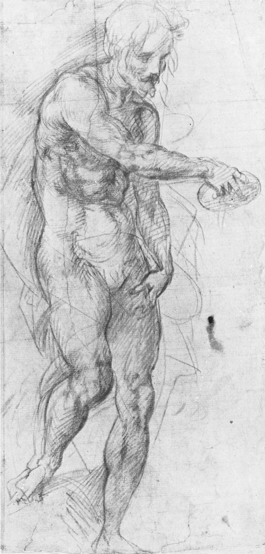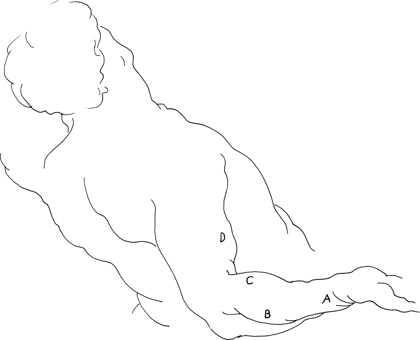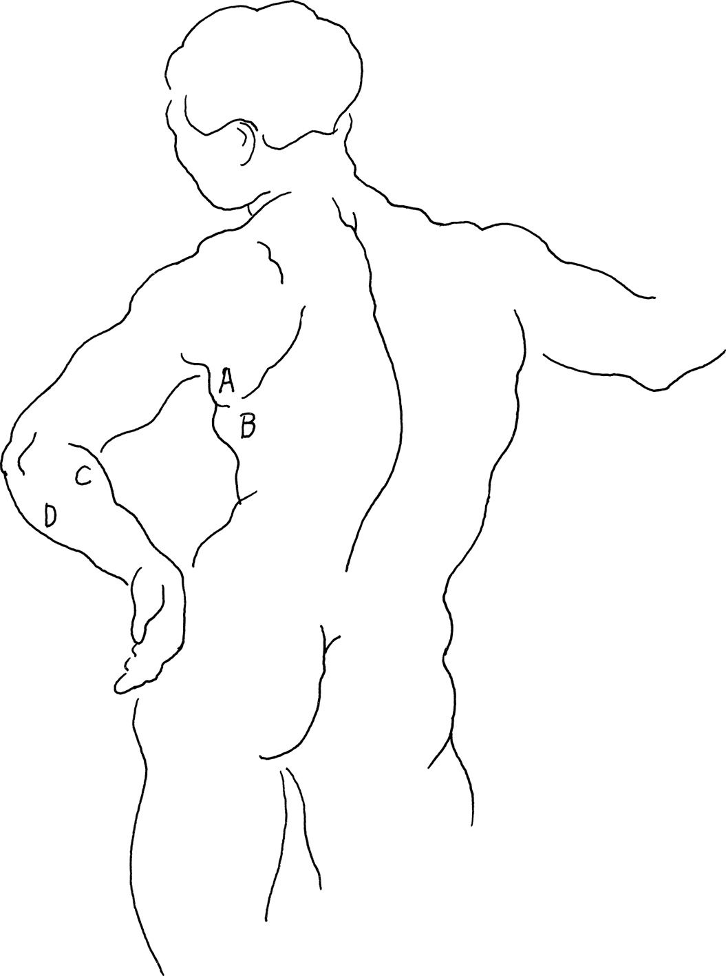
When the arm moves forward, away from the body, in a movement known as “flexion,” a small portion of the hollow under the arm becomes visible. This is the axilla or armpit, where the arm emerges from the trunk.
This pyramidal area is partly formed by the muscles that pass from the trunk to the humerus bone of the arm. The posterior fold or wall of the armpit is formed by the latissimus dorsi (A) and the teres major (B). They go from their origins in the pelvic and shoulder girdles, respectively, to insert (C,D) in the humerus bone. In this view, the hollow of the armpit is concealed by the long head of the triceps (E). You can also see the serrations of the serratus anterior (F) that, with the rib cage, form the inner wall of the axilla. The edge of the pectoralis major (G), which forms its anterior or front wall, is just barely visible beneath the triceps.
Notice the contour lines of the latissimus dorsi (A), teres major (B), the infraspinatus (H), and the posterior (I) and middle (J) portions of the deltoid. Their lines converge as lines and wedgelike pointers from their inner bases to the outstretched arm. You can easily follow the rhythmical flow of these transitions of line and mass from the trunk to the arm.
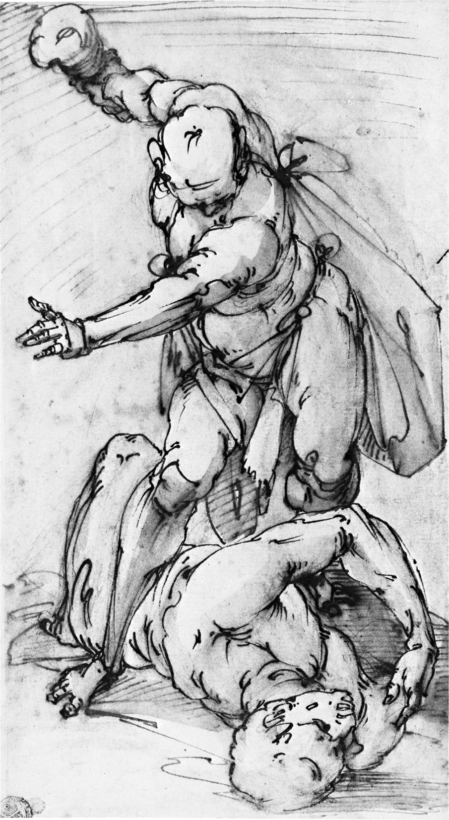
Andrea del Sarto (1486-1531)
STUDY OF ST. JOHN THE BAPTIST FOR BAPTISM OF THE MULTITUDE
red chalk
Felton Bequest, 1936
National Gallery of Victoria, Melbourne
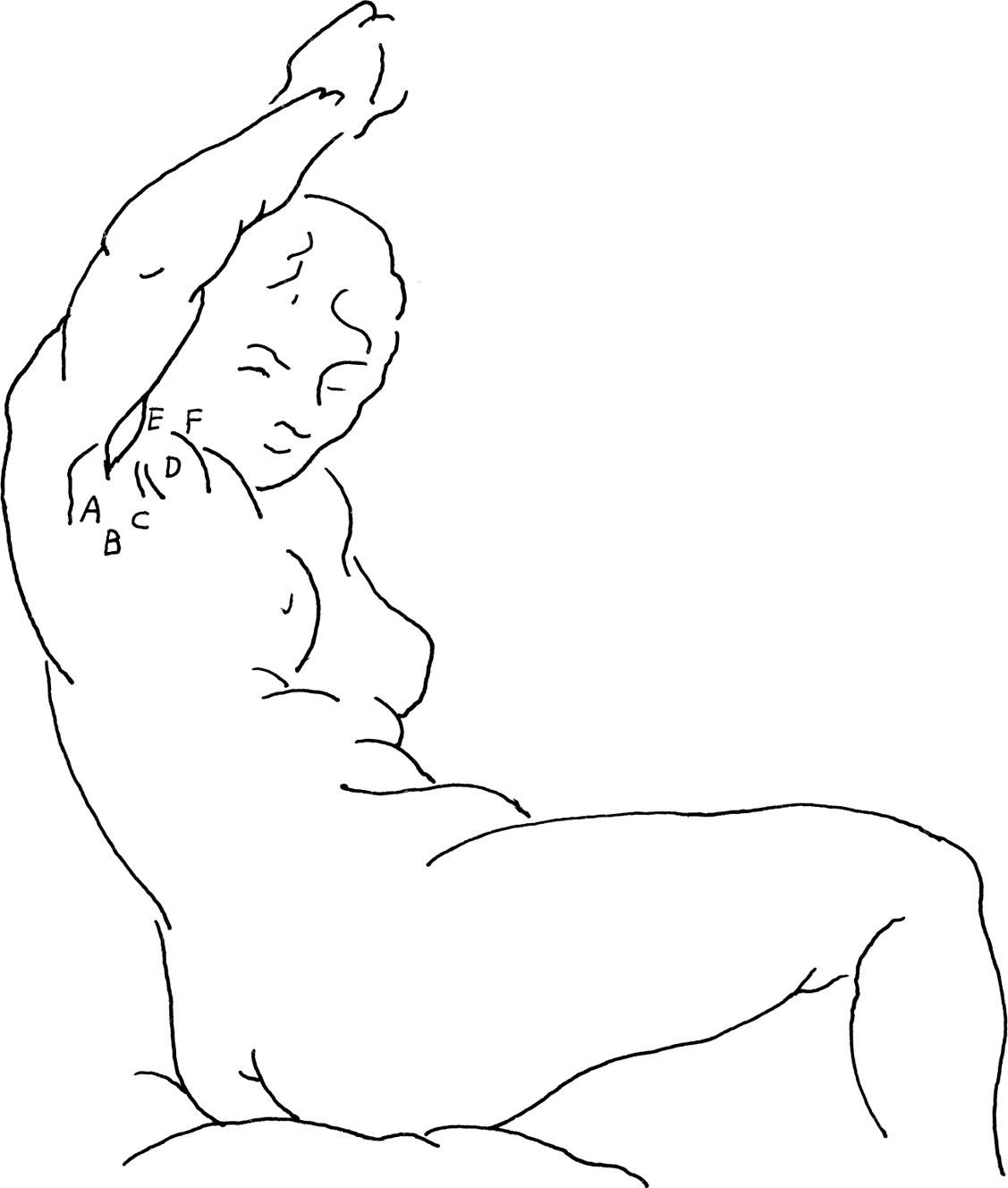
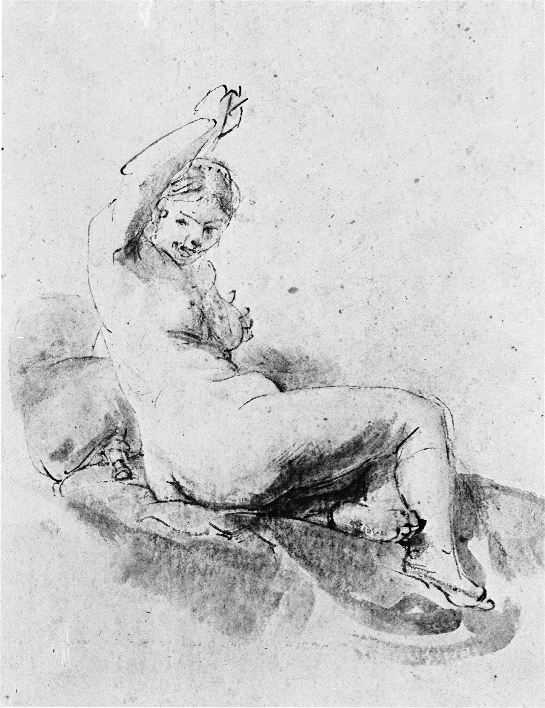
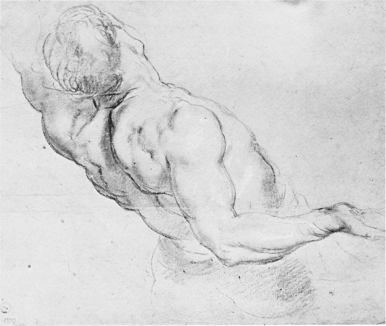
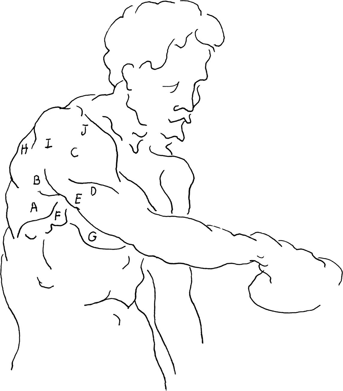
The shape and size of the axilla, or armpit, varies with each movement of the arm. In this figure by Rembrandt, with the arm raised above the head, we see more of the hollow of the armpit.
The masses of the teres major (A) and the latissimus dorsi (B) constitute the posterior wall of the axilla and conceal the medial wall (C). Two little oblique lines, called the anterior furrow, separate the masses of pectoralis major (D) from the latissimus dorsi (B). This furrow twists around the coracobrachialis (E) and follows the edge of the pectoralis major (F) which forms the anterior wall of the armpit.
Students often make use of memory devices. Scottish medical students use one to learn the placement and order of muscles about the axilla from back to front. The sounds of the first syllables of the names of the muscles are made into a sentence: triceps, teres major, latissimus dorsi, corocobrachialis, biceps, pectoralis, which is read as, “Try to let corbie pet.” “Corbie” is Scottish for “crow.” This is an imaginative use of positional order that, when combined with a word order, creates an easily remembered mental image.
Rembrandt van Rijn (1606-1669)
STUDY OF A NUDE
pen and brush with bistre wash on brown paper
9 1/8″ × 7 1/8″ (233 × 178 mm)
Collection of Tiffany and Margaret Blake
Art Institute of Chicago
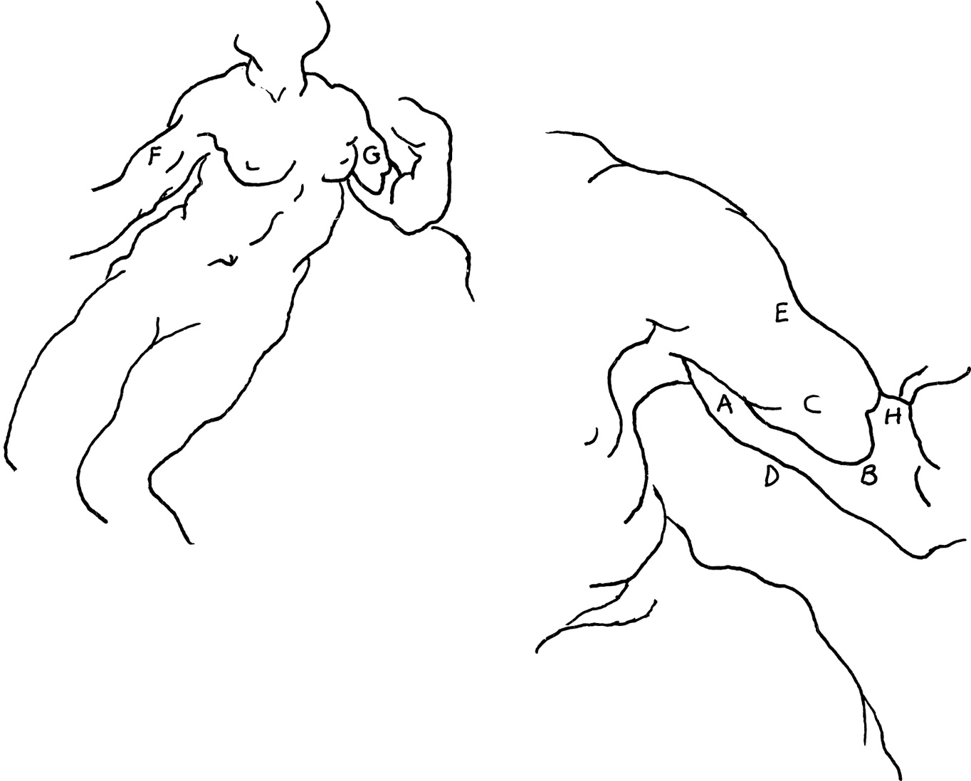
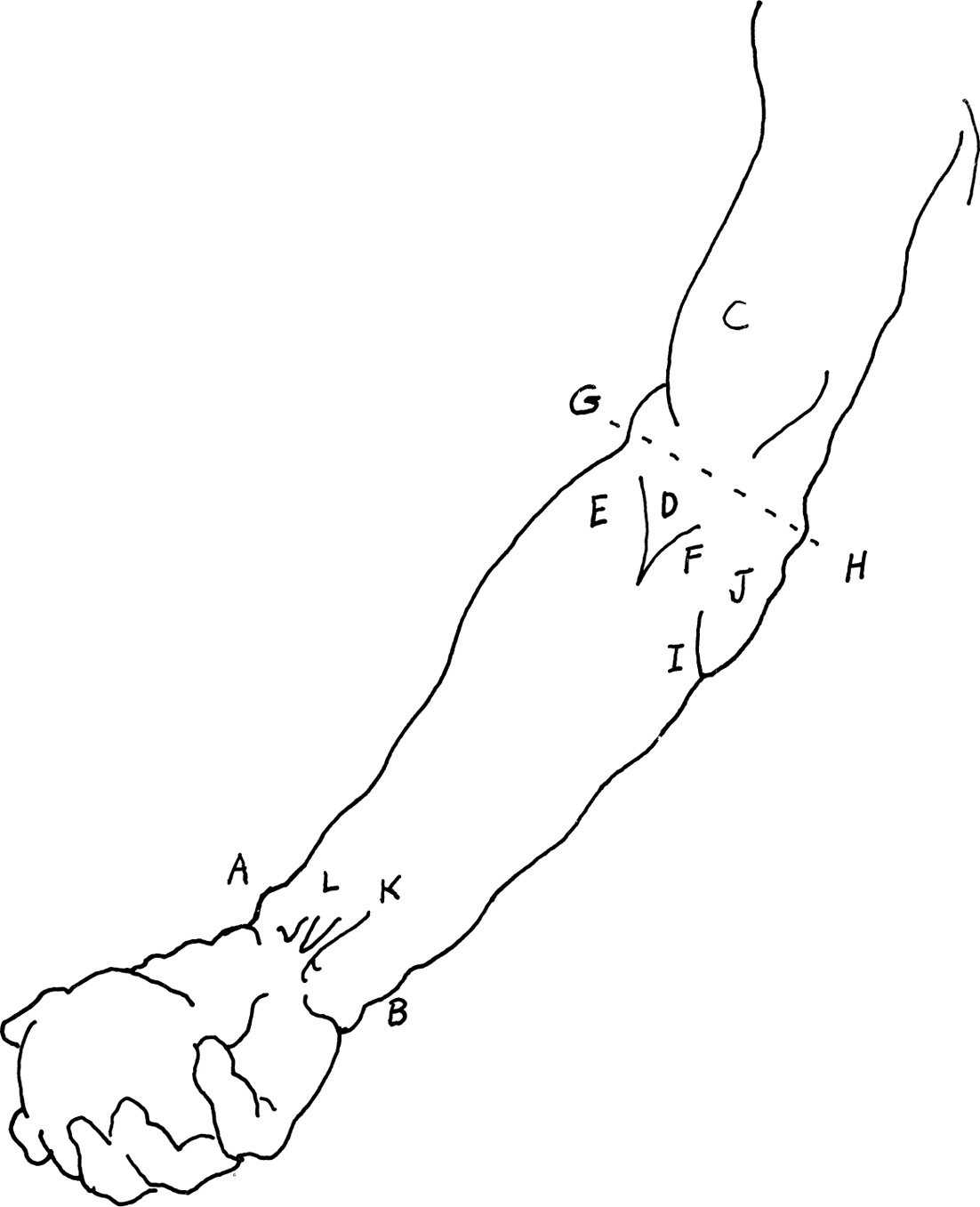
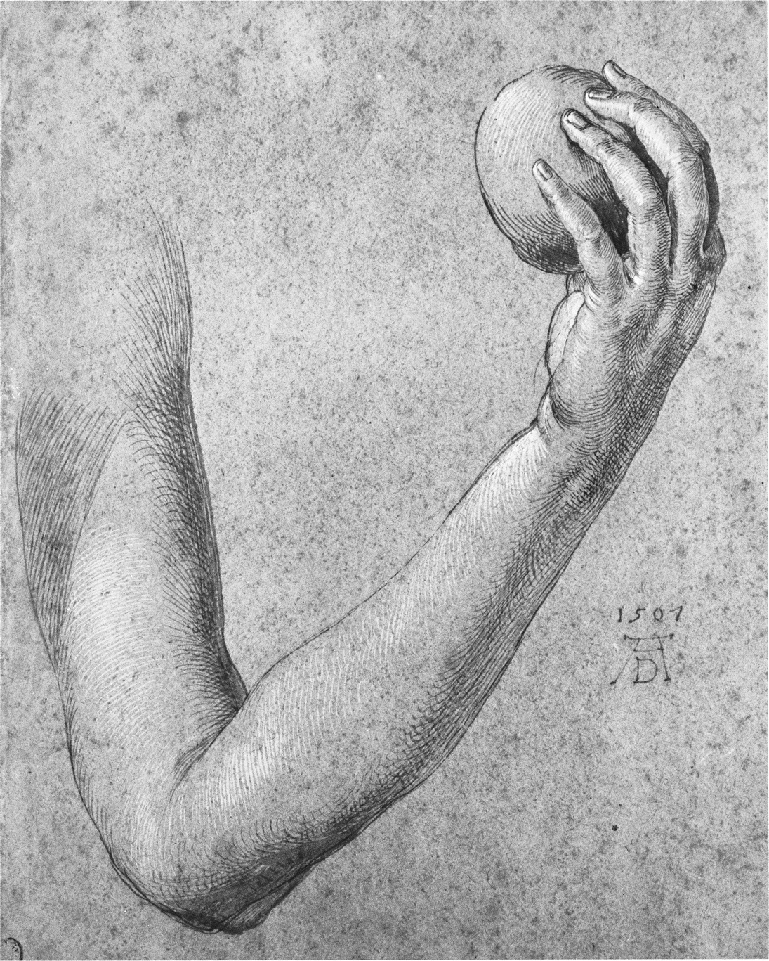
The fleshy masses of the upper arm lie along both sides of the humerus, the bone of the upper arm. From its ball and socket joint in the shoulder blade, the humerus extends downward about two shoulder blade or sternum lengths. Its upper cylindrical shaft curves slightly to accommodate the rib cage. Its lower end is flattened and widened into two condyles, which receive the radius and ulna at the elbow.
Two muscles help fill out the inside of the arm in the figure on the right: the coracobrachialis (A) and the brachialis (B), both of which lie in the groove between the short or inner head of the biceps (C), and the medial head of the triceps (D). The long outer (E) and short (C) heads of the biceps lie upon the brachialis (B) or “pillow muscle” below.
Together with the coracobrachialis (A) and brachialis (B), the biceps flex the forearm. These muscles bulge when they contract, becoming shorter and thicker as they pull upon the radius bone of the forearm when the elbow is bent. In the drawing, compare the surface changes of the biceps during its different phases: from its relaxed extension position (F), to its flexion at right angle (G).
Observation and knowledge of anatomical structure can help you to harmonize the breaks between forms when you draw the body. For example, the small curved plane created by the tendon of the biceps (H) as it inserts into the forearm, softens the right angle between upper arm and forearm. As another example, the distinct separation and varied lengths of the long (E) and short (C) heads of the biceps adds an interesting contrast of size and shape to these two masses, as they move between scapula and radius.
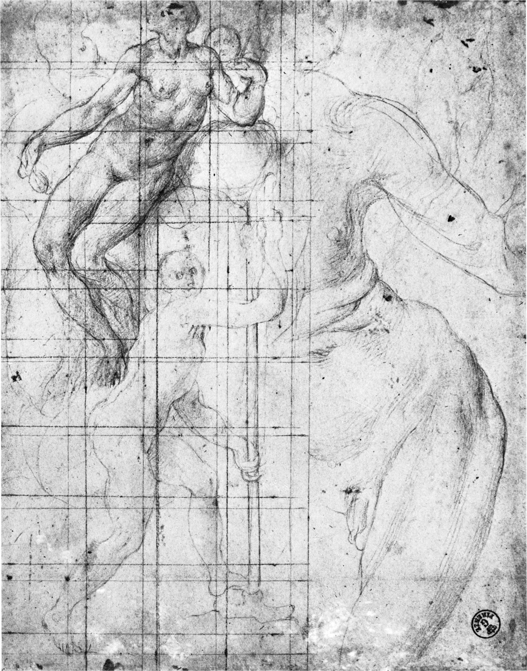
Jacopo Pontormo (1494-1556)
STUDY OF FIGURES
black and red pencil blended on white paper
8″ × 6 1/4″ (204 × 158 mm) Uffizi, Florence
Function creates form. The form of the fish allows it to flow easily through water, maneuvering like an airfoil. The limbs of a running animal must move clear of the body, so the curve of the humerus bone performs a practical function. In man, the dominant muscular masses of the upper arm follow the slightly curving shape of the humerus beneath.
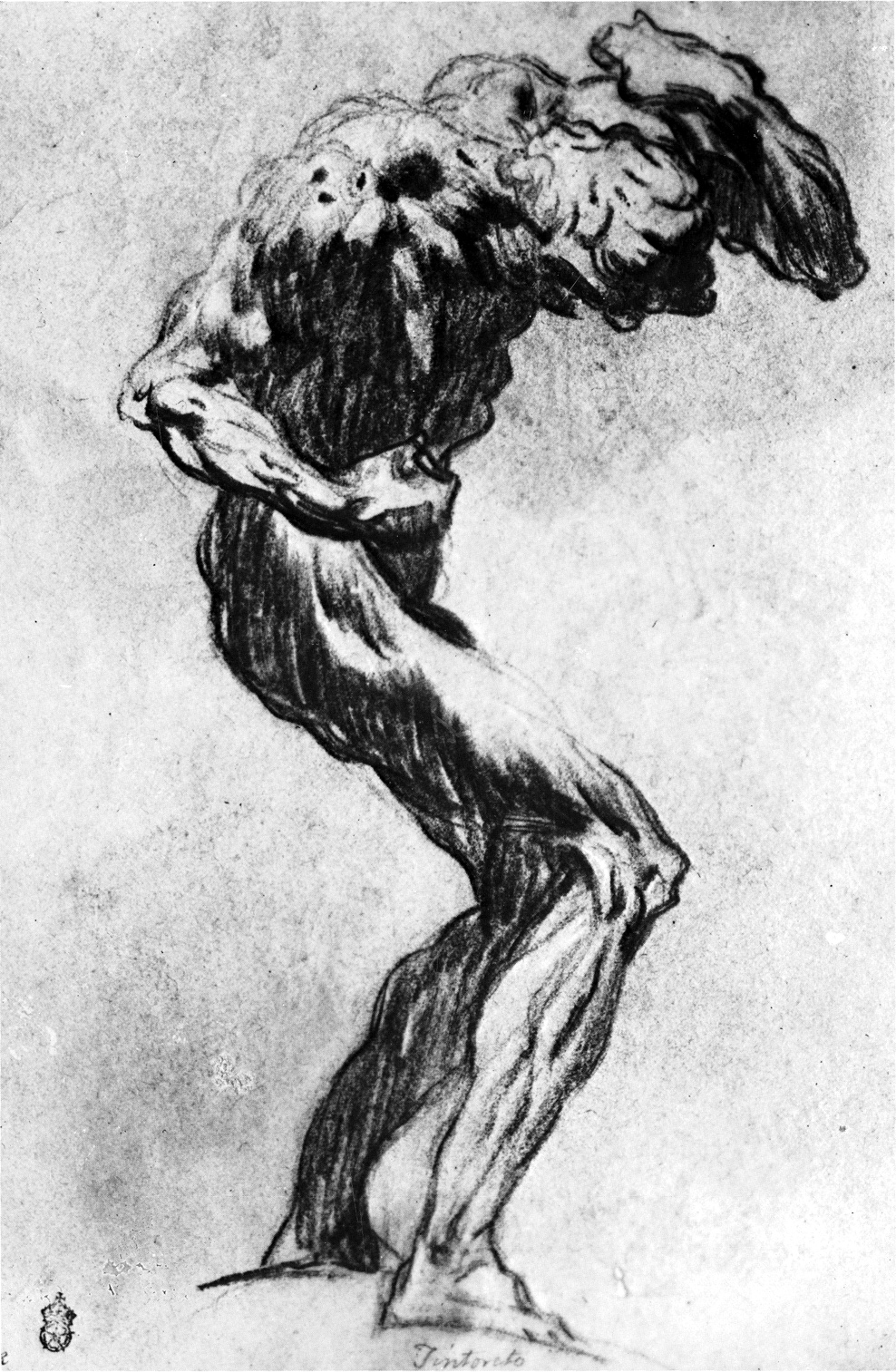
Draw the muscles of the upper arm from their origins in the bone of the shoulder to their insertion in the bones of the lower arm. First locate the bump of the outer edge of the clavicle (A). This leads you to the summit of the shoulder (B) and the acromioclavicular joint, where the clavicle rises above the acromion process of the scapula. The acromion process, in turn, hovers protectively over the glenoid cavity (C), which holds the head of the humerus. The rounded highlight on the deltoid reflects the head of the humerus below that fits into this cavity. Put your pencil at the top of the glenoid cavity (C) and follow the edge of the long head of the biceps (D) to its inserting tendon (E) in the radius. Now place your pencil at the bottom of the glenoid cavity (C) and move it down to the outside contour line of the triceps (F). Follow the triceps to its insertion in the olecranon process of the ulna (G).
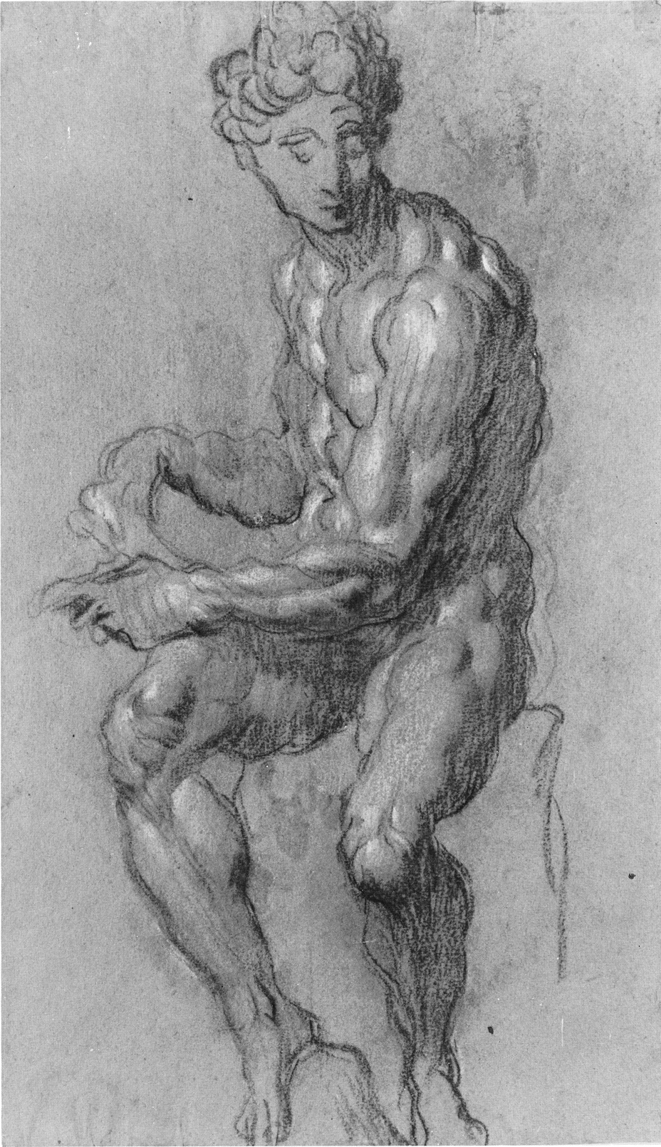
Observe in particular the mass of the deltoid (H) as it moves into the groove between the biceps and triceps enroute to its insertion. There it goes between the fibers of the brachialis (I), and bulges with the biceps as it helps flex the arm. Tintoretto has placed the highlight (J) and the strong dark (K) of the big cylinder of the arm well away from the groove of the brachialis (I), which forms a line between biceps and triceps. He thus makes certain that the mass conception (of the arm as a cylinder) dominates over the individual forms.
Jacopo Tintoretto (1518-1594)
STUDY OF A MODEL OF THE GIULIANO DE’ MEDICI BY MICHELANGELO
black and white chalk
The Governing Body of Christchurch, Oxford
Flexion or bending of the forearm upon the upper arm is carried out by the biceps brachii (A) and the brachialis anticus (B). At rest, when the arm hangs limply at the elbow, the biceps is a long, tapered, cylindrical shape. Here, Tintoretto shows it contracting to flex the forearm upon the upper arm. He renders it short and globular-shaped, with its maximum thickness front to back, at about the middle of the upper arm. The upper portion of the biceps is covered by the deltoid (C), which moves into the intermuscular groove between the triceps (D) and the biceps (A).
The triceps muscle is the antagonist to the biceps. The biceps flexes or bends the arm and the triceps bulges slightly in counterbalancing resistance. The flexed elbow stretches the common tendon of the triceps (E) from mid-arm to its insertion into the posterior and superior part of the olecranon process (F). The flat surface of the tendon is distinct from the swelling fleshy heads of the surrounding muscles.
Tintoretto knows that the upper back of the olecranon process (F) of the ulna forms the tip of the elbow, and that it becomes very prominent when the arm is bent. Notice how he uses curved rather than flat lines around the point of the elbow, where ulna meets humerus.
The supinator mass (G) emerges from between triceps and brachialis, about one third of the way up the arm. When the radius crosses over the ulna, as it does here, it carries the supinator mass over these two underlying bones, widening the upper forearm. At the back of the lower arm, Tintoretto has emphasized the extensors (H), which contract synergetically, that is, working together as a counterbalance or mild opposition, when the wrist and fingers are flexed.
Jacopo Tintoretto (1518-1594)
STUDY OF A STATUE OF ATLANTIS
black chalk heightened with white, on blue paper
10 11/16″ × 7 1/8″ (268 × 181 mm)
Museum Boymans-van Beuningen, Rotterdam
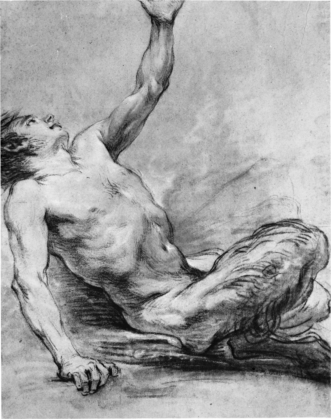
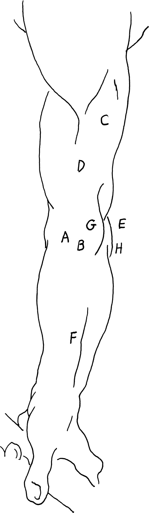
The triceps, virtually three muscles in one, covers the entire posterior surface of the upper arm. All three heads of this muscle are inserted through a large tendon (A) to the end of the olecranon process of the ulna (B).
The triceps is the chief extensor of the lower arm. Its long or middle head (C) crosses the shoulder joint from its origin in the humerus, and also aids the extension and adduction of the upper arm.
Just above the curve of the lateral head of the biceps (D), the mass of the outer head of the triceps (E) moves out from under the deltoid (F), where it originates in the upper lateral surface of the humerus. The inner head of the triceps (G) also arises from the humerus, but from the lower medial portion, so that it covers the entire posterior surface of the lower part of the bone.
A characteristic of the triceps, which becomes more prominent in extension, is its outward slanting line (H). There the rounded fibers of the muscle meet the long, low, flat plane of the common tendon.
If you see a side view of the insertion of the common tendon at the olecranon process of the ulna, you will see the small triangular relief of the anconeus (I). This little muscle fills the gap between the back of the lateral epicondyle of the humerus (J) where it originates, the outer border of the olecranon process, and the posterior surface of the ulna (K) into which it inserts. In extension of the forearm, the anconeus (I) initiates the action and stabilizes the elbow joint. When more strength is needed, the inner, outer, and then the long heads of the triceps must come into play.
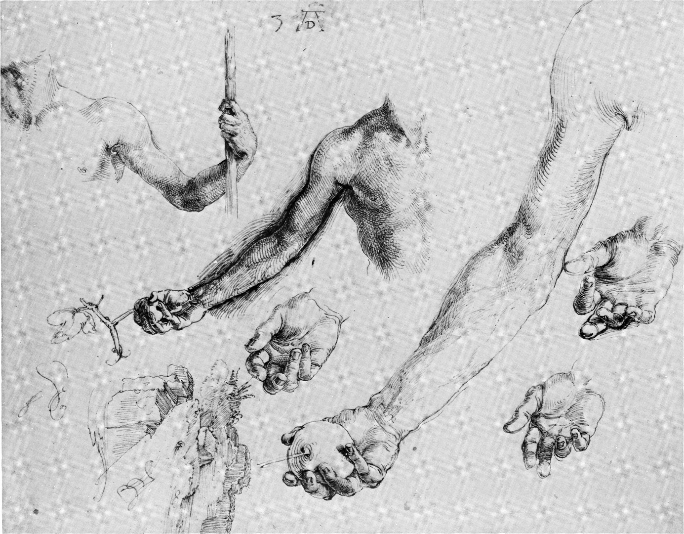
Raphael Sanzio (1483-1520)
NUDE MAN SITTING ON STONE
black chalk
13 5/8″ × 10 7/16″ (345 × 265 mm)
Ashmolean Museum, Oxford
It helps to know that the big cylinder of the arm, seen from the side in this drawing by Boucher, might be broken up into the smaller cylinders of the outer head of the triceps (A) and the biceps (B). The downward end of the triangular bulb of the deltoid (C) plunges into the groove between these two cylinders to its insertion in the mid-humerus (D).
At about this midpoint in the upper arm, you can see a dip (E) in the outline of the triceps, where muscles meet tendon. It is through this long, flat, common tendon (F) of the triceps that its three heads are inserted into the olecranon process (G) of the ulna. In the upraised arm, you can see the furrow separating the long head (H) and the inner head (I) of the triceps.
The inner head (I) of the triceps might be compared to the brachialis (J), which is the workhorse of the flexors of the arm. The outer (A) and long (H) heads of the triceps are reserves in the action of extension, as the two heads of the biceps are reserves in the action of flexion. Different muscles of the body work together to complement each other’s actions by steadying and supporting the dominant movement. The muscles also follow an order of action required by the demands imposed upon the body. This insures functional efficiency and conserves energy.
François Boucher (1703-1770)
RECLINING SATYR
charcoal heightened with white
19 11/16″ × 15 3/8″ (500 × 390 mm)
Woodner Family Collection II, New York
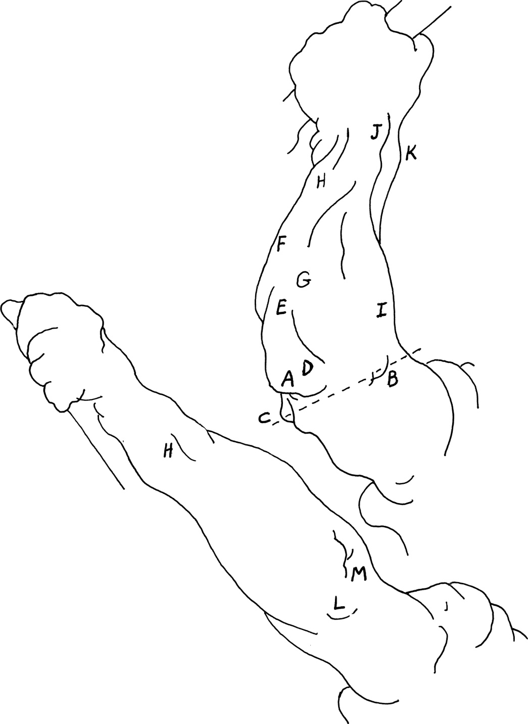
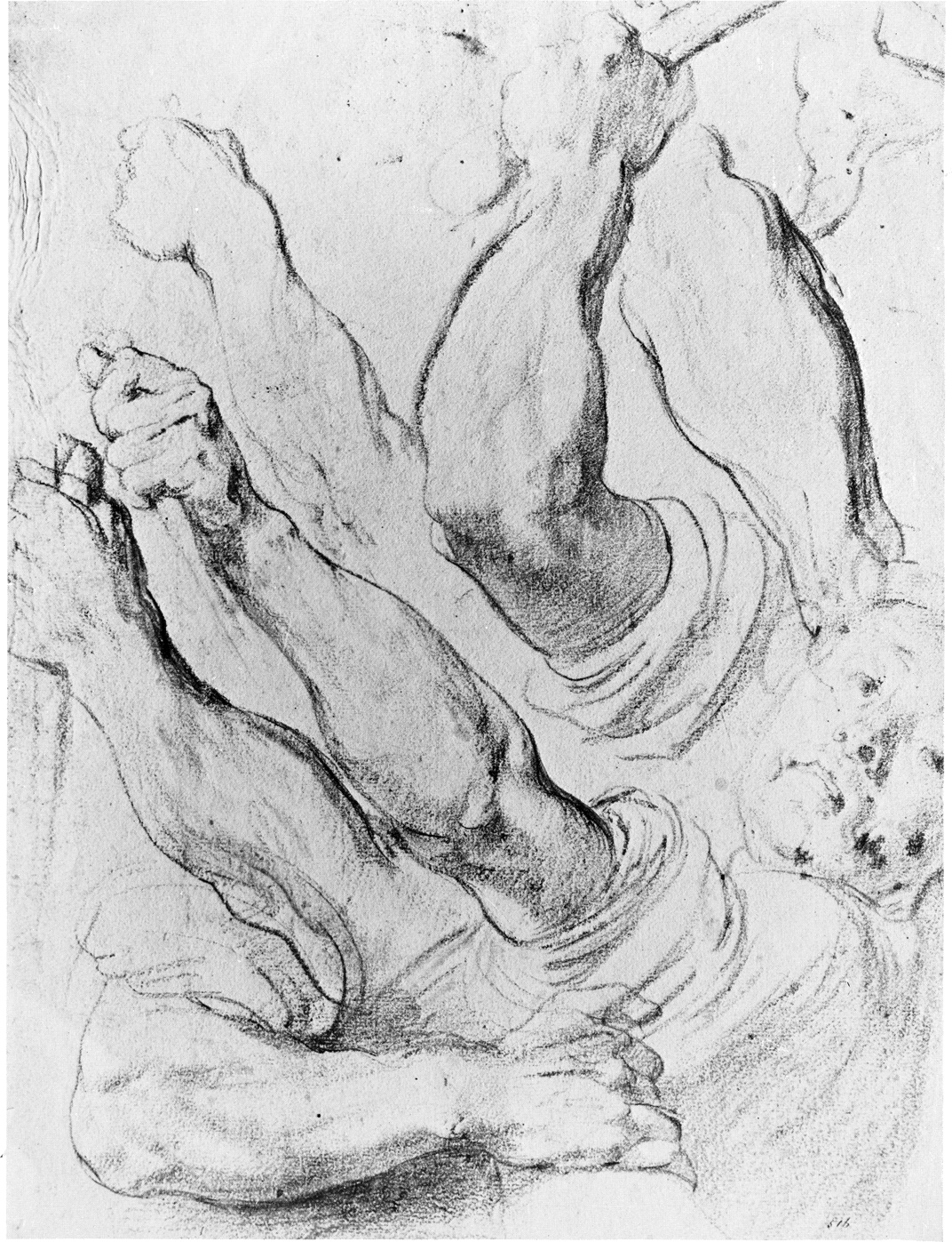
Extension is the opposite of, or antagonistic to, flexion. Its movements make the angles between joints larger rather than smaller. Extensions are straightening or stretching movements rather than bending movements. In this example, the fingers are extended upon the hand, the wrist is extended upon the forearm, and the lower arm is extended upon the upper arm. Extension of the forearm upon the upper arm is produced by the anconeus (A) and triceps brachii (B).
In this back view of the extended arm, you can see three knobs of bone at the elbow: the inner (C) and outer (D) condyles of the humerus, with the olecranon process (E) of the ulna between them (marked by the two little extension wrinkles). You can easily follow the direction of the ulna bone from the olecranon process (E) down to the ulnar furrow (F), which lies between the flexors (G) and extensors (H), to its lower extremity in the styloid process (I) on the little finger side of the wrist.
Like many artists who reduce anatomical forms to simple geometrical shapes in order to clarify their direction in perspective, Cambiaso visualizes the arm and hand as a block or cube. He blocks in the hand simply, starting with the extension fold (J) of the annular ligament of the wrist. From there, the eye is led back into the picture, moving from volume to volume. From the radius (K) at the thumb side of the blocklike wrist, you move back along the mass of the extensor digitorum (L), to the suggestion of the extensor carpi radialis brevis (M), to the two overlapping muscles of the supinator mass (N) that invade the upper arm, and then to the biceps (O), which in turn overlaps the deltoid (P).
Luca Cambiaso (1527-1585)
CAIN AND ABEL
pen and wash drawing
11 1/4″ × 6 1/4″ (286 × 159 mm)
Woodner Family Collection I, New York
In Dürer’s drawing, the arms of the two figures on the right are extended at the elbow, and in supination with the fingers flexed. In this anatomical position, the two bones of the forearm lie side by side. You can verify their position by locating and identifying the superficial landmarks made by them and the muscles that relate to them.
At the wrist, the styloid process of the radius (A) on the thumb side and the styloid process of the ulna (B) opposite give us the lower extremities of these two bones.
The biceps (C) overlook the front of the elbow joint, and the furrows on either side of this muscle converge to a triangular hollow (D) on the front of the elbow. This triangle points downward, its outer side bordered by the supinator mass (E), its inner side by the pronator teres (F), and its base determined by a line drawn between the exterior (G) and the interior (H) condyles of the humerus. This line is not at right angles to the axis of the upper arm, but is lower on the inside. The angle between the axis of the upper and lower arm is called the carrying angle.
By way of its aponeurotic tendon, the biceps produces a shallow oblique furrow (I) in the upper fleshy body of the flexor mass (J). The large forearm muscles—the supinator mass (E) on the outside and the flexor mass (J) on the inside—merge into narrow tendons midway between elbow and wrist. The flexor tendons of the palmaris longus (K) and the flexor carpi radialis (L) stand out at the base of the forearm. If you place your fingers on the flexor carpi radialis, you can feel the beat of the radial artery where the doctor takes your pulse.
Albrecht Dürer (1471-1528)
THREE STUDIES FROM NATURE FOR ADAM’S HAND
engraving
8 1/2″ × 10 13/16″ (216 × 274 mm)
British Museum, London
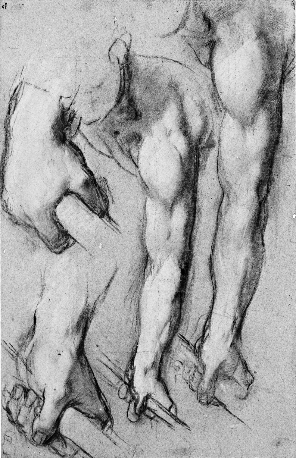
At the back of the arm, the olecranon process (A) of the ulna forms the tip of the elbow. In the flexed arm on the right, it forms the apex of a triangle of three bony protrusions. A construction line between the external (B) and internal (C) condyles of the humerus, forms the base of this triangle of bony landmarks. Rubens has stressed the more prominent internal condyle, and emphasized the protrusion of the olecranon process in this flexed position of the arm.
A greater awareness of shapes can help you more quickly locate bone and muscle forms. The triangular relief of the anconeus (D) originates in the external epicondyle (B) of the humerus and points downward to its insertion (E) into the posterior surface of the ulna. The flexor carpi ulnaris (F) and the extensor carpi ulnaris (G) border the sides of the anconeus (D) and the shaft of the ulna bone, and help create the ulnar furrow (H).
The mass of the supinators (I) swell the upper half of the forearm. The tendon of the extensor digitorum (J) helps you to block in the wrist. The slight swelling at the side indicates the extensors of the thumb (K).
When the arm is extended, the olecranon process (L) moves upward and inward, creating a deep furrow (M) toward the outside. If you extend your arm and place your fingers in this furrow, you will feel the bony mass of the external condyle (B) of the humerus. If you rotate your arm back and forth from supination to pronation, just below the external condyle you will feel the head of the radius articulating with the capitulum or radial head of the humerus at the elbow.
Peter Paul Rubens (1577-1640)
STUDIES FOR A PAINTING OF THE DEATH OF DECIUS MUS
black chalk, heightened with white
15 15/16″ × 12 1/4″ (405 × 310 mm)
Victoria and Albert Museum, London
The most prominent muscular mass of the lateral arm is that of the adjacent spiral muscles, called the supinator longus or brachioradialis (A) and the extensor carpi radialis longus (B). These two spiral muscles are massed together under the name of the supinator group.
The supinator group emerges from between the triceps (C) and brachialis anticus (D) muscles, about a third of the way up the upper arm. It swells to its greatest width just below the external condyle (E) of the humerus. The forearm narrows into tendons of the extensor group (F) at about the middle of the forearm.
Between the bulge of the interior border of the extensor carpi radialis longus (G) and the anconeus (H), you can see the deep furrow of the back of the extended arm at the position of the underlying external condyle and styloid process of the radius (see anatomical plate). This is a larger version of the dimples that are found throughout the body wherever a bone protrudes. You can find such dimples appearing at the sacrum (at the base of the spine) and by the bulge of the great trochanter of the femur (at the side of the hip). The dimple is caused by the bulging of muscles surrounding a bone. The bulging muscles create a prominence, the bone appears as an indent, and the result is a dimple.
Federico Barocci (c. 1535-1612)
STUDIES FOR THE MARTYRDOM OF SAN VITALE
chalk
Staatliche Museen, Berlin
In Dürer’s drawing of the medial aspect of the forearm, the ulnar furrow (A) lies well below the dark shadow of the plane break (B). This plane break follows the mass of the flexor carpi ulnaris (C). The ulnar furrow moves down from the styloid process (D), and separates the mass of the flexors (C) at the front and side of the forearm, from the extensor group (E) at the back.
What more convenient, yet often overlooked, model can you find than your own body? Look carefully in a mirror to see and feel your muscles in action, so they become more than mere words to memorize. Extend and turn your right forearm upward. Place the thick upper portion of your forearm in the palm of your left hand. Clench and relax your right fist, and flex your wrist upon your forearm, trying to touch the front of your arm with your fingers. You will see and feel the contraction and relaxation of the flexors (C), moving around from the ridge of the ulna bone at the back to the front half of the lower arm.
To feel the extensors at the back of your hand, move your wrist as far back as you can. This position is called hyperextension or dorsiflexion. Alternate this movement with a side-to-side movement of the wrist. You will feel the changing forms of the long muscular bundles of the extensors (E) as they contract and expand. The motion created by this muscular movement is transferred to the wrist and fingers by long, cordlike tendons in the lower, more narrow half of the forearm.
Albrecht Dürer (1471-1528)
THE ARM OF EVE
brush and brown ink
13 3/16″ × 10 1/2″ (335 × 267 mm)
Cleveland Museum of Art
Here are examples of two opposite stages in the rotation of the forearm. The right arm of the figure is in pronation, or turned downward, and the left arm that is extended behind the trunk is in supination, or turned upward.
The downward movement of pronation is carried out by two muscles in the front of the forearm, the pronator teres (A) and the pronator quadratus (B). From their inner origins, they pull the radius, together with the hand, across the ulna. The forearm is moved back into supination by the supinator longus (C), the deep supinator brevis, and the biceps (D), pulling in an opposite direction from a different vantage point.
In all muscular actions, the same laws of leverage that apply to simple machines also apply to the human body as it overcomes gravity and inertia. In pronation, the contracting pronator teres (A) and pronator quadratus (B) pull by their tendons on the lever of the radius. The radius pivots on the axis or fulcrum of its joint at the base of the humerus. Against the resistance of gravity and its own weight, the hand is carried inward with the radius as it crosses the ulna.
Giovanni Battista Piazzetta (1683-1754)
ACADEMIC STUDY OF A MALE FIGURE
black chalk on gray paper heightened with white
21 1/8″ × 15 3/16″ (537 × 386 mm)
National Gallery of Canada, Ottawa
Spiral contour lines are linear symbols of rotation. The lines that curve over the extensors of the thumb (A) in the lower forearm suggest that Rubens’ model has just rotated the forearm from a palm-down or pronated position to this palm-upward, supinated position.
The contracting of the extensor digitorum (B), together with the activity at the extensors of the thumb (A), are followed by the spreading of the fingers and thumb. Try these movements on your own forearm and feel the changes in the different masses as you move your thumb and fingers.
Supination is carried out by the supinator longus (C) and its deeper muscle, the supinator brevis. In sudden movements or in supinating against resistance—such as when, in turning the key in your door lock it sticks a little—the biceps (D) comes to the rescue. By their turning action, the supinators and the biceps pull the pronated radius back across the ulna, so that both bones are again parallel with the palm upward. (This is the position your hand would be in if, for example, you were holding a bowl of soup.) The biceps shorten as they contract in supination. You can feel them move if you place one hand on your biceps as you rotate your other hand.
In such simple everyday activities as tightening a screw or turning the cap of a jar, you can experience a practical application of two anatomical factors—that the right hand is usually dominant and that supination is stronger than pronation.
Peter Paul Rubens (1577-1640)
STUDY OF A NUDE MALE TORSO
charcoal and white chalk
12 7/16″ × 14 7/16″ (315 × 367 mm)
Ashmolean Museum, Oxford
In the demiprone position of the forearm, the palm faces inward. This position has been described as the natural position of the forearm, or the position of rest. This is the position that the forearm and hand most naturally assume when you place your hand across your chest. If you drop your arms to your sides, your palm will neither face forward in supination nor backward in pronation, but will face inward in demipronation. This is also the position of greatest mechanical advantage for most functions of the upper limb.
The figure in Pontormo’s drawing is holding a squirming child. The biceps (A) and supinators (B) are contracted and, together with the opposing pronator teres (C), they stabilize the arm in this demipronated position. The flexor mass (D) bulges in contraction as it flexes the wrist and fingers.
Pontormo’s use of several lines of action at the supinator mass (B) suggests agitation of the upper forearm. Below, he breaks the long outline of the inner forearm and flexor mass (E) with an area of contrast to the hard unbroken line. At the same time he fuses values into a single tone in order to unify front and back masses.
Jacopo Pontormo (1494-1556)
YOUNG MAN HOLDING A SMALL CHILD
black chalk
15 3/8″ × 8 15/16″ (389 × 227 mm)
National Gallery of Scotland, Edinburgh
The movement of pronation, or rotating the hand so as to bring the palm downward, is accompanied by some rotation of the humerus, even in the initial stages. In the extreme position of forced pronation, rotation of the humerus is considerable. This inward and forward rotation of the humerus is brought about by the action of the teres major (A) and the latissimus dorsi (B) from the back, and by the pectoralis major from the front all pulling on the anterior or front surface of the humerus.
Notice the change in the form of the lower forearm as it moves from its generally flatter form in supination to a very cylindrical shape in this position of forced pronation. The mass of the supinators (C) follows the radius to the inside of the arm and adds to the thickness of the rounded form of the upper forearm. The bulging of the flexor mass (D) on the opposite side of the forearm is followed by flexion in the wrist and fingers.
Raphael Sanzio (1483-1520)
PRELIMINARY STUDY FOR THE DISPUTÀ
pen and ink
11″ × 16 3/8″ (280 × 415 mm)
Stadelsches Kunstinstitut, Frankfurt-am-Main