Chapter 5:
You will need to have an understanding of the structures and functions of the human body in your work as a medical assistant. In this chapter, you will review the major body systems, how they function in the normal patient, and the key changes that disease and illness cause in the human body.
Listed in the following table are the major body systems, their unique function in maintaining a state of wellness, and the organs that make up each system. All systems are needed to maintain a state of homeostasis for normal body function.
| System | Function | Sample of Involved Organs |
|---|---|---|
| Musculoskeletal | Support and shape/movement | Bones, muscles, tendons, ligaments |
| Nervous/Sensory | Translate input and make decisions | Brain, spinal cord, nerves/sensory organs |
| Circulatory/Cardiovascular | Transportation of oxygen, hormones, and nutrients | Blood vessels, capillaries, heart |
| Endocrine | Turn functions on and off | Glands such as pituitary, thymus, thyroid, adrenal, pancreas |
| Reproductive | Continuation of species, human sexual function | Ovaries, testes, uterus, penis, fallopian tubes, prostate |
| Integumentary | Protection of internal organs | Skin, hair, nails |
| Lymphatic | Protection from infection | Lymph vessels, spleen, tonsils |
| Digestive | Intake and absorption of nutrients | Mouth, teeth, esophagus, stomach, intestines, liver, gall bladder, pancreas |
| Respiratory | Intake of oxygen and output of waste | Mouth, nose, trachea, bronchi, lungs, alveoli |
| Excretory | Output of waste | Kidney, ureter, urethra, bladder |
The human body is made up of 10 body systems, each of which has a specific function. Each body system is made up of organs that work together toward that function. Each organ is made of various tissues. Tissues are made of cells. The smallest unit of the body is the cell, which is visible only under a microscope. Cells are made up of organelles.
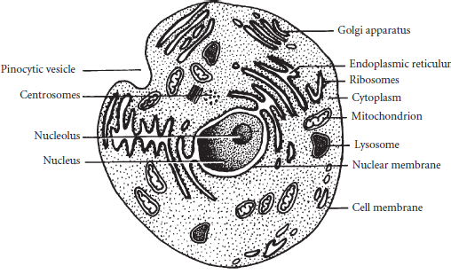
The typical cell has a variety of organelles that carry out the processes necessary for cellular life. Every cell has a covering called the cell membrane. Each cell has an individual shape and is filled with a colloidal filling called cytoplasm. The key organelle in the cell is the nucleus. It is where the genetic material resides and proteins are made. These proteins are called ribosomes. They are responsible for the growth and development of the cell. They move away from the nucleus and out into the cell with the assistance of a folded membrane called the endoplasmic reticulum. Energy for this and other operations in the cell is produced by mitochondria. The mighty mitochondria are the cell’s power plants. These multifunctional units move throughout the entire cell to devour foreign particles or other dead organelles. They move to the edges of the cell and open up the cell membrane to complete pinocytosis (cell drinking) and phagocytosis (cell eating).
Like people, cells move. The Golgi apparatus (also called as Golgi body) is another folded membrane that is used for transportation within the cell. The tips of a Golgi body pinch off and transport mucus through the cytoplasm. Cilia are hair-like appendages that wave back and forth pushing along secretions or particles. Flagella are tail-like appendages that whip through fluids much like swim fins and help propel the cell. A sperm cell is a good example of a cell with flagella.
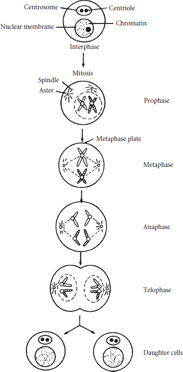
Reproduction in the cell is carried out through the centriole. When reproduction takes place, the centriole separates and each part goes to an opposite end of the cell. Spindle fibers form between the two centriole pieces and chromosomes gravitate to either end of the cell. Gradually, the cell divides into two identical daughter cells. This process is called mitosis.
The organs of the body can be comprised of four layers of tissues. The layers are described in Table 5.2.
| Type | Function | Description |
|---|---|---|
| Epithelial | Covers and lines | Found as skin, glands, and lining of organs |
| Connective | Joins together | Identified by type of matrix:
|
| Muscle | Contracts | Types:
|
| Nervous | Carries electric potential | Neurons pick up potential and pass it on to subsequent neurons |
There are times when aberrant tissue growth appears. That tissue is called a neoplasm or tumor. If the tissue does not spread beyond the original site, it is benign. However, if the abnormal tissue growth spreads to other areas of the body, the neoplasm is malignant. Malignant epithelial tissue is called a carcinoma. Malignant connective tissue is called a sarcoma. Cancer is detected through tests such as MRI (magnetic resonance imaging), CT scan (computer tomography), lab tests such as PSA (prostatic specific antigen), or biopsy and cell studies (pap smear). Treatment for cancer may require chemotherapy, radiation, surgery, or immunotherapy. The aim of treatment is to destroy the aberrant cell growth.
These seven warning signs of cancer should alert a person to seek a physician’s assessment:
To communicate with other members of the health care team, health care professionals use standard terms to describe the body. Anatomical position is the standard visualization of the human body to delineate direction and relative position of body planes.
| Term | Meaning | Example |
|---|---|---|
| Superior | Above | The nose is superior to the chin |
| Inferior | Below | The lips are inferior to the nose |
| Ventral/Anterior | Belly side | The breasts and knees are on the ventral/anterior side of the body |
| Dorsal/Posterior | Back side | The buttocks and knuckles are on the dorsal/posterior side of the body |
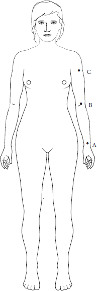
| Distal | Greatest distance away from a point of insertion | Lesion A is distal from the shoulder |
| Proximal | Closest to a point of insertion | Lesion C is proximal to the shoulder |
| Lateral | Away from the midline | The ears are lateral to the nose |
| Medial | Toward the midline | The nose is medial to the ears |
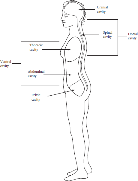
The body is often divided into body cavities.
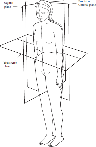
Additionally, the body is divided into planes. Pictures and views of the body are presented through these planes to show different organs.
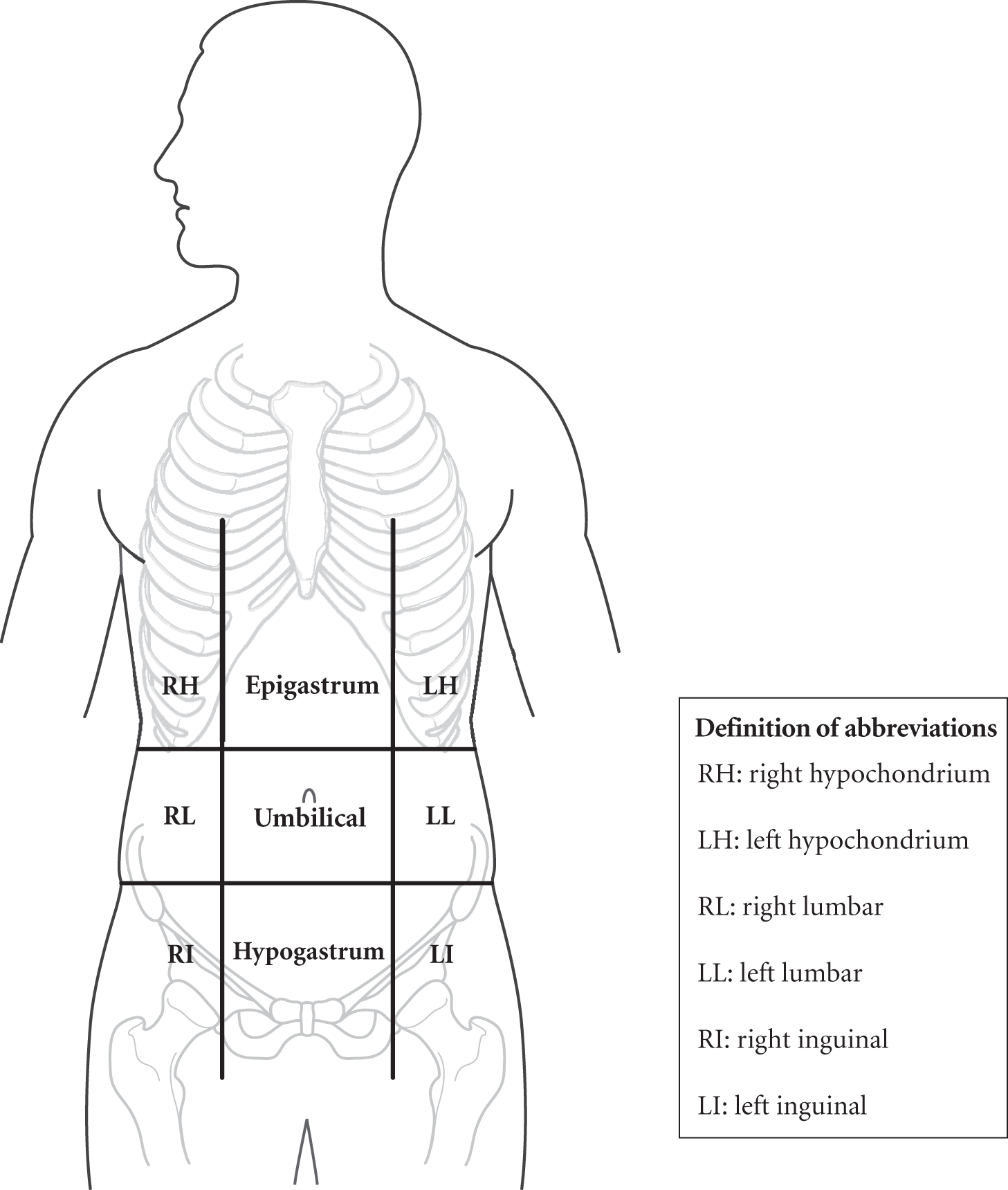
The abdomen is divided into regions to help identify the location of pain or lesions. Remember right and left are always oriented from the patient’s perspective.