39 Nervi Craniales
Nervi Craniales: Overview
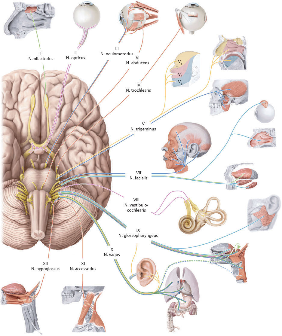
Fig. 39.1 Nervi craniales
Inferior (basal) view. The 12 pairs of nn. craniales (CN) are numbered according to the order of their emergence from the truncus encephali. Note: The sensory and motor fibers of the nn. craniales enter and exit the truncus encephali at the same sites (in contrast to nn. spinales, whose sensory and motor fibers enter and leave through radices posterior and anterior, respectively).
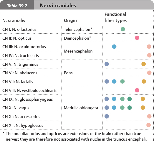
 The nn. craniales contain both afferent (sensory) and efferent (motor) axons that belong to either the somatic or the autonomic (visceral) nervous system (see pp. 682–683). The neurofibrae somaticae allow interaction with the environment, whereas the neurofibrae autonomicae regulate the autonomic activity of internal organs. In addition to the general fiber types, the nn. cranialis may contain special fiber types associated with particular structures (e.g., auditory apparatus and caliculi gustatorii). The neurofibrae craniales originate or terminate at specific nuclei, which are similarly classified as either general or special, somatic or visceral, and afferent or efferent.
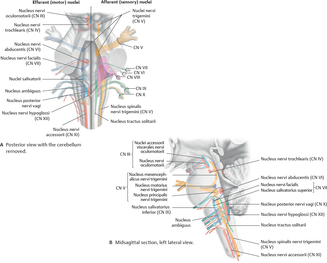
Fig. 39.2 Nuclei nervorum cranialium
The sensory and motor fibers of nn. craniales III to XII originate and terminate in the truncus encephali at specific nuclei.
CN I & II: Nervus Olfactorius & Nervus Opticus
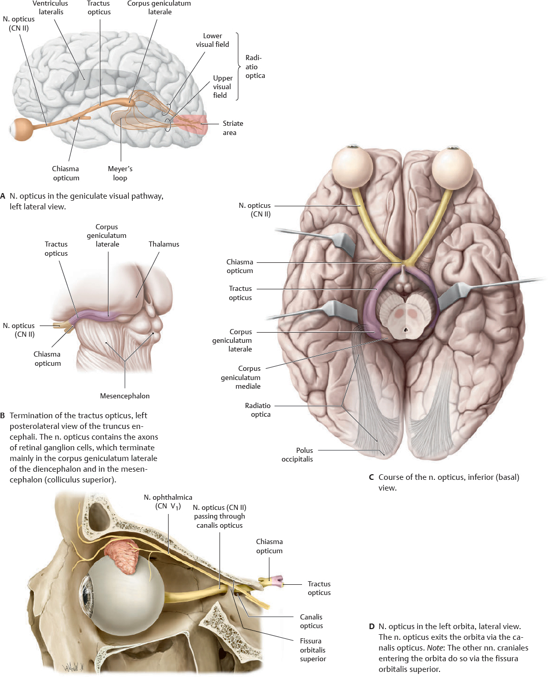
 The nn. olfactorius and opticus are not true peripheral nerves but extensions (tracts) of the telencephalon and diencephalon, respectively. They are therefore not associated with nuclei nervorum cranialium in the truncus encephali.
Fig. 39.3 Nervus olfactorius (CN I)
Fiber bundles in the pars olfactoria tunicae mucosae nasi pass from the cavitas nasi through the lamina cribrosa of the os ethmoidalis into the fossa cranii anterior, where they synapse in the bulbus olfactorius. Axons from second-order afferent neurons in the bulbus olfactorius pass through the tractus olfactorius and stria olfactoria medialis or lateralis, terminating in the cortex cerebri of the prepiriform area, in the corpus amygdaloideum, or in neighboring areas.
Fig. 39.4 Nervus opticus (CN II)
The n. opticus passes from the bulbus oculi through the canalis opticus into the fossa cranii media. The two nn. optici join below the base of the diencephalon to form the chiasma opticum, before dividing into the two tractus optici. Each of these tracts divides into a radix lateralis and radix medialis. Many retinal cell ganglion axons cross the midline to the contralateral side of the brain in the chiasma opticum.
CN III, IV & VI: Nervus Oculomotorius, Nervus Trochlearis & Nervus Abducens
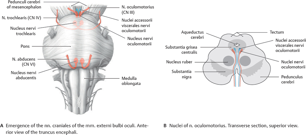
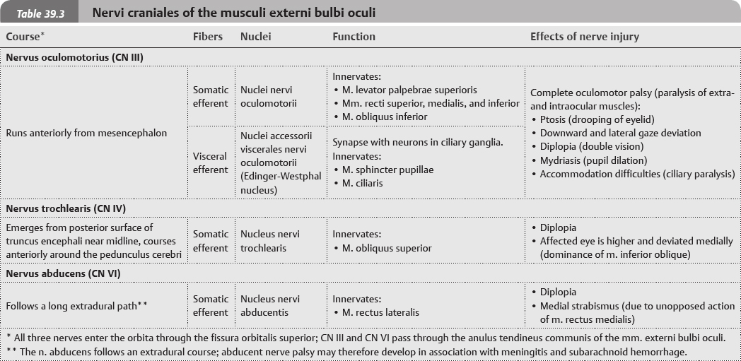
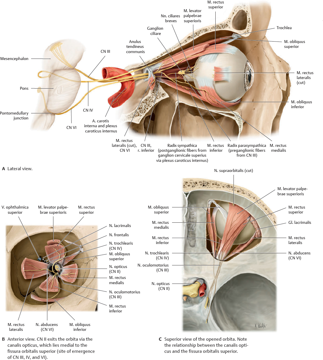
 Nn. craniales III, IV, and VI innervate the extraocular muscles (see p. 569). Of the three, only the n. oculomotorius (CN III) contains both somatic and visceral efferent fibers; it is also the only n. cranialis of the mm. externi bulbi oculi to innervate multiple extra-and intraocular muscles.
Fig. 39.5 Nuclei of the nn. oculomotorius, trochlearis, and n. abducens
The n. trochlearis (CN IV) is the only n. cranialis in which all the fibers cross to the opposite side. It is also the only n. cranialis to emerge from the dorsal side of the truncus encephali and, consequently, has the longest intradural (intracranial) course of any n. cranialis.
 Note: The n. oculomotorius supplies parasympathetic innervation to the intraocular muscles and somatic motor innervation to most of the mm. externi bulbi oculi (also the m. levator palpebrae superioris). Its parasympathetic fibers synapse in the ganglion ciliare. Oculomotor nerve palsy may affect exclusively the parasympathetic or somatic fibers, or both concurrently.
Fig. 39.6 Course of the nerves innervating the musculi externi bulbi oculi
Right orbita.
CN V: Nervus Trigeminus
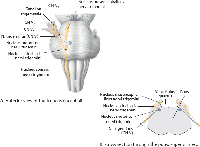
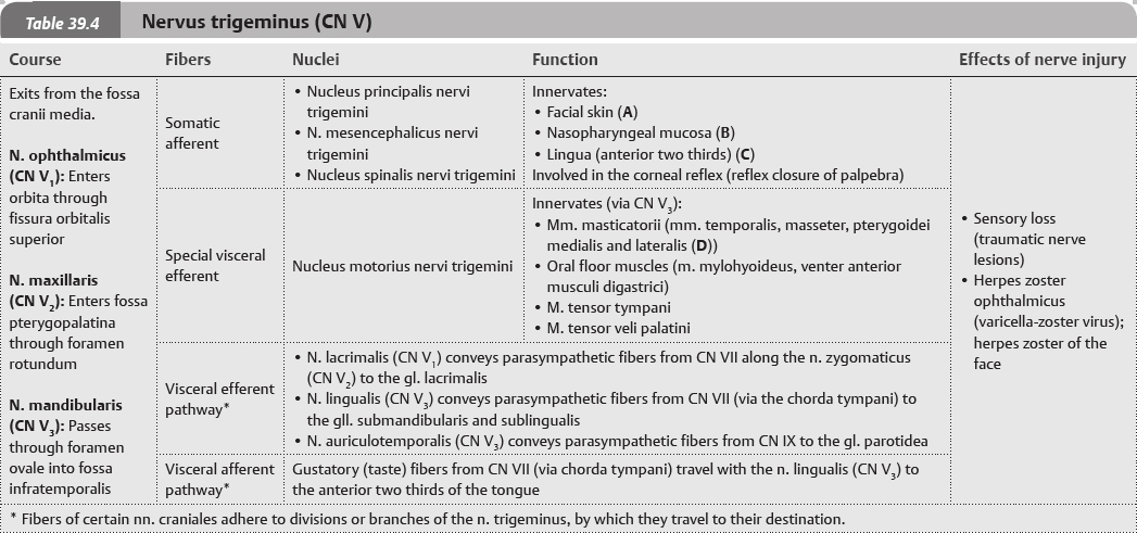
 The n. trigeminus, the sensory nerve of the head, has three somatic afferent nuclei: the nucleus mesencephalicus nervi trigemini, which receives proprioceptive fibers from the mm. masticatorii; the nucleus principalis nervi trigemini, which chiefly mediates touch; and the nucleus spinalis nervi trigemini, which mediates pain and temperature sensation. The nucleus motorius nervi trigemini supplies motor innervation to the mm. masticatorii.
Fig. 39.7 Nuclei nervi trigemini
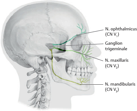
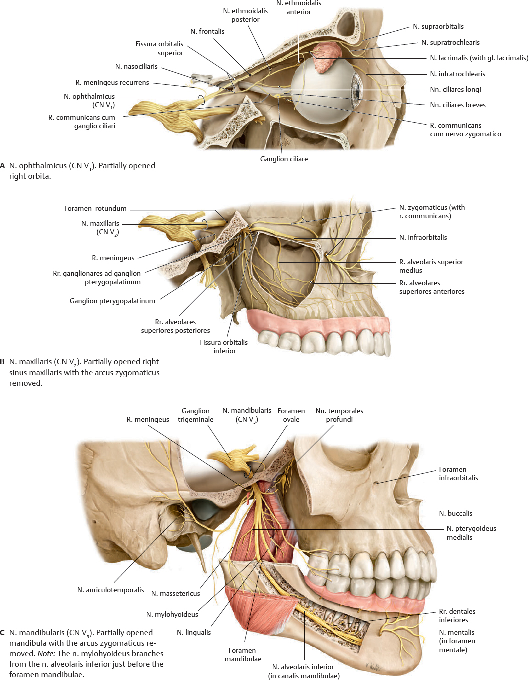
Fig. 39.8 Divisions of the nervus trigeminus (CN V)
Right lateral view.
Fig. 39.9 Course of the nervus trigeminus divisions
Right lateral view.
CN VII: Nervus Facialis
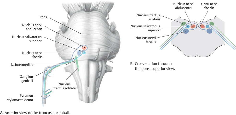
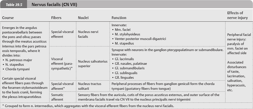
 The n. facialis mainly conveys special visceral efferent (branchiogenic) fibers from the nucleus nervi facialis to the mm. faciei. The other visceral efferent (para-sympathetic) fibers from the nucleus salivatorius superior are grouped with the visceral afferent (gustatory) fibers to form the n. intermedius.
Fig. 39.10 Nuclei nervi facialis
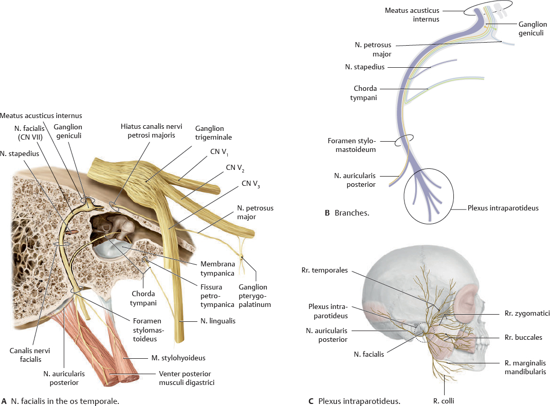
Fig. 39.11 Branches of the nervus facialis
Right lateral view.
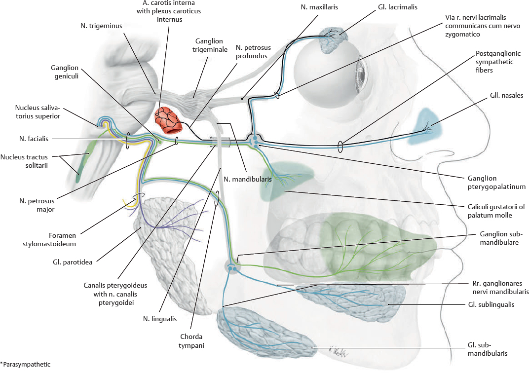
Fig. 39.12 Course of the nervus facialis
Right lateral view. Visceral efferent (parasympathetic) and special visceral afferent (taste) fibers shown in black.
CN VIII: Nervus Vestibulocochlearis
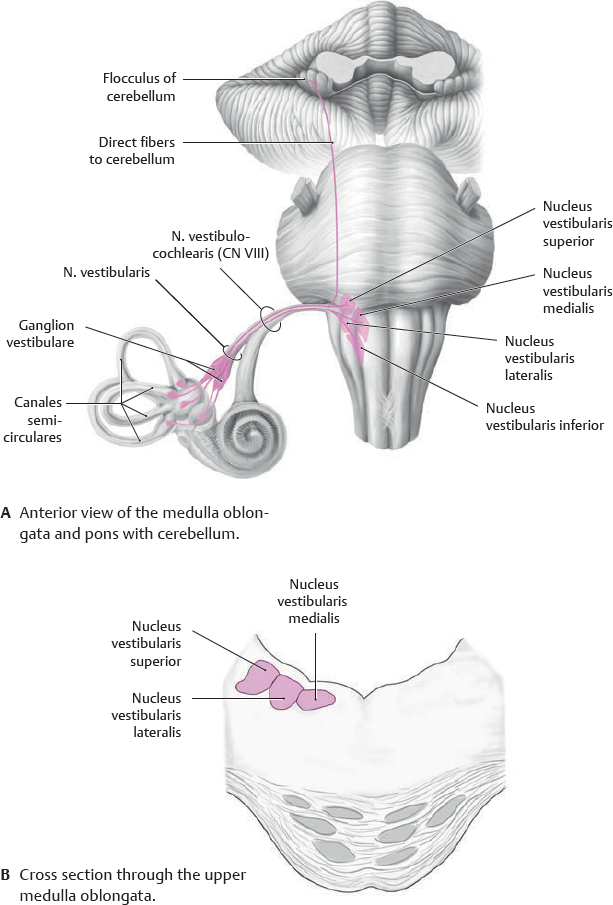
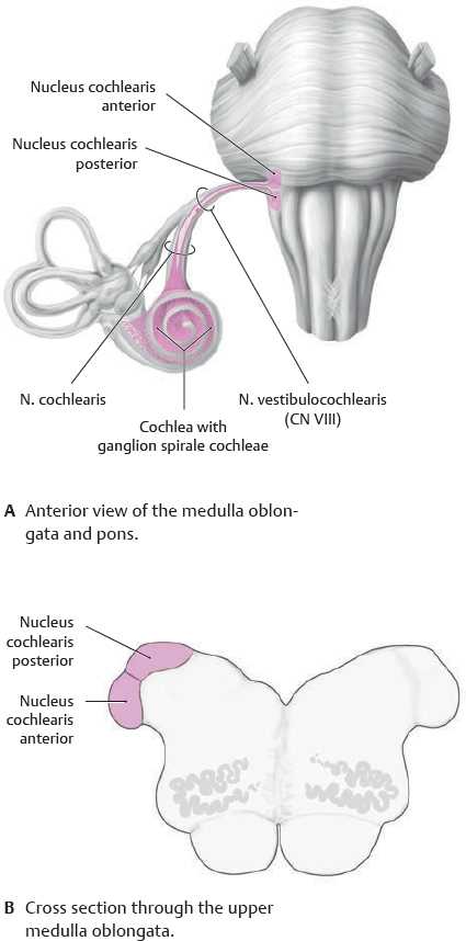
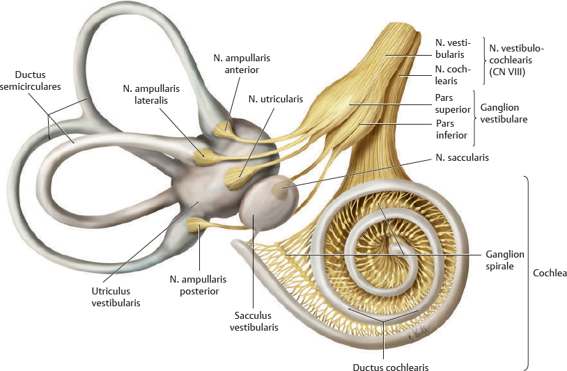
 The n. vestibulocochlearis is a special somatic afferent nerve that consists of two roots. The n. vestibularis transmits impulses from the vestibular apparatus; the n. cochlearis transmits impulses from the auditory apparatus.
Fig. 39.13 Nervus vestibulocochlearis: Nervus vestibularis
Fig. 39.14 Nervus vestibulocochlearis: Nervus cochlearis
Fig. 39.15 Ganglion vestibulare and ganglion spirale cochleae
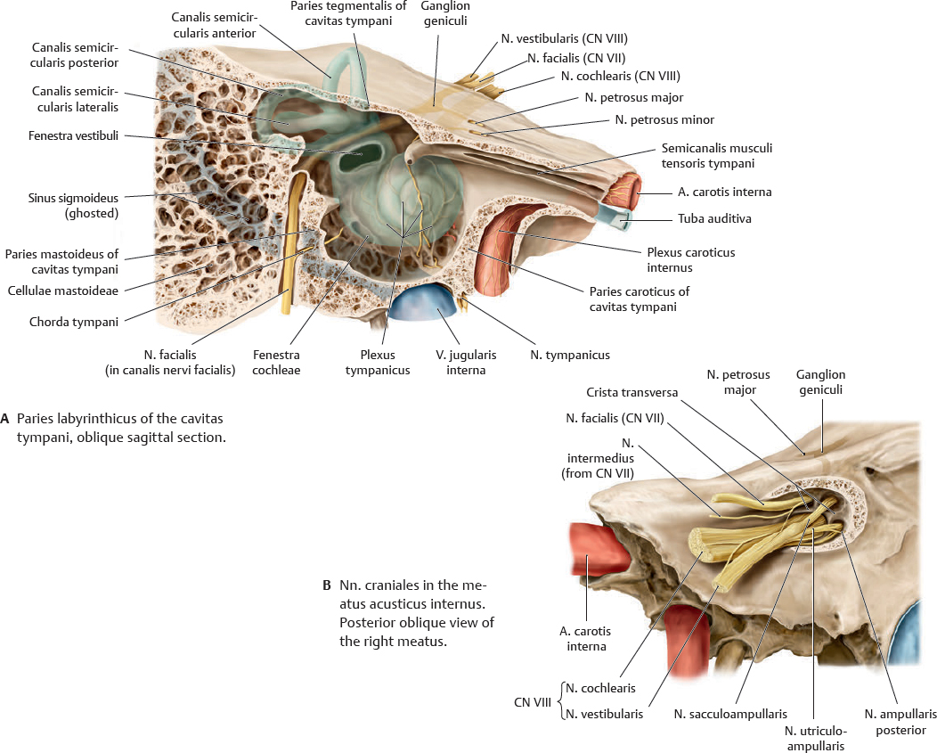
Note: The nn. vestibularis and cochlearis are still separate structures in the pars petrosa ossis temporalis.
Fig. 39.16 Nervus vestibulocochlearis in the os temporale
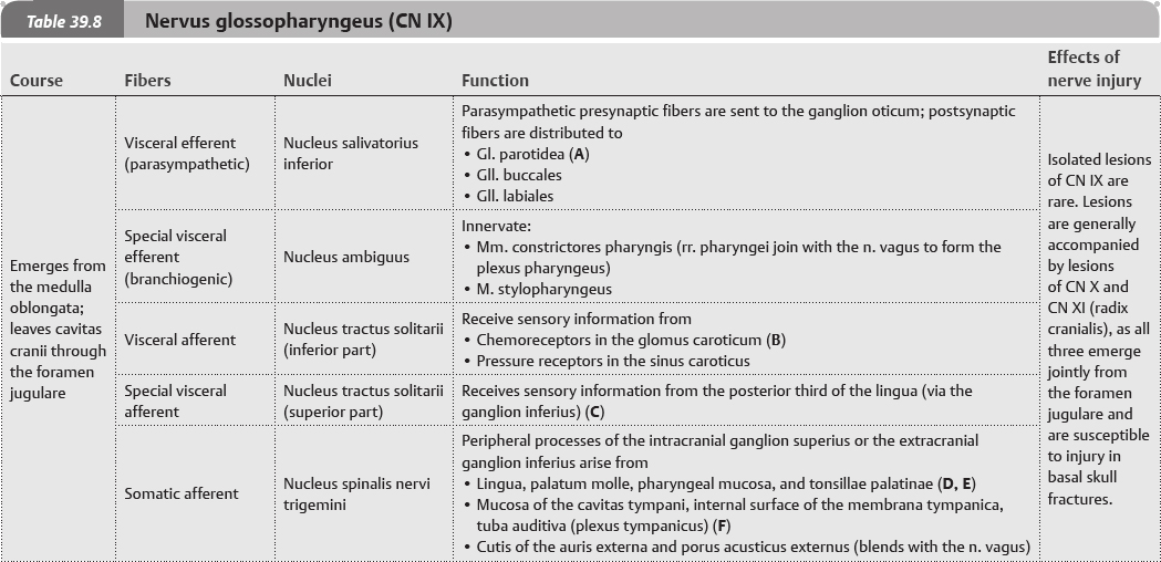
CN IX: Nervus Glossopharyngeus
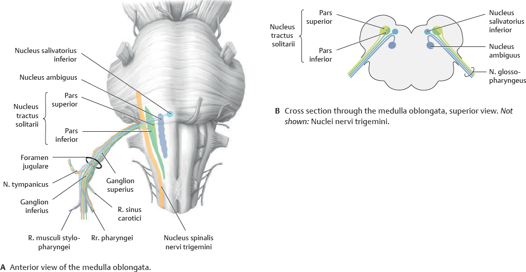
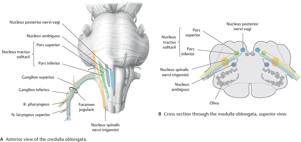
Fig. 39.17 Nuclei nervi glossopharyngei
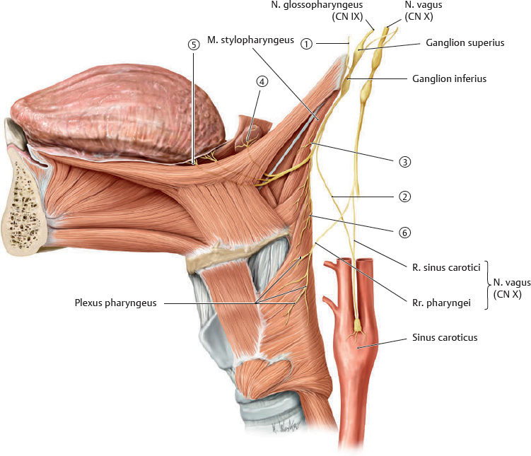
Fig. 39.18 Course of the nervus glossopharyngeus
Left lateral view. Note: Fibers from the n. vagus (CN X) combine with fibers from the n. glossopharyngeus (CN IX) to form the plexus pharyngeus and supply the sinus caroticus.
Table 39.7 Branches of nervus glossopharyngeus

|
N. tympanicus |

|
R. sinus carotici |

|
R. musculi stylopharyngei |

|
Rr. tonsillares |

|
Rr. linguales |

|
Rr. pharyngei |
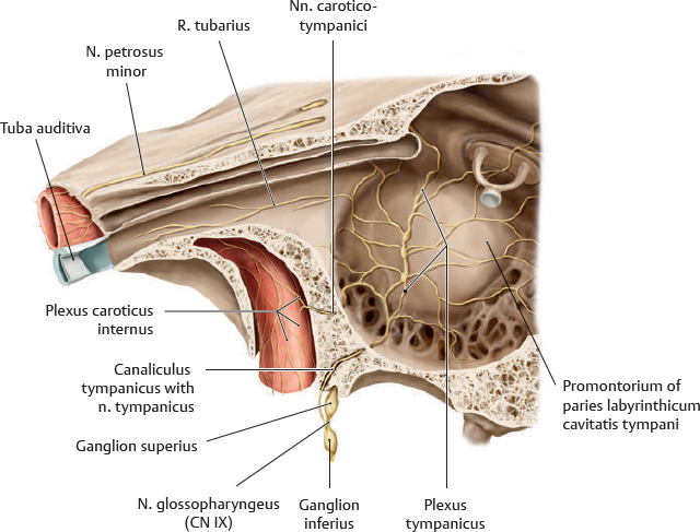
Fig. 39.19 Nervus glossopharyngeus in the cavitas tympani
Left anterolateral view. The n. tympanicus contains visceral efferent (presynaptic parasympathetic) fibers for the ganglion oticum, as well as somatic afferent fibers for the cavitas tympani and tuba auditiva. It joins with sympathetic fibers from the plexus caroticus internus (via the nn. caroticotympanici) to form the plexus tympanicus.
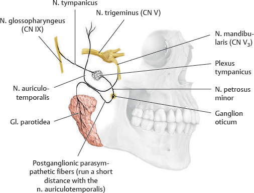
Fig. 39.20 Visceral efferent (parasympathetic) fibers of CN IX
CN X: Nervus Vagus
Fig. 39.21 Nuclei nervi vagi
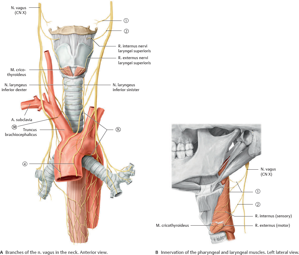
Fig. 39.22 Course of the nervus vagus
The n. vagus gives off four major branches in the neck. The nervi laryngei inferiores are the terminal branches of the nn. laryngei recurrentes. Note: The left n. laryngeus recurrens hooks around the arcus aortae, while the right n. laryngeus recurrens hooks around the a. subclavia.
Table 39.10 Branches of nervus vagus in the neck

|
R. pharyngeus |

|
N. laryngeus superior |

|
N. laryngeus recurrens dexter |

|
N. laryngeus recurrens sinister |

|
R. cardiaci cervicales |
CN XI & XII: Nervus Accessorius & Nervus Hypoglossus
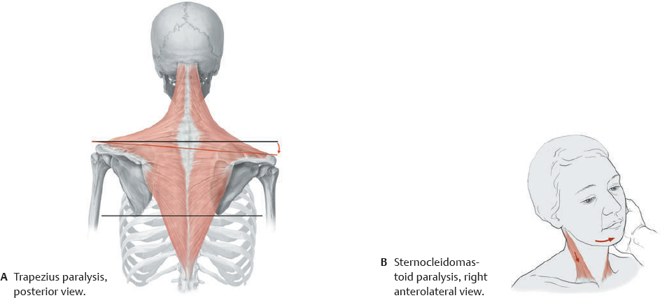
 The traditional “radix cranialis” of the n. accessorius (CN XI) is now considered a part of the n. vagus (CN X) that travels with the radix spinalis for a short distance before splitting. The radix cranialis fibers are distributed via the n. vagus while the radix spinalis fibers continue on as the n. accessorius (CN XI).
Fig. 39.23 Nervus accessorius
Posterior view of the truncus encephali with the cerebellum removed. Note: For didactic reasons, the muscles are displayed from the right side.
Fig. 39.24 Nervus accessorius lesions
Lesion of the right n. accessorius.
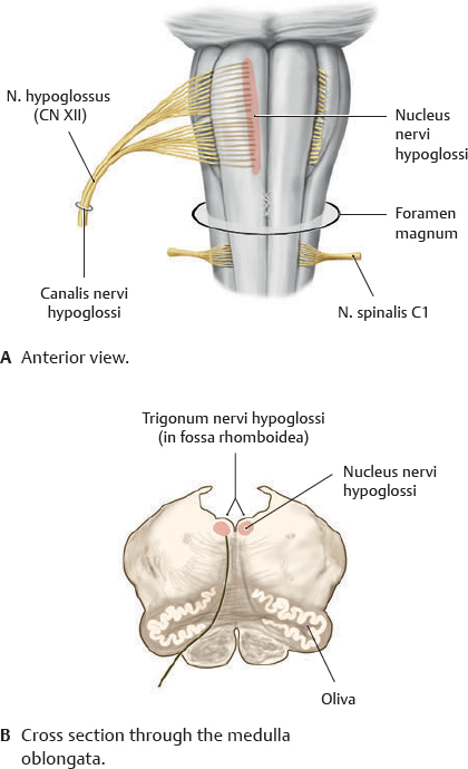
Fig. 39.25 Nervus hypoglossus
Posterior view of the truncus encephali with the cerebellum removed. Note: N. spinalis C1, which innervates the mm. thyrohyoideus and geniohyoideus, runs briefly with the n. hypoglossus.
Fig. 39.26 Nuclei of the nervus hypoglossus
Note: The nucleus nervi hypoglossi is innervated by cortical neurons from the contralateral side.
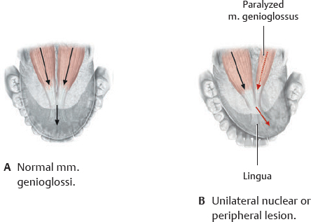
Fig. 39.27 Nervus hypoglossus lesions
Superior view.
Autonomic Innervation
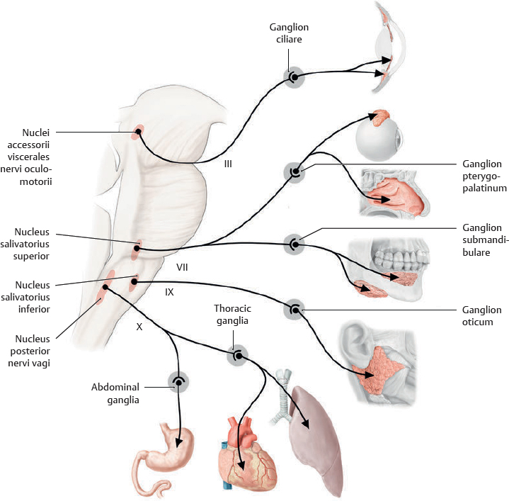
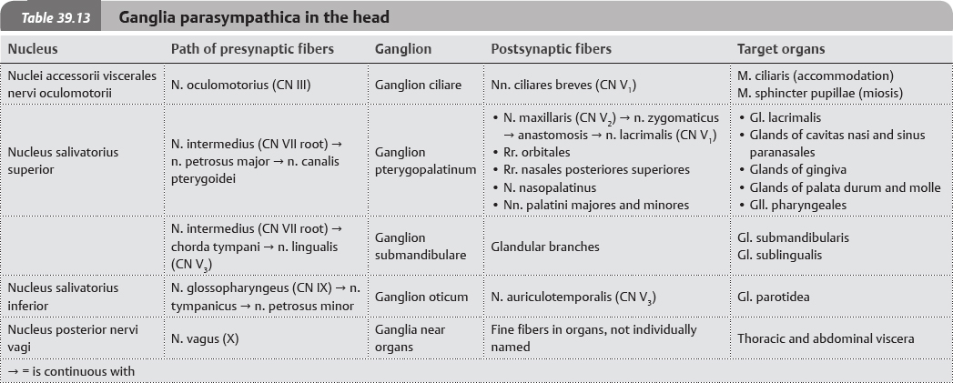
Fig. 39.28 Pars parasympathica divisionis autonomica (pars cranialis): Overview
There are four parasympathetic nuclei in the truncus encephali. The visceral efferent fibers of these nuclei travel along particular nn. craniales, listed below.
- Nuclei accessorii viscerales nervi oculomotorii (Edinger-Westphal nucleus): n. oculomotorius (CN III)
- Nucleus salivatorius superior: n. facialis (CN VII)
- Nucleus salivatorius inferior: n. glossopharyngeus (CN IX)
- Nucleus posterior nervi vagi: n. vagus (CN X)
The presynaptic parasympathetic fibers often travel with multiple nn. craniales to reach their target organs. The n. vagus supplies all of the thoracic and abdominal organs as far as a point near the flexura coli sinistra.
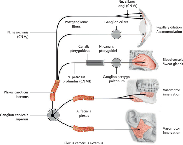
Note: The sympathetic fibers to the head travel along the arteries to their target organs.
Fig. 39.29 Sympathetic innervation of the head
Sympathetic preganglionic neurons of the head originate in the cornu laterale of the medulla spinalis (TI–L2). They exit into the truncus sympathicus and ascend to synapse in the ganglion cervicale superius. Post-ganglionic neurons then travel with arterial plexuses. Postganglionic fibers that travel with the plexus caroticus internus (on the a. carotis interna) join with the n. nasociliaris (of CN V1) and then the nn. ciliares longi to reach the m. dilatator pupillae (pupillary dilation); other post-ganglionic fibers travel through the ganglion ciliare (without synapsing) to reach the m. ciliaris (accommodation). Still other postganglionic fibers from the plexus caroticus internus leave with the n. petrosus profundus which joins with the n. petrosus major (CN VII), to form the n. canalis pterygoidei (vidian nerve). This nerve travels to the ganglion pterygopalatinum where it distributes fibers via branches of the n. maxillaris to the glands of the cavitas nasi, sinus maxillaris, palata durum and molle, gingiva, and pharynx, and to sweat glands and blood vessels in the head. Postganglionic fibers from the ganglion cervicale superius that travel with the a. facialis plexus pass through the ganglion submandibulare (without synapsing) to the gll. submandibularis and sublingualis. Other post-ganglionic fibers travel with the a. meningea media plexus, through the ganglion oticum (without synapsing), to the gl. parotidea.
 The nn. craniales contain both afferent (sensory) and efferent (motor) axons that belong to either the somatic or the autonomic (visceral) nervous system (see
