15.1 Introduction
Systemic lupus erythematosus (SLE) is a chronic inflammatory condition that can affect various organ systems. The version of this disorder, which occurs in childhood, is labeled as childhood-onset systemic lupus erythematous (cSLE).
15.2 Epidemiology
Childhood-onset SLE (cSLE) has its peak incidence in early adolescence. The average age of presentation of cSLE is 12–14 years. M:F ratio is 1:5 prior to puberty. Following puberty, the ratio is 1:9. Although no registry exists, estimates of the incidence are typically quoted as 3.3–8.8 per 100,000 individuals. It has been noted that cSLE affects children of Black, Hispanic, and Asian ancestry more often, and they have a worse prognosis.
15.3 Clinical Findings

SLE 3 Erythema of the malar areas and nose

DLE 1 Somewhat annular lesion with scale, hypopigmentation, and atrophy
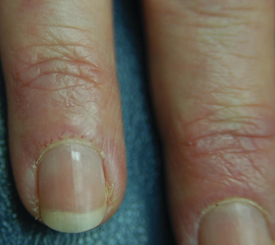
Cuticles show dilated capillaries

(a) Scle Erythematous scaly patches of the back. (b) Scle2 Scaly patch in close up
15.4 Laboratory
In 99% of children with cSLE, the antinuclear antibody (ANA) is positive. If the titer is greater than 1:1280 or higher, cSLE is a likely diagnosis.
The anti-dsDNA antibodies can be used to monitor disease activity. The anti-dsDNA and cardiolipin antibodies are more frequent in cSLE as compared to aSLE (adult-onset SLE); however, a positive rheumatoid factor is less common.
Urinalysis is an important tool to evaluate any renal involvement that may develop.
15.5 Treatment
Treatment options for cSLE include oral hydroxychloroquine, glucocorticosteroids, and other immunosuppressants, including biologics. The cutaneous lesions may be treated with topical or intralesional steroids or with topical calcineurin inhibitors.
15.6 Prognosis
Adolescents with cSLE have a good prognosis when there is no renal involvement. Those adolescents with renal involvement require more aggressive therapies and close follow-up.